Endodontic Management of Bilateral Radix Entomolaris Third Root- a Rare Endoexperience: A Case Report
Department of Conservative Dentistry & Endodontics, K.D. Dental College & Hospital, Mathura
ABSTRACT
Success of endodontic treatment depends on the proper identification of all the canals, thorough chemomechanical preparation followed by three-dimensional obturation with hermetic seal. Failure of any of these steps may occur due to unusual tooth morphology. Usually mandibular molars have two roots with three canals but in few teeth, the number of roots and canals vary. The variation in the number of roots, if extra root located lingually called radix entomolaris (RE) or located bucally called radix paramolaris (RP). This article presents successful endodontic treatment of bilateral mandibular first molars with extra root with radix entomolaris which is a rare microstructures.
Keywords: Mandibular first molar, Anatomical variations, Endodontic treatment, Radix endomolaris.
INTRODUCTION
A thorough knowledge of dental anatomy and an understanding of the potential for variations from the normal are required to achieve success in endodontics. Incomplete instrumentation and cleaning of the root canal space and faulty obturation are the main reasons for failure of endodontic treatment. Root canals are often left untreated because the operator fails to recognize their presence, especially in teeth exhibiting anatomic irregularities or accessory or aberrant root canals.1,2
Anatomical variations are an acknowledged characteristic of mandibular permanent molars. Permanent mandibular first molars usually have 2 roots placed mesially and distally and 3 root canals, but variations in the number of roots and in canal morphology are not uncommon. The presence of a third root in the permanent first molar is the major variant in this group.3
This additional third root, first mentioned in the literature by Carabelli (1844), is called the radix entomolaris (RE), located distolingually in the mandibular molars, mainly first molars. An additional root at the mesiobuccal side is called the radix paramolaris (RP).4 The permanent mandibular first molar is the earliest permanent posterior tooth to erupt, responsible for development of occlusion and important physiologic functions like chewing. Commonly, it is the most frequently in need of endodontic treatment. Thus, it is of utmost importance that the clinician be familiar with variations in the root and root canal anatomy of the mandibular first molar.5,6
The frequency of radix entomolaris is less than 5% in Caucasian, African, Eurasian, and Indian populations, whereas its bilateral occurrence is less than 2.19%. This extra root is typically smaller and more sharply curved than the distobuccal (DB) root, requiring special attention when root canal treatment is being considered.
CBCT is a noninvasive technique to determine the occurrence of distolingual root and reveals the true nature of microstructure three-dimensionally and its curvature and angulation.7,8 CBCT shows the exact position of distolingual root and hence it helps in tracking the curvature and prevents iatrogenic event that might occur in relation to canal curvature like instrument separation, perforation, ledge formation, and so forth.
CASE PRESENTATION
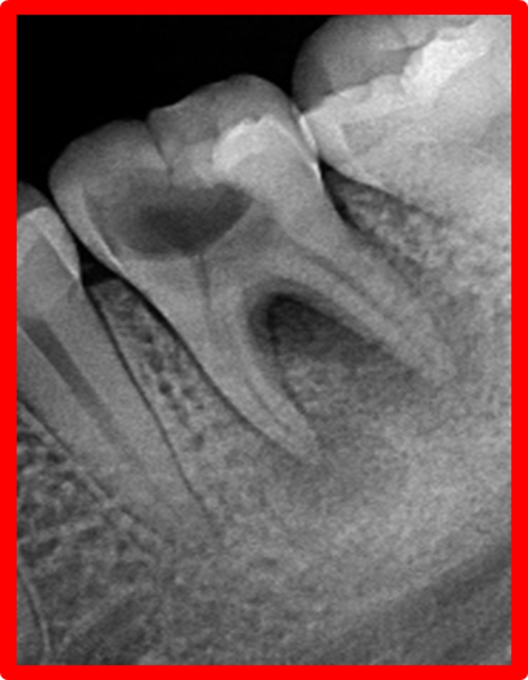
A 27-year-old male patient reported to the Department of Conservative Dentistry and Endodontics, K.D.Dental College and Hospital, with the chief complaint of pain in lower right and left back tooth region for last five days. The pain was aggravated on mastication. The clinical examination revealed the carious right and left mandibular first molars. His medical history was noncontributory. Intraoral periapical radiograph of #46 & #36 (Figure 1) revealed deep carious lesion involving the pulp with periapical radiolucency at the root apices and presence of an extra root in #46 & #36. Based on clinical and radiographic examination, diagnosis of acute apical abscess with #46 and symptomatic irreversible pulpitis with acute apical periodontitis of #36 was established.
A cone beam computerized tomography was advised to confirm the diagnosis of distolingual root. CBCT (Planmeca Promax, Planmeca, Finland) was done and determined the exact position, angle, and curvature of distolingual root of the permanent mandibular first molars. The morphology of teeth was obtained in sagittal (Figure 7), axial (Figure 8), curve slicing and coronal section.
With informed consent, after adequate anaesthesia and isolation with rubber dam, access cavity was prepared using endoaccess kit in the mandibular right first molar. The cavity design was slightly modified by extending more distolingually using ultrasonic tips for exploring extra distal canal orifice (Figure 5). Working length was determined radigraphically (Figure 2). Cleaning and shaping of canals was performed with protaper rotary instruments in a hybrid manner upto F1 (Dentsply Maillefer,Switzerland). Glyde lubricant (Dentsply Maillefer,Switzerland) was used and the canals were irrigated using sodium hypochlorite solution (Prevest Denpro Limited, India) and normal saline solution (Prevest Denpro Limited, India) and final irrigation was done with chlorhexidine (Septodont Parcan, India). Calcium hydroxide (ApexCal, IvoclarVivadent) was used as an intracanal medicament and closed dressing was given for 1 week.
In second visit, the canals were irrigated and dried. Obturation was done by cold lateral compaction technique using guttapercha and AH Plus sealer (DentsplyMaillefer, Switzerland). Post obturation restoration was done with silver amalgam (Figure 4).
After completion of treatment of #46 root canal treatment was also performed on #36 in the same manner as of #46 done.
Postoperative instructions were given to the patient and then both the molars were prosthetically rehabilitated with porcelain fused metal crowns.
The patient was followed up at 6 months and 1 year. The patient was completely asymptomatic and radiographic examination showed absence of any periapical lesion, suggestive of favourable treatment outcome.
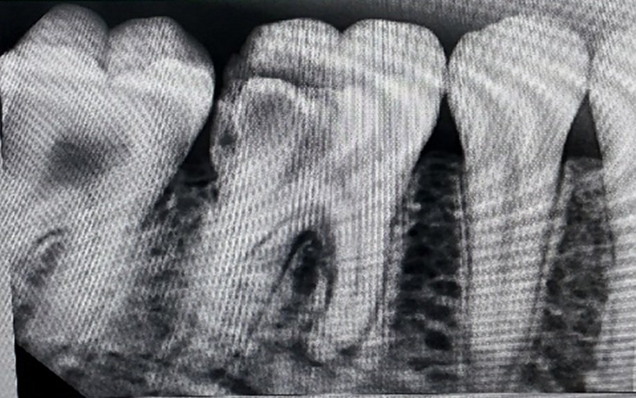

Figure 1:- IOPA of #46 & #36


Figure 2:- Working length of #46 & #36
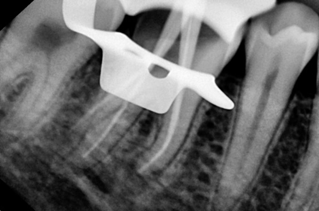

Figure 3:- Mastercone of #46 & #36


Figure 4:- Post obturation and with prosthesis in respect to #46 and #36.


Figure 5 : Access opening showing root canal orifice of #46 and #36.

Figure 6 : 3D images of #46 and #36.

Figure 7 : Saggital cross section of #46 and #36.

Figure 8 : Axial cross section of #46 and #36.
DISCUSSION
The success of endodontic therapy depends on the root canal morphology to some extent, so the clinician should have a thorough understanding of normal anatomy as well as its unusual anatomical configurations. The root canals of the mandibular permanent first molars have several typical anatomical features and anomalies. The presence of four canals is relatively frequent9, but the presence of two distal roots is uncommon and bilateral presence of three rooted mandibular molar is a rare case. This additional third root, first mentioned in the literature by Carabelli (1844), is called the radix entomolaris (RE), located distolingually in the mandibular molars, mainly the first molars.
The coronal third of distolingual root of RE is fixed partially and completely to the distal root. Based on buccolingual orientation, De Moor et al.10 have classified RE into three types. Type I refers to straight root or canal. Type II refers to an initially curved entrance which continues as a straight root/root canal. Type III refers to an initial curve in the coronal third of the root canal and a second curve beginning in the middle and continuing to the apical third. In this case, distolingual root of #46 & 36 is classified as Type I. Distolingual root of RE is mostly in buccolingual plane as in conventional radiography (two-dimensional); tooth is visualized in mesiodistal plane only. CBCT is a three-dimensional imaging overcoming these major limitations by visualization of the third dimension and ascertaining the exact location and anatomy of RE.11,12
The location of distolingual canal orifice has an implication for access cavity preparation. The orifice of RE is located far distally from the main distal canal. Hence, the modification of the classical triangular access cavity to a trapezoidal form is required to locate the additional canal orifice and establish the straight line access. The interorifice distance between extra distolingual canal and remaining canals can be measured with CBCT, serving as a useful guideline to locate and treat RE. The axial sectioning of CBCT allows for the exact visualization of distolingual orifice in relation to other canals. Ultrasonic tips help in careful removal of dentin and thus prevent weakening of tooth structure.13
The determination of root canal curvature is a key procedure for endodontic planning. Knowledge of root curvature radius allows for selection of NiTi rotary files and accurate planning of root canal instrumentation. NiTi rotary files undergoe hundreds of cycles of alternating compression and flexure when placed in curved canal. Radius of curvature is the most important factor influencing the cyclic fatigue. The more severe the angle of curvature with small radius of curvature, the greater the cyclic fatigue and thus, lower its life expectancy. Instrument separation increased as radius of curvature decreased. Thus, file design features such as NiTi core diameter, cross-sectional shape, and flute depth should be taken into account while selecting the NiTi rotary instrument for curved canals. In this case, new set of controlled memory flexible NiTi files has been used as they have high resistance to cyclic fatigue and reduce the incidence of instrument separation.14
CONCLUSION
Unlike in other races, Radix Entomolaris in mandibular first molar is not a frequent finding in the Indian population. However, Dental clinician should be aware of the bilateral occurrence of radix entomolaris as an anatomical variant. The detection and its thorough cleaning, shaping and obturation would contribute significantly toward the success of primary endodontic treatment. Further, mandibular first molars have lower success rate following root canal treatment due to factors like missed canal, etc, and awareness about extra root helps in the diagnosis and to better the overall prognosis for endodontic retreatment.
For the above reasons, molars also have high rate of extraction and early identification of extra distolingual root will minimize complications related to exodontia like root breakage. This case report also highlights the role of CBCT alone in the early identification and endodontic management of bilateral Radix Entomolaris. Additional aids that help in location of orifices are following law of symmetry and orifice location, DG 16 probe, microopener, sodium hypochlorite “bubble” test, long shank burs, ultrasonic instruments, cbct, and dental operating microscope.
REFERENCES
- Prabhu NT, Munshi AK. Additional distal root in permanent mandibular first molars: report of a case. Quintessence International 1995;26(8):567-569.
- Segura-Egea JJ, Jimenez-Pinzon A, Rios-Santos JV. Endodontic therapy in a 3-rooted mandibular first molar: Importance of a thorough radiographic examination. J Can Dent Assoc 2002; 68(9):541-544.
- Tu MG, Huang HL, Hsue SS, Hsu JT, Chen SY, Jou MJ, et al. Detection of permanent three-rooted mandibular first molar by cone-beam computed tomography imaging in Taiwanese individuals. J Endod 2009;35:503-507.
- Bolk L. The importance of endodontic in maxillary and mandibular molar canals. J Can Dent Assoc 1994;60:527-532.
- Barker BCW, Parson KC, Mills PR, Williams GL. Anatomy of root canals. III. Permanent mandibular molars. Aust Dent J 1974; 19:403-413.
- Vertucci F. Root canal anatomy of the human permanent teeth. Oral Surg Oral Med Oral Pathol 1984;58:589-599.
- M. E. Curzon, “Three-rooted mandibular permanent molars in English Caucasians,” Journal of Dental Research, vol. 52, no. 1, p. 181, 1973.
- C. L. Sabala, F. W. Benenati, and B. R. Neas, “Bilateral root or root canal aberrations in a dental school patient population,” Journal of Endodontics, vol. 20, no. 1, pp. 38–42, 1994.
- T. P. Cotton, T. M. Geisler, D. T. Holden, S. A. Schwartz, and W. G. Schindler, “Endodontic applications of cone-beam volumetric tomography,” Journal of Endodontics, vol. 33, no. 9, pp. 1121–1132, 2007.
- K. Gulabivala, T. H. Aung, A. Alavi, and Y.-L. Ng, “Root and canal morphology of Burmese mandibular molars,” International Endodontic Journal, vol. 34, no. 5, pp. 359–370, 2001.
- R. J. G. De Moor, C. A. J. G. Deroose, and F. L. G. Calberson, “The radix entomolaris in mandibular first molars: an endodontic challenge,” International Endodontic Journal, vol. 37, no. 11, pp. 789–799, 2004.
- M.-G. Tu, H.-L. Huang, S.-S. Hsue et al., “Detection of permanent three-rooted mandibular first molars by cone-beam computed tomography imaging in Taiwanese individuals,” Journal of Endodontics, vol. 35, no. 4, pp. 503–507, 2009.
- G. Plotino, C. H. Pameijer, N. Maria Grande, and F. Somma, “Ultrasonics in endodontics: a review of the literature,” Journal of Endodontics, vol. 33, no. 2, pp. 81–95, 2007.
- J. P. Pruett, D. J. Clement, and D. L. Carnes Jr., “Cyclic fatigue testing of nickel-titanium endodontic instruments,” Journal of endodontics, vol. 23, no. 2, pp. 77–85, 1997.
Disclaimer from Guident
The views and opinions expressed in articles published in Guident are solely those of the authors and do not necessarily reflect the views of the editors, the editorial board or the publisher, Ivory India. Guident does not endorse any product, technique or treatment described. Clinical content is intended for qualified dental and medical professionals and is not a substitute for individual clinical judgement. Guident, its editors and the publisher accept no liability for any consequence arising from the use of information published in the journal.
How to cite this article
Dr. Priyanka Priyadarshni, Dr. Ajay Nagpal, Dr. Mutiur Rahman. Endodontic Management of Bilateral Radix Entomolaris Third Root- a Rare Endoexperience: A Case Report. Guident. 2023 Nov;16(12). Available from: https://ivoryindia.in/articles/endodontics/endodontic-management-of-bilateral-radix-entomolaris-third-root-a-rare-endoexperience-a-case-report.html