Management of Open Apex Using Mineral Trioxide Aggregate
Department Of Conservative Dentistry & Endodontics, K. D. Dental College & Hospital, Mathura
ABSTRACT
The completion of root development and closure of the apex occurs up to 3 years after the eruption of the tooth. The treatment of pulpal injury during this period provides a significant challenge for the clinician. The most commonly advocated medicament was calcium hydroxide, although recently considerable interest has been expressed in the use of mineral trioxide aggregate (MTA). We report a case with MTA used successfully for one step apexification in teeth with open apex
Keywords: open apex, mineral trioxide aggregate, apexification
INTRODUCTION
Apexification is defined as a method to induce a calcified barrier in a root with an open apex or the continued apical development of an incompletely formed root in teeth with necrotic pulp tissue. Trauma or infection to a tooth at an early age causes interruption in root development leading to incomplete root formation with a wide funnel shaped canal.1 Young people are more prone to injuries at the time when the root development is incomplete. These injuries are associated with a significant risk of teeth losing their vitality.1 The maxillary central incisor is the most commonly affected tooth in both dentitions. Lack of root development can provide a challenging clinical situation since cleaning, shaping, and obturation during root canal therapy may become difficult and unpredictable.2 Apexification is indicated for nonvital immature permanent teeth which induces apical barrier which can be confirmed by clinical and radiographic assessment. The traditional approach to the treatment of nonvital teeth with incompletely developed roots has been apexification using calcium hydroxide.3 The barrier formed facilitates the placement of an appropriate root-filling with a seal within the root canal. The introduction of mineral trioxide aggregate (MTA) led the way to a more definitive treatment and prognosis of apexification procedures.3,4
Case report
28 year old male patient reported to department of endodontics, KD dental college Mathura with the chief complaint of broken tooth in the upper front tooth region of mouth since 20 years. Patient gives a history of trauma in maxillary anterior tooth region of mouth due to fall on the floor 20 years back. Intra-Oral Examination revealed Ellis Class IV fracture irt 11 and tooth was not tender on percussion. Cold test and electric pulp test showed negative response irt 11. Radiographically periapical radiolucency with widening of PDL space and loss of lamina dura and presence of an open apex was seen irt 11 (Fig 1), so based on clinical and radiographic findings diagnosis of pulpal necrosis with apical periodontitis irt 11 was made and treatment plan was apexification followed by placement of crown irt 11
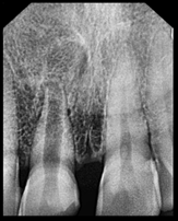
Fig 1: Preoperative radiograph

Fig 2: Working length
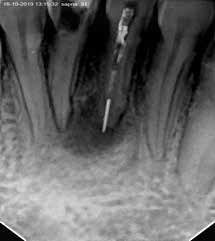
Tooth was isolated using rubberdam and access cavity was prepared by using an endo access bur and 90K-file was introduced into the canal to ensure the patency. Working length was determined 20 mm (Fig 2). Gentle instrumentation was done in circumferential filing motion with 90K- file. Root canal debridement was done using alternate irrigation with 3% NaOCl and saline throughout cleaning and shaping procedure. Canal was dried with absorbent paper points and calcium hydroxide paste (AveuCal) was placed into the root canal (Fig 3). Access cavity was closed with a cotton pellet and temporary restoration. The patient was scheduled for a second visit after 1 week
SECOND VISIT
The tooth was asymptomatic during the entire postoperative period, and the temporary restoration was intact. After isolation with rubber dam, the temporary restoration and cotton pellet were removed from the access cavity. Canal was irrigated with 3% NaOCl followed by 17% EDTA and final rinse with 2% chlorhexidine and saline. The canal was dried with absorbent paper points and a small piece of resorbable collagen membrane(Colla plug) was placed at the apical portion of the canal. The Colla plug was introduced into the root canal and gently compacted by using prefitted hand pluggers slightly beyond the apex to achieve a matrix. MTA was mixed with distilled water according to the manufacturer’s instructions and carried to the canal

Fig. 3: Calcium hydroxide

Fig. 4: MTA Plug
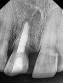
Fig. 5: Obturation
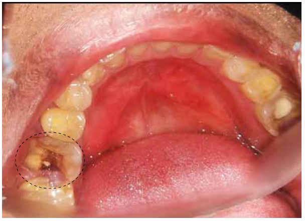
Apical plug of about 3-5 mm of MTA was placed and confirmed radiographically.(Fig 4) A sterile cotton pellet moistened with sterile water was placed over the canal orifice and the access cavity was sealed with Cavit. In next visit, the hard set of MTA was confirmed and the remainder of the root canal was obturated with zinc-oxide eugenol sealer and injectable thermoplasticized gutta-percha (Calamus) and confirmed radiographically.(Fig 5) The access cavity was sealed with GIC followed by composite resin. After one week tooth preparation was done followed by placement of crown.( Fig 6 & 7)

Fig 6: Preoperative

Fig 7: Postoperative
DISCUSSION
Apexification treatment of immature permanent teeth with pulp necrosis is an endodontic procedure to achieve apical closure. For many years, calcium hydroxide paste was used to induce a calcified barrier followed by root canal therapy. It has several disadvantages such as difficulty of the patient’s recall management and delay in the treatment.5,6 Furthermore, there is a risk of tooth fracture after dressing with calcium hydroxide for extended periods. Mineral trioxide aggregate (MTA) became the chosen material to induce the formation of the apical barrier because of its sealing properties & biocompatibility.7 Several studies demonstrated its capacity to induce odontoblastic differentiation, good radiopacity, low solubility, high pH, expansion after setting, & antimicrobial activity. Regardless of the technique used, a critical step in treatment of pulpless teeth with open apices is proper debridement and disinfection of the canal space. Complete disinfection of root canal is mandatory before obturation.7-9 Root canals with open apices have more communication compared to completely closed apex. So disinfection of canal was done using 3% sodium hypochlorite and calcium hydroxide dressing was given for periapical healing and eliminating the survived bacteria after cleaning and shaping.10 Placement of MTA is done and condensed using endodontic pluggers followed by obturation. Kusgoz et al stated that necrotic pulp in teeth with open apices in which MTA is used as a filling material is effective with shorter treatment time and better sealing ability.10,11 The apical plug created with MTA can be interpreted as an artificial barrier to condense the subsequent root canal filling material, in order to prevent reinfection of the canal system. The biggest drawback of MTA is expensive, difficult handling and long setting time.12
Conclusion
Apexification with a novel biocompatible material like MTA is a boon in effective management of teeth with open apex. This innovative procedure is predictable and less time consuming. Mineral trioxide aggregate showed clinical and radio-graphic success as a material used to induce root-end closure in necrotic immature permanent teeth.
REFERENCES
- Glossary of Endodontic Terms, ed 7, Chicago, IL, 2003, American Association of Endodontists.
- Morse et al; “Apexification: review of the literature,” Quintessence international, vol. 21, no. 7, pp. 589–598, 1990
- JOE 2013. Treatment Options: Apexogenesis and Apexification, Shahrokh Shabahang
- JOE — Volume 39, Number 3S, March 2013. Treatment Options: Apexogenesis and Apexification, Shahrokh Shabahang
- Apexification: review of literature, Quintessence International; 1990
- Int J Dent Med Res. 2015,VOL 1, ISSUE 6 Paulindraraj S et al: Apexification-Then and Now
- Huang G.T.-J. Apexification: the beginning of its end. International Endodontic Journal, 42, 855–866, 2009.
- JOE 2016. Apical Closure in Apexification: A Review and Case Report of Apexification Treatment of an Immature Permanent Tooth with Biodentine Karla Vidal et al
- Abu-Hussein Muhamad,Abdulgani Azzaldeen,Abdulgani Mai ( 2016); Single-Step Apexification with Mineral Trioxide Aggregate (MTA) –Case ReportsJournal of Dental and Medical SciencesVolume 15, Issue 2, 49-53 DOI: 10.9790/0853-15284953
- Goomer P, Jain RL. Comparison Of pulp revascularization & MTA apexifi cation in adjacent central incisors with immature apex – A case report. Indian J Dent Sci 2014; 6: 92-1.
- Kubasad GC, Ghivari SB. Apexifi cation with apical plug of MTA – Report of cases. Arch Oral Sci Res 2011;1:104-7.
- Witherspoon DE, Small JC, Regan JD, Nunn M. Retrospective analysis of open apex teeth obturated with mineral trioxide aggregate. J Endod 2008; 34: 1171-6.
Disclaimer from Guident
The views and opinions expressed in articles published in Guident are solely those of the authors and do not necessarily reflect the views of the editors, the editorial board or the publisher, Ivory India. Guident does not endorse any product, technique or treatment described. Clinical content is intended for qualified dental and medical professionals and is not a substitute for individual clinical judgement. Guident, its editors and the publisher accept no liability for any consequence arising from the use of information published in the journal.
How to cite this article
Dr. Mansi Luthra, Dr. Ajay Nagpal, Dr. Rohit Paul. Management of Open Apex Using Mineral Trioxide Aggregate. Guident. 2020 Apr;13(5). Available from: https://ivoryindia.in/articles/endodontics/management-of-open-apex-using-mineral-trioxide-aggregate.html