Central Ossifying Fibroma a Case of Diagnostic Dilemma
Department of Oral Medicine and Radiology, Oral and Maxillofacial Surgery, Swargiya Dadasaheb Kalmegh Smruti Dental College & Hospital, Hingna Nagpur
ABSTRACT
Mixed lesions of jaw are radiolucent and radiopaque images of the lesions on the radiographs. The mixed lesion denotes an intermediate stage in the development of the lesion. Fibro-osseous lesions are most common lesions which appears mixed on the radiograph. Central ossifying fibroma is benign fibro-osseous neoplasm of jaw which is rarely encapsulated and consists of fibrous tissue with variable amounts of mineralized material similar to bone/cementum. Here we report a case of 40 years old female patient who presented with a mixed lesion in periapical region.
Keywords: Central Ossifying Fibroma, Fibro-Osseous Lesions, Ossifying fibroma, Cement ossifying fibroma, mixed.
INTRODUCTION
Certain disease states produce mixed radiopaque and radiolucent images on the radiographs. Some pathologies may be present as an osteolytic lesion, which appears as radiolucency in radiograph. During it’s development, foci of calcified material may form within the osteolytic area. When these foci become large and mineralized, they become radiographically apparent. Thus, mixed radiolucent & radiopaque condition frequently represents an intermediate stage in development of lesion. Mineralization continues until most of the lesion with a possible exception of a thin radiolucent rim becomes opaque. On histologic examination all radiolucent-radiopaque stage contain calcified area of hard tooth tissue or nondescript mineralized material. The most common mixed lesions of the jaw include periapical cemental dysplasia, cemento-ossifying fibroma, odontoma, adenomatoid odontogenic tumor, Calcifying epithelial odontogenic tumor, Calcifying epithelial odontogenic cyst, Fibrous dysplasia, Pagets disease, chronic osteomyelitis.1
Central ossifying fibroma is a fibro-osseous lesion of jaw which originates from the periodontal membrane. This bone tumor consists of highly cellular, fibrous tissue that contains varying amounts of calcified tissue resembling bone, cementum or both. It most commonly occurs in mandibular posterior region and has a female predilection.1 Radiographically, Central ossifying fibroma presents as a well-defined, unilocular lesion which contains varying amounts of radiopaque material.2 Here we report a case of 40 years old female patient who presented with a mixed lesion in periapical region.
CASE REPORT
A 40 years old female patient reported to our department with a chief complaint of decayed teeth in lower right posterior region of jaw since
1 year. Patient also gives history of pain in the same region since 1 month. Pain was dull aching, intermittent and of non-radiating type. There was no history of bleeding and pus discharge and trauma in lower right posterior region. Patient had undergone thyroidectomy 7-8 years ago and was not on medications for thyroid. On examination patient was having deep occlusal caries with permanent right first molar with which tenderness on vertical percussion was positive. No other clinical abnormality was detected. On an intra-oral periapical radiograph a mixed radiolucent and radiopaque lesion was seen in the periapical region of first molar.
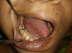
Fig 1 : Clinical examination showing carious tooth no 46

Fig 2: Intraoral periapical radiograph showing mixed lesion in periapical area of tooth no 46.

Fig 3: CBCT (Sagittal section)

Fig 4: CBCT (Coronal section)

Fig 5: CBCT (Axial section)

Fig 6 : CBCT (3D view)

Fig 7: Post extraction radiograph showing extraction of 46 with surgical curettage of the lesion.

Fig 4: Specimen excised
Further a cone beam computed tomography was taken to determine the association of the lesion with the carious teeth. CBCT revealed a radiopaque lesion surrounded by a thick radiolucent capsule lined by a thick radiopaque border. Margin was well defined and surrounding structures were normal. The lesion was in close approximation with the carious teeth. A differential diagnosis of odontoma, chronic osteomyelitis, focal cementosseous dysplasia was considered. A surgical extraction of carious teeth along with curettage of the lesion was performed and send for histopathological examination. The H and E section showed connective tissue devoid of epithelial linning. The connective tissue was composed of collagen fiber bundles arranged irregularly throughout the tissue with many small mature bony trabeculae with osteocytes in the lacunae. Also many small areas of osteoid tissue were visible with few areas of extra vasculated blood elements. The overall histopathological features were suggestive of central ossifying fibroma.
DISCUSSION
Central Ossifying Fibroma (COF) is a benign fibro-osseous neoplasm which consists of highly cellular, fibrous tissue with varying amounts of calcified tissue, which resembles the bone, the cementum or both. WHO in 1972 classified it in two types as ossifying fibroma and cementifying fibromas; but in 1992 WHO considered it in one heading as cementoossifying fibroma. Further, the term “cementossifying fibroma” was replaced by “ossifying fibroma” in 2005 under the new WHO classification. It was first described by Menzel in 1872.4
The etiology of ossifying fibroma is unknown but it is thought to be due to odontogenic, developmental and traumatic origins to periodontal ligament may that lead to production of cementum and osteoid material. Other possible etiologies include developing from the multipotential mesenchymal cells of periodontal ligament origin which are able to form both bone and cementum or mutation in tumor suppressor gene HRPT2, a protein product known as parafibronin which leads to tumor formation. Few studies have also reported chromosomal abnormalities such as translocation and interstial deletion of codings in chromosome2.5
The central ossifying fibroma occurs in third to fourth decades of life with a female predilection. The most common site is in mandibular premolar – molar region. Clinically, the ossifying fibroma presents as a painless, slowly growing mass in the jaw where displacement of teeth may be the only early clinical feature. The tumor is well-circumscribed from its surrounding bone and will continue to grow bigger, slowly or actively, with larger lesions occasionally leading to facial deformity. Large lesions may show facial asymmetry, epiphora, nasal stuffiness, visual disturbances, exophthalmos due to compression of tumour.2,4
Radiologically the borders of central ossifying fibroma are well defined and a thin radiolucent line may separate it from surrounding bone. Internal structure is mixed radiopaque & radiolucent with a pattern which depends on amount of calcified material. The lesions may be either unilocular or multilocular. In the early stages, the ossifying fibroma appears as a radiolucent lesion with no evidence of internal radiopacities. With increasing tumor maturity radiographs show increase in the appearance of radiopaque masses which may coalesce to form a large radiopaque foci surrounded by radiolucent border.3,4,6 The ossifying fibroma presents a radiolucent appearance in 53%, a sclerotic radio density in 7% and mixed or mottled appearance in 40% of the cases. Cortical expansion may be present and displacement of inferior alveolar canal may be present.5
An important diagnostic feature radiographically is that there is a centrifugal growth pattern rather than a linear one and therefore the lesions grow by centripetal direction and present as a round tumor mass. Another important radiographic feature of COF which helps in distinguishing it from other benign fibro-osseous lesions is that it is more well circumscribed.5
There are three different patterns of radiographic borders of cemento-ossifying fibroma which includes a defined lesion without sclerotic border (40%), defined lesion with sclerotic border (45%) and lesion with ill-defined border (15%) indicating a rapidly growing tumor.5
The differential diagnosis of central ossifying fibroma is considered according to the stage of the lesion. In initial stages it appears as completely radiolucent lesion therefore a differential diagnosis of ameloblastoma, periapical cemental dysplasia (initial stage) can be considered. Ameloblastoma is usually multilocular, occurs in posterior part of mandible and may cause paresthesia of lip. Periapical cemental dysplasia occurs in lower anterior region and affects the older age group.7
The mixed lesion stage can be related with calcifying odontogenic cysts, calcifying epithelial odontogenic tumor, odontoma, osteogenic sarcoma, periapical cemental dysplasia, Paget’s disease and fibrous dysplasia. Calcifying odontogenic cyst and calcifying odontogenic tumor is associated with one or more missing teeth. Osteogenic sarcoma have ill defined borders with periosteal bone formation. They have a rapid growth and do not have a fibrous capsule. Periapical cemental dysplasia has a predilection for mandibular anterior and seldom attains a diameter above 1cm whereas central ossifying fibroma usually attains a diameter more than 2-4cm. In odontoma, pattern of calcification resembles that of tooth structure. Fibrous dysplasia shows margins which blend with the surrounding bone and ossifying fibroma shows a concentric expansion of bone around a definite epicenter which leads to alteration in bone morphology, displacement of teeth and also resorption of tooth root. On contrary to this, fibrous dysplasia causes minimum changes maintaining the normal bone morphology and rarely causes root resorption.8,9
The mature stage of central ossifying fibroma consists of completely radiopaque lesion so the differential diagnosis includes hypercementosis, condensing osteitis and idiopathic osteosclerosis. Hypercementosis is attached to part of root and is separated from the periapical bone by a radiolucent periodontal ligament space which surrounds the entire tooth. Condensing osteitis occurs around the periapex of non-vital teeth and do not have a radiolucent band around the lesion. Idiopathic osteosclerosis is usually irregular in shape without a radiolucent rim and associated with periapical region of vital teeth.10
Treatment of central ossifying fibroma includes surgical enucleation of the lesion. Eversole et al reported recurrence rate of 28% following curettage.11
REFFERENCES
- Reddy BP, Reddy SB, Prasad N, Kiran G, Patlolla K. Central ossifying fibroma of the mandible: An unusual case report. J Clin Diag Res. 2012;6(3):539-41.
- Gambhir A,Rani G.Central ossifying fibroma of the Mandible – Report of a rare case. American Journal of Advances in Medical Science. 2013;1(2):42-47.
- Saravanakumar B, Sarumathi T, Manigandan T, Manisundar N, Aarthinisha V. Central Ossifying Fibroma of the Mandible: A Case Report with Review of Literature. Academic Journal of Cancer Research.2014;7(1): 46-49.
- Mohapatra M, Banushree CS, Nagarajan K, Pati D. Cemento-ossifying fibroma of mandible: An unusual case report and review of literature. Journal of oral and maxillofacial pathology: JOMFP. 2015 Sep;19(3):405.
- Swami AN, Kale LM, Mishra SS, Choudhary SH. Central ossifying fibroma of mandible: A case report and review of literature. Journal of Indian Academy of Oral Medicine and Radiology. 2015 Jan 1;27(1):131.
- Acharya S, Lohe VK, Bhowate RR. A Case Report: An Unusual Case of Central Ossifying Fibroma. Oral health case Rep. 2016;2(116):2.
- Wood NK, Goaz PW, Jacobs MC. Periapical radiolucencies. In: Wood Nk, Goaz PW, editors. Differential Diagnosis of Oral and Maxillofacial Lesions. 5th ed. New Delhi: Mosby; 2011. p. 252-78.
- Wood NK, Goaz PW, Jacobs MC. Mixed Radiolucent-Radiopaque lesions associated with teeth. In: Wood Nk, Goaz PW, editors. Differential Diagnosis of Oral and Maxillofacial Lesions. 5th ed. New Delhi: Mosby; 2011. p. 415-32.
- White SC, Pharoah MJ. Diseases of bone manifested in jaws. In: White SC, Pharoah MJ, editors. Oral Radiology: Principle and Interpretation. 6th ed. New Delhi: Mosby; 2004. p. 500-1.
- Wood NK, Goaz PW, Jacobs MC. Periapical Radiopacities. In: Wood Nk, Goaz PW, editors. Differential Diagnosis of Oral and Maxillofacial Lesions. 5th ed. New Delhi: Mosby; 2011. p. 457-76.
- MacDonald-Jankowski, D.S., 1998. Cemento-ossifying fibromas in the jaws of the Hong Kong Cemento-ossifying fibroma—a rare case. J. Indian Chinese. Dentomaxillofac Radiol., 27: 298-304.
Disclaimer from Guident
The views and opinions expressed in articles published in Guident are solely those of the authors and do not necessarily reflect the views of the editors, the editorial board or the publisher, Ivory India. Guident does not endorse any product, technique or treatment described. Clinical content is intended for qualified dental and medical professionals and is not a substitute for individual clinical judgement. Guident, its editors and the publisher accept no liability for any consequence arising from the use of information published in the journal.
How to cite this article
Dr. Rucha Pandharipande, Dr. Rakhi Chandak, Dr. Krishna Kurawar. Central Ossifying Fibroma a Case of Diagnostic Dilemma. Guident. 2020 Apr;13(5). Available from: https://ivoryindia.in/articles/oral-medicine/central-ossifying-fibroma-a-case-of-diagnostic-dilemma.html