Carcinoma LEFT Buccal Mucosa
Department of Oral Medicine & Radiology, Meenakshi Academy of Higher Education & Research, Chennai
CHIEF COMPLAINT
Patient complains of a swelling in his left cheek region for the past 2 months.

Fig. 1
HISTORY OF PRESENT ILLNESS
Patient gives h/o swelling that was initially small in size and following trauma before 5 months gradually attained the present size. He also gives h/o trauma to that region 15 days back following which he was under medication
(T. Paracetamol: 500mg, C. Amox: 500 mg, T. Riboflavin) and the swelling didn’t subside even after the medication. Pt gave no h/o recurrence, no h/o associated pain, and h/o interference of the swelling during chewing food occasionally.
PAST MEDICAL HISTORY
Diabetic: No H/o DiabetesHypertension: No H/o Hypertension
Asthma: No H/o Asthma
Cardiac illness: No H/o Cardiac illness
Bleeding Disorders: No H/o bleeding disorders
Hospitalization: No H/o hospitalization
Any regular medications: No H/o medication undertaken regularly
Drug allergy: No H/o drug allergy
PAST DENTAL HISTORY
Pt had visited a dentist previously 1 week back for which medication was given, following which the swelling didn’t subside.
PERSONAL HISTORY
Diet: Mixed
Brushing Habits: Once daily
H/o hans chewing since 2 years(son gave h/o that habit was present since 10 years), chews 3 packets/ day, stores the quid in his left cheek region and spits it out after 5 – 10 minutes. H/o cessation of habit since 20 days.
H/o occasional alcohol consumption since 5 years.
Marital Status: married
Family History: All family members are alive and apparently healthy
CLINICAL EXAMINATION
Conscious, co-operative, well- oriented to time and place and responds well to questions
Appearance: Normal and well built
Stature: short
Nutritional Status: well nourished
Deformities: Not present
Structural Alterations:
Hair: body
Scalp
Skin: color and texture: fair complexion
Vital signs: Temperature: 98.7 degree
Pulse: 68 beats/min
Respiratory rate: 10 cycles/ minute
Blood pressure: 110/70mm of Hg
Anemia: Not anemic
Clubbing: No clubbing
Cyanosis: No Cyanosis
Icterus: No icterus
Pedal edema: No pedal edema present.
REVIEW OF SYSTEMS: SCREENING QUESTIONNAIRE
CNS: No H/o headache, vomiting, loss of memory with normal vision and speech.
CVS: No H/o chest pain, palpitations, breathlessness on exertion, dyspnoea, with normal heart sounds
RS: Normal breathing pattern with no cough or expectoration, dyspnoea
GIT: Normal appetite and normal bowel movements. No H/o gastric irritation, stomach pain with no abdominal swellings and hepatospleenomegaly.
Genitourinary System: Normal micturition pattern, no abnormal discharge/burning sensation in micturition
Musculoskeletal System:
Gait: Normal gait
Muscular pain/ spasm: no muscular pain/spasm
Movement of joints/ Stiffness: Normal with no stiffness elicited
LOCAL EXAMINATION
Extra - Oral
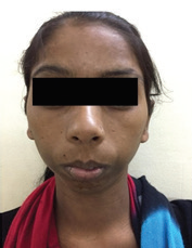
Facial symmetry: facial asymmetry in relation to the left lower third of face was present.
Scar/ Sinus: Absent
Ear Eyes nose: Appear normal
No paresthesia in the area of distribution of the inferior alveolar/ mental nerve
TMJ EXAMINATION
Jaw movements- Normal
Deviation on opening/closing-Normal
TMJ sounds- No abnormal sounds
Other abnormalities:-
Lips: Competent
Maxillary sinus tenderness
Not present.Lymph node status: presence of a single palpable submandibular lymph node in the Lt side measuring approx 2×2cm in dimension, which was stony hard in consistency, mildly tender, and immovable on all palpation.
INTRA ORAL
Size of mouth: Normal
Mouth Opening 4.2 cms
Hard Tissue Examination
No. Of teeth present: 32
Root stumps: 48; Calculus: mildly present.
Grossly decayed: 26 with tenderness on percussion;
Pit caries: 27, 28; DC: 11, 38
Palatally placed: 13; lingually placed: 32
Soft Tissue Examination
Gingiva: Coral pink, with normal contour and consistency
Labial Mucosa: NADs
Alveolar Mucosa: NAD
Buccal Mucosa: NAD.
Hard Palate: NAD
Soft Palate: No abnormalities detected. Movements of the soft palate and uvula are normal.


Intra Oral Pictures
TONGUE
Dorsal surface: Normal
Ventral surface: frenal attachments normal, normal tortuous venules present. NAD
Lateral borders: No abnormalities detected
Fauces: NAD
Tonsils: NAD
Floor of mouth: opening of salivary ducts normal. No abnormalities detected.
LOCAL EXAMINATION
Extra Orally
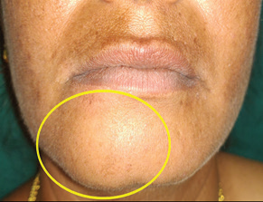
Inspection: presence of a single diffuse swelling in relation to the left lower third of face, approximately of size 3×2.5 cm, extending anteriorly approx 1cm away from the comissure of mouth upto approx 4cm anterior to the angle of mandible, superiorly 2cm below the ala tragal line and inferiorly 1cm below the lower border of mandible. The skin over the swelling was of normal colour as that of adjacent region, with no other evident secondary changes.
Palpation: on palpation, the inspectory findings such as site, size and extent were confirmed. The swelling was hard in consistency, tender on palpation, with no local rise in temperature.
INTRA ORALLY
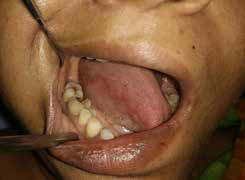
Inspection: presence of an ulcerproliferative growth in the left buccal mucosa, measuring approx 3× 3.5 cm in dimension, extending anteriorly from the mucosa adjacent to 36, posteriorly approx 0.5 cm before the pterygomandibular raphe, superiorly approx 1.5 cm below the upper buccal vestibule and inferiorly upto the lower buccal vestibule. The growth is present with ill defined borders, irregular surface texture and is elevated from the normal mucosal surface level. Presence of multiple papillary like projections from the surface of the growth is seen. Towards the inferior and posterior aspect, presence of psudomembranous slough covering the surface of the growth is seen. There is no interference of the growth with the occlusal tips of 26, 27,28 & 36, 37, 38 during occlusion.
Palpation: on palpation, the inspectory findings such as size, site, shape and extent are confirmed. The growth is tender, rough on palpation, firm in consistency, present with raised and indurated margins, with no bleeding during palpation, no paresthesia. Pseudo membranous slough was scrappable in the posterior aspect during palpation, which left behind erythematous regions.
SUMMARY
A 58 year old male patient came to the department with the chief complaint of a swelling in his left cheek region for the past 2 months. History revealed that, the swelling that was initially small in size and gradually attained the present size, h/o trauma to that region 15 days back following which he was under medication(T. Paracetamol: 500mg, C. Amox: 500 mg, T. Riboflavin), no h/o associated pain, and h/o interference of the swelling during chewing food.
On clinical examination, there was presence of a single diffuse extra oral swelling in relation to the left lower third of face, approximately of size 3×2.5 cm, extending anteriorly approx 1cm away from the comissure of mouth upto approx 4cm anterior to the angle of mandible, superiorly 2cm below the ala tragal line and inferiorly 1cm below the lower border of mandible. The skin over the swelling was of normal colour as that of adjacent region, with no other evident secondary changes. On palpation, the inspectory findings were confirmed. The swelling was hard in consistency, tender on palpation, with no local rise in temperature. Presence of a single palpable submandibular lymph node in the Lt side measuring approx 2×2cm in dimension, which was stony hard in consistency, mildly tender, and immovable on all palpation.
Intra orally, there was presence of an ulcerproliferative growth in the left buccal mucosa, measuring approx 3× 3.5 cm in dimension, extending anteriorly from the mucosa adjacent to 36, posteriorly approx 0.5 cm before the pterygomandibular raphe, superiorly approx 1.5 cm below the upper buccal vestibule and inferiorly upto the lower buccal vestibule. The growth was present with ill defined borders, irregular surface texture and is elevated from the normal mucosal surface level. Presence of multiple papillary like projections from the surface of the growth was seen. Towards the inferior and posterior aspect, presence of psudomembranous slough covering the surface of the growth was seen. There is no interference of the growth with the occlusal tips of 26, 27,28 & 36, 37, 38 during occlusion. On palpation, the inspectory findings such were confirmed. The growth is tender, rough on palpation, firm in consistency, present with raised and indurated margins, with no bleeding during palpation, no paresthesia. Pseudo membranous slough was scrappable in the posterior aspect during palpation, which left behind erythematous regions.
PROVISIONAL DIAGNOSIS
Malignant growth in relation to the Left Buccal Mucosa – T2N1M0
OTHER FINDINGS
Chronic Generalised Gingivitis
Root stumps: 48 Dc with partial loss of crown structure and apical periodontitis: 26
Dc:11, 38 (early enamel caries:27, 28)
INVESTIGATIONS
Cytology: Numerous polygonal squamous epithelial cells with few cels showing cellular pleomorphism, hyperchromatism and increased nuclear cytoplasmic ratio. Few cells show intracytoplasmic inclusions with few anucleated cells, suggestive of CLASS IV CYTOLOGY.

Cytology

IOPA

OPG
lt buccal mucosa.

Histopathology
FINAL DIAGNOSIS
WELL DIFFERENTIATED SQUAMOUS CELL CARCINOMA: LEFT BUCCAL MUCOSA.
Disclaimer from Guident
The views and opinions expressed in articles published in Guident are solely those of the authors and do not necessarily reflect the views of the editors, the editorial board or the publisher, Ivory India. Guident does not endorse any product, technique or treatment described. Clinical content is intended for qualified dental and medical professionals and is not a substitute for individual clinical judgement. Guident, its editors and the publisher accept no liability for any consequence arising from the use of information published in the journal.
How to cite this article
Dr. K. Saraswathi Gopal, Dr. R. Anitha Assist., Dr. S. Srividhya Associate. Carcinoma LEFT Buccal Mucosa. Guident. 2019 Nov;12(12). Available from: https://ivoryindia.in/articles/oral-medicine/carcinoma-left-buccal-mucosa.html