Central Giant Cell Granuloma
Department of Oral Medicine and Radiology, Faculty of Dentistry, Meenakshi Academy of Higher Education & Research, Chennai
ABSTRACT
Central giant cell granuloma (CGCG) is a relatively uncommon benign intraosseous lesion of clinical behavior ranges from nonaggressive to aggressive type, accounting for less than 7% of all benign lesions of the mandible. The actual etiology of CGCG is still unclear, although inflammation, hemorrhage, and local trauma have all been suggested. It usually associated with periodontium of jaw bones. The presence of distinctive multinucleated giant cells (MGCs) in a prominent fibrous stroma is histological and unilocular radiolucency radiographical hallmark for central giant cell granuloma (CGCG). The present case focuses on the case of a 38-year-old female with CGCG in the mandibular region.
Keywords: Central giant cell granuloma, Giant Cells, Periodontium
INTRODUCTION
Central giant cell granuloma (CGCG) of the jaw is a benign osteolytic lesion. It was first described by Jaffe in 1953, as a central giant cell reparative granuloma.1 It is defined as a benign,reactive and intraosseous developmental anomaly or a benign neoplasm in the jaws. It has often been compared with the giant cell tumor of long bones. Etiology of which is still completely unknown but thought to be of a reactive process to some unknown stimuli or internal bony hemorrhage and inflammation, may become aggressive leading to the expansion and perforation of cortex resulting into mobility and displacement of teeth with root resorption. Jaffe separated Central giant cell granuloma from Giant Cell Tumor of the bone on clinical and histologic grounds and suggested that Multinucleated Giant Cells in Central giant cell granuloma represent a phagocytic response to hemorrhage.2,3 Neville et al. consider this entity to be a non-neoplastic lesion and WHO also classifies it has not a tumor, although its clinical behavior and radiographical features are often associated with benign tumor. The etiology is unknown, but genetic abnormalities are implicated. Giant cell lesions of the maxillofacial area can vary from asymptomatic slowly growing to aggressive tumors showing high recurrence rate as well as the rapid expansive progression of jaw characterized by root resorption, osseous destruction, cortical perforation, adjacent tooth mobility and pain. Most commonly seen in adolescents and young adults. Radiographically, The lesion commonly presents as a solitary, cyst like radiolucency with a multilocular appearance or less commonly, a unilocular appearance.4
CASE REPORT
A 38 year old female reported to the department of Oral Medicine with the chief complaint of swelling in her lower front teeth region for the past 5 months. The swelling was initially small in size and gradually enlarged to reach the present form. There was paresthesia in the mucosa over the swelling for the past 2-3 months. Teeth in the region were mobile for the past 2-3 months. No relevant medical history on history taking.
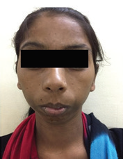
Upon clinical examination, a single diffuse bony hard extra-oral swelling which was causing mild facial asymmetry and measuring 2.5 x 3 cm2 in it’s greatest dimension was evident on the right para-symphyseal region. The swelling extended supero-infriorly from underneath the lip to the inferior border of the mandible, medially till the midline of face and laterally till the region corresponding to the angle of the mouth ( fig: 1).
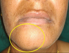

Fig. 1
The swelling was tender along the lower border of the mandible.
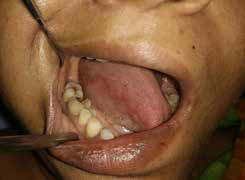
Intra-oral examination revealed, a roughly ovoid, bony hard and tender swelling measuring 4 x 2cm2 in its greatest dimension and involving the marginal and attached gingiva and alveolar mucosa of 43, 42; attached gingiva and alveolar mucosa irt 44,41, 31 and alveolar mucosa irt 32. The swelling extended inferiorly from the alveolus and caused obliteration of the labial and buccal vestibule irt 32, 31, 41, 42, 43, 44. The swelling was fixed and non-fluctuant and the surface of the swelling was slightly irregular on palpation. There was parasthesia on the region of the swelling (fig: 2).

Fig. 2
The blood reports of the patient revealed that her WBC count, serum alkaline phosphatase, SGOT and SGPT levels were raised.
IOPA (Fig 3) shows evidence of a ill-defined radiolucent lesion in the periradicular region of 42, 43 and 44 which causes mesial displacement of 43 and 42. The radiolucency appears to involve the crest of the alveolar bone also in this region. There is evidence of sepatations within the radiolucent lesion. Features suggestive of lytic lesion of bone. Evidence of a partially well-defined radiolucent lesion in the periradicular region of 33, 32, 31, 41 and 42 which causes distal displacement of 42. The radiolucency appears to involve the crest of the alveolar bone also in this region. There is evidence of sepatations within the radiolucent lesion. Features suggestive of lytic lesion of bone.

Fig. 3
OPG revealed a partially well-defined radiolucent lesion in the periradicular region of 31, 41, 42, 43 and 44 and causing displacement of 43 and 42 with evidence of intralesional septations which was suggestive of a lytic lesion irt. 31, 41, 42, 43.(Fig 4)

Fig. 4
CBCT revealed a partially well defined, lytic lesion measuring
30.4mm x 22.02mm x 19.20mm in its greatest dimension was evident in the periradicular region of 31, 41, 42, 43, 44. The lesion has caused destruction of the labial cortical plate and has expanded anteriorly. The lesion appears to have sepations within it and is causing distal displacement of roots of 43 and mesial displacement of root of 42. The features are suggestive of a benign bony pathology.
CT revealed ill defined lytic lesion measuring 2.1 x 1.9 cm2 noted along the anterior aspect of right hemi mandible with few septations within the lesion which is suggestive of benign bony pathology. Further the incisional biopsy of the lesion confirmed central giant cell granuloma.(Fig 5)

Fig. 5
Based on the history, clinical presentation, radiographic appearance and histopathological features, the lesion was diagnosed as Central Giant Cell Granuloma of the mandible irt 32, 31, 41, 42, 43, 44. Further patient underwent surgical curettage along with extraction.(fig 6)

Fig. 6
Pic courtesy: Dr. Manikandan, PROFESSOR & HEAD of the Department.
Department of oral and maxilla- facial surgery.
Meenakshi Ammal Dental College, Chennai.

Fig. 7
Pic 7 4x Microscopic view of Central Giant Cell Granuloma.
Pic courtesy: Dr. Sivapathasundaram Professor and Head of the Department
Department of oral pathology
Meenakshi ammal dental dental. Chennai.
DISCUSSION
Central giant cell granuloma (CGCG) is an uncommon, benign, and proliferative lesion. The central giant cell granuloma occurs most frequently in female patients under 30 years of age, and approximately two of three lesions are found in the anterior mandible with frequently lesion crossing midline.5 The incidence of CGCG in the general population is estimated to be 0.0001% with 60% of cases occurring before the age of 30. Occurrence in mandible predominates 3:1 over that in maxilla. Anterior to the mandibular molar region is the usual site of involvement with tendency for bony expansion. Paresthesia is more common in this lesion. It may probably be reactive or responsive in nature. Speculation suggests it may represent a developmental anomaly.6
Depending on clinical and radiographic features, central giant cell granuloma can be classified into two types. The first type of lesion is nonaggressive, slow growing, does not show root resorption or cortical perforation, and often shows new bone formation. Such lesions are typically discovered during routine radiographic examination or as a result of painless expansion.
The second type is an aggressive type which grows quickly, shows pain, cortical perforation, and root resorption. Extension occurs into soft tissue and ulceration of overlying mucosal surface.7 Develop in somewhat younger patients and exhibit greater recurrence potential.
Radiographically appear as a unilocular or multilocular radiolucency with well defined radiographic margin. in most cases periphery shows no evidence of cortication. The lesion may vary from 5 mm in as incidental radiographic finding to destructive lesion greater than 10 cm. The lesion appearing in maxilla may have ill defined, almost malignant appearing borders. The internal structure shows subtle granular pattern of calcification. Occasionally this granular bone is organized into ill defined, whispy septa. Granular septa emanate at right angles from the periphery of the lesion.8 In some instances the septa are better defined and divide the internal aspect into compartments creating multilocular appearance. Often displaces tooth and root resorption is not a common feature. loss of lamina dura of the involved tooth is seen. The mandibular nerve canal is displaced in inferior direction. Uneven expansions occurs which gives a appearance of double boundary when viewed using occlusal film.
Low-to-intermediate signal intensity at T1- and T2-weighted images and avid heterogeneous enhancement are seen as MRI findings. CT imaging helps to better delineate the extent of the lesion and also reveal the extension of the lesion into adjacent soft tissue.9
The histopathological lesions of the jaws show a variety of features. Common to all is the presence of few to many multinucleated giant cells in a background of ovoid to spindle shaped mesenchymal cells. The giant cells may be focally aggregated in the lesional tissue or may be diffusely present throughout the lesion. In some cases, the stroma is loosely arranged & edematous and quite cellular. Stromal cells- are of 2 types, such as resembling fibroblast ( oval/ spindled with cigar shaped nuclei) and Macrophage( small round hyperchromatic nuclei). Areas of erythrocyte extravasation and hemosiderin deposition may be prominent, some lesion show fibrosis of stroma. Foci of osteoid and newly formed bone may be present in the lesion. These features may be identical with those seen in cherubism and in brown tumour of hyperparathyroidism. Stromal cells- swirls, storiform or herring bone pattern.Giant cells such as foreign body or osteoclast like cells. Immunohistochemical findings are noted such as, giant cells express phenotypic markers for both macrophages and osteoclasts.
Differential diagnosis may include ameloblastoma, odontogenic myxoma, aneurysmal bone cyst and ossifying fibroma. if granular internal structure present, ossifying fibroma is considered. Ameloblastoma commonly tends to occur in older age group and posterior mandible and have a curved course, well defined trabaculae. odontogenic myxoma occurs in older age group, appear as sharper and straight septa. Aneurysmal bone cyst occur in posterior aspect of jaw and cause profound expansion.2
The treatment of the CGCG ranges from curettage to resection. Surgical treatment such as simple curettage, curettage with peripheral ostectomy, enucleation, and en bloc resection can be carried out. Aggressive lesions should be corrected and treated with curettage. Intralesional injection of corticosteroids (triamcinolone hexacetonide 20 mg/ml diluted in anesthetic solution of 2% of lidocaine twice weekly for 6 weeks) has been used successfully. Recurrence rates have been reported to range between 11% and 49% for curettage alone and can be as high as 72% for aggressive subtypes.
CONCLUSION
Central Giant Cell Granulomas are benign, but occasionally they can be aggressive lesions developing in the jaws. Although simple curettage is effective in treatment for the majority of Central Giant Cell Granuloma of the jaws, aggressive lesions should be treated by surgical resection along with curettage.
REFERENCES
- Jadu FM, Pharoah MJ, Lee L, Baker GI, Allidina A. Central giant cell granuloma of the mandibular condyle: A case report and review of the literature. Dentomaxillofac Radiol 2011;40:60-4.
- White S, Pharoah M. Diseases of bone manifested in the jaws. In: White S and Pharoah M. Oral radiology: principles and interpretation (5th edn). St. Louis, MO: Mosby, 2004: 501–503. 3
- Neville B, Damm D, Allen C, Bouquot J. Bone pathology. In: Oral and maxillofacial pathology (2nd edn). Philadelphia, PA: W.B. Saunders Company, 2002: 544– 547. 6
- De Lange J, van den Akker HP. Clinical and radiological features of central giant-cell lesions of the jaw. Oral Surg Oral Med Oral Pathol Oral Radiol Endod 2005; 99: 464– 470
- Shafer WG, Hine MB, Levy BM. A Text Book of Oral pathology. 4th ed. Philadelphia: Saunders; 1983. p. 146-9.
- Kramer, Pindborg JJ, Shear M. Histological typing of odontogenic tumours. 2nd ed. Berlin: Springer-Verlag; 1991. p. 31.
- Wood NK, Goaz PW, Kallal RH. Multilocular radiolucencies. In: Differential Diagnosis of Oral and Maxillofacial Lesions. 5th ed. St. Louis: Mosby; 1997. p. 333-55.
- Freny R.karjodkar . essential of oral. Max. rad.
- Anita Gohel, BDS, PhDa, *, Alessandro Villa, DDS, PhD, MPHb , Osamu Sakai, MD, PhDc,d, dental clinics of nrth America.
- Abu-El-Naaj, Ardekiam L, Leberman R, Peled M. Central giant cell granuloma of the mandibular condyle: A rare presentation. J Oral Maxillofac Sur 2002;50:939-41.
Disclaimer from Guident
The views and opinions expressed in articles published in Guident are solely those of the authors and do not necessarily reflect the views of the editors, the editorial board or the publisher, Ivory India. Guident does not endorse any product, technique or treatment described. Clinical content is intended for qualified dental and medical professionals and is not a substitute for individual clinical judgement. Guident, its editors and the publisher accept no liability for any consequence arising from the use of information published in the journal.
How to cite this article
Dr. K. Saraswathi Gopal, Dr. V. Poongodi, Dr. Sosa George. Central Giant Cell Granuloma. Guident. 2019 Dec;13(1). Available from: https://ivoryindia.in/articles/oral-medicine/central-giant-cell-granuloma.html