Cephalometric Evaluation of Symphysis Morphology and Antegonial Notch in Predicting Direction of Mandibular Growth
(Professor & HOD), Department of Orthodontics and Dentofacial Orthopedics Inderprastha Dental College & Hospital, Sahibabad, Ghaziabad
INTRODUCTION
Growth and development are integral processes, which define the existence of life. Growth of an organism is the interplay between its genetic constitution and the environment in which it thrives. While the genotype provides the basic architecture, the environment acts to alter it to the form which may be best suited to the existing conditions. At the macroscopic or clinical level, growth is exemplified by an increase in height and weight, while at the microscopic level, it is accompanied by an increase in the number of cells and their size.1,2
The mandible develops from the mandibular process of the first branchial arch. Mandibular growth occurs as a result of the combined processes of proliferation and ossification of secondary cartilage at the condyle, as well as differential formation and remodeling of bone along the entire surface of the mandible. The direction of mandibular growth is expressed in a downward and forward direction relative to the rest of the cranium.
It is important to predict the magnitude and direction of mandibular growth in any case of orthodontic treatment. Mandibular growth is very critical in the development of balanced dentofacial structures and hence, influence the therapeutic result of orthodontic treatment. With the knowledge of mandibular growth, better therapeutic decisions can be made regarding timing and duration of the treatment, extraction pattern and possible need for surgery.
Prediction of mandibular growth pattern has been done by various parameters like condylar morphology3, shape of lower border of mandible4, anterior facial height5, cervical vertebrae6, craniocervicalangle7, frontal sinus8with varying degree of success.
Many studies have been done to assess the reliability of various mandibular anatomic structures to predict mandibular growth direction. Some authors found a significant association between mandibular morphology and growth direction3,5,9,10,11 whereas others have annulled any correlation between the two.12,19
Among the mandibular structures, two major factors have been studied widely, the mandibular symphyseal morphology and antegonial notch depth.
The mandibular symphysis is the area of the lower jaw where the two halves that comprise the mandible, join or articulate. The mandibular symphysis separates at its base as it travels downward, enclosing a triangular projection called the mental protuberance. Mandibular symphysis serves as a reference anatomical landmark for esthetics and beauty of the face in general and of the lower part in particular.13,14 Mandibular symphysis morphology is a complex phenotype that results from different genetic, nongenetic, and adaptive factors.15,16 Functional environment can affect the shape and size of mandibular symphysis demonstrating an adaptive morphological response to the biomechanical loads experienced at various points in the masticatory cycle.17,18 Other factors that affect the morphology or dimension of mandibular symphysis are vertical jaw relationship19 and inclination of the lower incisors.20 Inclination of the lower incisors indirectly affect the shape of mandibular symphysis during the growth period. Vertical growth direction may have an indirect effect on the anteroposterior position of the mandible and subsequently, on mandibular symphysis.
Mandibular antegonial notch is upward curving or concavity of the inferior border of mandible just anterior to the angular process (Gonion), it lies at the junction of the body and the ramus. The notch tends to be longer in the congenital than in the acquired state, and the ascending ramus is at a more obtuse angle to the body.
Ricketts21 (1960) did research whether symphysis morphology may be used to predict the direction of mandibular growth and associated a thick symphysis with an anterior growth direction.
Aki et al5 (1994) studied whether symphysis morphology could be used as a predictor of the direction of mandibular growth. It was concluded that there was a relationship of mandibular growth direction with symphysis and found that a mandible with an anterior growth direction was associated with a small height, large depth, small ratio, and large angle of the symphysis.
Bremen et al19 (2005) studied whether Bjork’s structural signs of mandibular growth rotation associated with hyperdivergent or hypodivergents kele to facial morphology and he found that it was difficult to categorize the present subjects as having either a hyper or a hypodivergents kele to facial morphology regardless of the age the subjects or the degree of hyper or hypodivergency.
Susan et al9 (2014) studied different anteroposterior jaw relationships and investigated whether craniofacial parameters have any correlation with its shape and dimensions and found that mandibular symphysis dimensions were strongly correlated to anterior facial dimensions.
Some studies have been done to find the correlation between antegonial notch depth and mandibular growth direction.3,10,12
Bjork3 (1969), described various structural signs which were seen in extreme types of mandibular rotators. He noted that the forward inclination of the condylar head was associated with forward mandibular rotators and a tendency towards backward mandibular rotation is associated with a pronounced apposition below the symphysis with more overall concavity of the lower mandibular border.
Singer et al10 (1987) attempted to determine if the antegonial notch depth indicated future mandibular growth direction or potential. Deep notch cases had more retrusive mandibles with a shorter corpus, smaller ramus height, and a greater gonial angle than did shallow notch cases. The lower facial height in the subjects with a deep mandibular notch was found to be longer, and both the mandibular plane angle and facial axis were more vertically directed.
Kolodziejet al12 (2002) took antegonial notch depth to predict the direction of facial growth. He found that antegonial notch depth fails to provide indication of facial growth.
Studies conducted so far to find the correlation between symphysis morphology or antegonial notch and mandibular growth direction have got varied conclusions with some of studies concluding the relationship of symphysis morphology or antegonial notch with mandibular growth direction while some of the studies concluding that there is no relationship between the two. Most of these studies have been done on American and Caucasians population. Only few studies have been conducted on Asian population and hence the present study is undertaken to assess the reliability of symphyseal morphology and antegonial notch depth as a predictor of direction of mandibular growth in Indian population.
AIMS AND OBJECTIVES
- The Aims and Objectives of the present study are-
- To determine if morphology of symphysis and antegonial notch could be used as a predictor of the direction of mandibular growth in Indian population.
- To determine if any gender variability exists in above study.
MATERIALS AND METHODS
Pretreatment lateral cephalograms of 30 adult patients (15 males and 15 females) aged 18 years and above reporting to the Department of Orthodontics and DentofacialOrthopaedics at Inderprashta Dental College and Hospital, Ghaziabad requiring orthodontic treatment were randomly selected.
MATERIALS:(Figure 1)
Lateral cephalograms, tracing sheet (Company- Gateway; Thickness- 90 Micron), 0.3mm lead pencil, metallic scale and set squares.

Figure 1- Armamentarium
Inclusion Criteria
- Good general and dental health
- Age-18 years and above
- Complete dental occlusion (except third molars)
Exclusion Criteria
- Patient with history of-
- Temporomandibular Joint Disorder
- Orthognathic Surgery
- Craniofacial anomaly or Facial Asymmetry
- History of trauma to mandible
- Previous Orthodontic Treatment
METHOD
Lateral cephalograms were taken in natural head position so that all landmarks were readily identifiable on lateral cephalogram. Lateral cephalograms were traced manually using acetate tracing paper and 0.3 mm lead pencil attached to the radiographs. Points and lines were marked with a lead pencil. The following cephalometric measurements had been used to assess direction of mandibular growth-
1. SN to Y-axis angle: It is the angle between SN plane and Y-axis
- SN Plane- It is the cranial line between the centre of sellaturcica (Sella) and the most anterior point of the fronto-nasal suture (Nasion).
- Y-axis- It is measured as the acute angle formed by the intersection of a line from the sellaturcica to Gnathion with the Frankfort horizontal plane.
- Mandibular Plane- Plane formed by a line joining Gonion and Menton.
- Palatal Plane- It is a plane formed by joining Anterior Nasal Spine (ANS) to Posterior Nasal Spine (PNS).
- Articulare- The point of intersection of the dorsal contours of the articular process of the mandible and the temporal bone.
- Gonion- The mostposteroinferior point on the angle of the mandible.
- Menton- The most inferior point on the chin.
- Saddle angle (N-S-Ar)- It is the angle between the anterior and posterior cranial base.
- Articulare angle ( S-Ar-Go)-It is the constructed angle between Sella, Articulare and Gonion.
- PFH (Posterior Facial Height)- It is a linear measurement from Sella to Gonion (S-Go).
- AFH (Anterior Facial Height)- It is defined as the linear measurement from Nasion to Menton (N-Me).
Cephalometric Evaluation of Symphysis:
1. Calculation of Symphysis Dimensions: A line tangent to point B was taken as the long axis of the symphysis. A grid was placed with lines of grid parallel and perpendicular to constructed tangent line. Superior limit of symphysis was taken at point B with inferior, anterior and posterior limits taken at most inferior, anterior and posterior borders of symphyseal outline respectively. Cephalometric evaluation of symphysis morphology taken with the following variables (Figure 2)-- Symphysis Height- Distance from superior to inferior limit on grid.
- Symphysis Depth- Distance from anterior to posterior limit on grid.
- Symphysis Ratio- Divide symphysis height by symphysis depth.
- Symphysis Angle-Postero superior angle formed by line through menton and point B and mandibular plane.
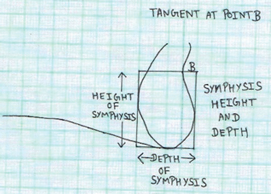
Figure 2- Measurement of Symphyseal Parameters
2. Calculation of Depth of Antegonial Notch:
- Antegonial notch is concavity on inferior border of mandible. Two points were traced on the inferior border of the mandible- inferior gonion and anterior convexity point. A line was drawn joining these two reference points.
- Anterior Convexity Point (ACP)- Point of greatest convexity along the anterior-inferior border of the mandible.
- Inferior Gonion (IGo)- Point of greatest convexity along the posterior-inferior border of the mandible.
- Antegonial Notch Depth- It is theperpendicular distance from the tangent to the lower border to the mandible to the greatest concavity on the antegonial notch (figure 3)

Figure 3- Measurement of Antegonial Notch Depth
Cephalometric Landmarks:
The following cephalometric landmarks are measured (Figure-4):
1. S.N to Y-axis angle
2. S.N to mandibular plane angle
3. Palatal plane –Mandibular plane angle
4. Gonial angle
5. Bjork sum
6. Percentage of lower facial height (ANS-Me) to total facial height (N-Me).
7. Posterior Facial Height (S-Go) to Anterior Facial Height (N-Me).

Figure 4- Measurement of Cephalometric Parameters
- A written consent was taken from patients regarding usage of his/her lateral cephalogram for the study
RESULTS
Lateral Cephalograms of 30 adult patients (15 males and 15 females) reporting to the Department of Orthodontics and Dentofacial Orthopaedics,Inderprastha
Dental College and Hospital, Sahibabad, Ghaziabad (U.P) aged 18 years and above were randomly selected and traced. Four symphyseal parameters (Height of Symphysis, Depth of Symphysis, Symphysis Ratio, Symphysis Angle) and Antegonial notch depth and seven cephalometric parameters (S.N to Y-axis angle, SN-Mandibular Plane Angle, Palatal Plane- Mandibular Plane Angle, Gonial Angle, Bjork Sum, Percentage of Lower Facial Height to Total Facial Height, Posterior Facial Height to Anterior Facial Height) were measured. The data was subjected to unpaired T-test to find any significant differences in the values between male and female groups and Pearson’s Correlation testto find any correlation between symphysis morphology and cephalometric parameters or antegonial notch depth and cephalometric parameters among overall, male and female group.
Unpaired t- Test (Table-1) indicated that there was a significant difference among male and female groups in Gonial angle with p value 0.013. Value for Gonial Angle for male group was found to be 124.20°± 4.39° and for female group was found to be 129.40°± 6.17°. There was no significant difference in all other values between male and female groups.
Pearson Correlation Test was done to find correlation between symphyseal morphology and antegonial notch depth and cephalometric parameters indicating mandibular growth direction variables.
Table 2 shows the correlation between symphysis height and cephalometric parameters. In the overall sample (Table 2A,Figure 5),there was a significant negative relationship between symphysis height and Bjork sum with p-value 0.016. There was no significant relationship between symphysis height and other cephalometric parameters. In the male group (Table 2B, Figure-6)posterior facial height / anterior facial height ratio andsymphysis height parameter showed a significant positive relationship with p-value 0.040 while all other values showed no significant relationship. In female group (Table 2 C),there was not a significant relationship between height of symphysis and cephalometric parameters.
Table 3 shows correlation between symphysis depth and cephalometric parameters. In the over-all sample (Table 3A, Figure-7), it was found that there is significant positive relationship between depth of symphysis and posterior facial height/ anterior facial height ratio with p-value 0.033. In the male and female groups (Table 3 B,Table 3 C), there was no significant correlation between depth of symphysis and cephalometric parameters.
Table 4 shows correlation between symphysis ratio and cephalometric parameters. In the overall group(Table 4A),there was not a significant relationship between symphysis ratio and cephalometric parameters. In the male group (Table 4B Figure-8), it was found that there was a significant positive relationship between symphysis ratio and posterior facial height to anterior facial height with p-value 0.022 while all other values showed no significant relationship. In the female group(Table 4C),there was not a significant relationship between symphysis ratio and cephalometric parameters.
Table 5 shows correlation between symphysis angle and cephalometric parameters. In the overall group, male and female groups, (Table 5A,Table 5B,Table 5C),there was no significant relationship between symphysis angle and cephalometric parameters.
Table 6 shows correlation between antegonial notch depth and cephalometric parameters. In the overall, male and female groups(Table 6A,Table 6B,Table 6C), there was no significant correlation between antegonial notch depth and cephalometric parameters.
TABLES AND GRAPHS
TABLE 1- UNPAIRED T-TEST


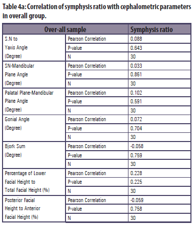


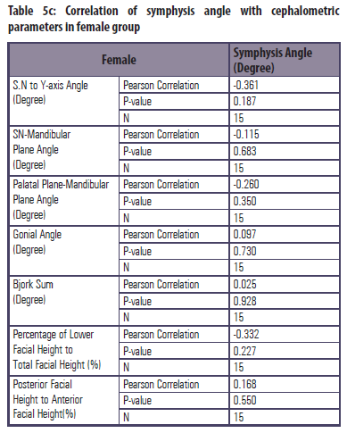


Figure 5: Correlation Of Height Of Symphysis (Mm) With Other Variables Among Over-All Group
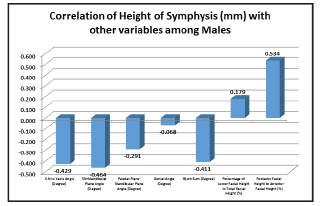
Figure 6: Correlation Of Height Of Symphysis (Mm) With Other Variables Among Male Group

Figure 7: Correlation Of Depth Of Symphysis (Mm) With Other Variables Among Over-All Group

Figure 8: Correlation Of Symphysis Ratio With Other Variables Among Male Group
DISCUSSION
The present study was undertaken to find the correlation if any between symphyseal morphological parameters or antegonial notch depth and mandibular growth direction.
Symphyseal parameters ( height, depth, ratio and angle), antegonial notch depth and seven cephalometric parameters ( S.N to Y-axis angle, SN-Mandibular plane angle, Palatal plane- Mandibular plane angle, Gonial angle, Bjork sum, Percentage of lower facial height to total facial height, Percentage of posterior facial height to anterior facial height) predicting growth directions were measured for 30 patients (15 Males, 15 Females).
The data was subjected to unpaired t-test and Pearson Correlation test.
Unpaired t-test results indicated that there is no significant difference among various parameters in Male and Female group except gonial angle. Gonial angle for females (129.40°) was found to be greater than males (124.20°).
Pearson correlation test was done to find any correlation between symphysis morphology or antegonial notch depth and cephalometric parameters among overall group and separately for male and female group.
Pearson correlation test indicated that in overall group, there was a significant negative relationship between symphysis height and Bjork sum. This further indicates that the decrease in symphyseal height is associated with vertical growth pattern. Also it was found that in overall group, there is a positive correlation between depth of symphysis and posterior facial height / anterior facial height ratio. This indicates that increase in depth of symphysis is associated with horizontal growth pattern.
In male group, Pearson correlation test indicated that there is significant positive correlation between symphysis height and posterior facial height / anterior facial height ratio. It denotes that increase in symphyseal height is associated with increase in Posterior Facial Height/ Anterior Facial Height ratio i.e. horizontal growth pattern.
Symphysis ratio and Posterior Facial Height/Anterior Facial Height was found to be having significant positive correlation in male sample which further indicates that increase in symphyseal ratio is associated with increase in Posterior Facial Height/ Anterior Facial Height i.e. horizontal growth pattern.
Thus, the present study results indicate that increase in height and depth of symphysis are associated with horizontal growth pattern in overall sample. In males increase in height and symphysis ratio is associated with horizontal growth pattern.
The results of this study are similar to the study done by Aki et al (1994)5in context to depth of symphysis where increase in depth was associated with horizontal growth pattern.
On the other hand, when we consider height of symphysis and symphysis ratio, the results of this study are not in synchronization with that of Aki et al5 (1994)where decrease in height and decrease in ratio are associated with horizontal growth pattern. This disparity may be because of difference in population group studied.
Antegonial notch depth was not found to be correlated with mandibular growth direction. This result is in accordance with the study done by Kolodziejet al12 (2002) who also found that antegonial notch depth fails to predict mandibular growth direction. On the other hand, Singer et al10 (1987) found that increase in depth was associated with horizontal growth pattern. This disparity may be because of different parameters used in this study to predict growth pattern.
CONCLUSION
The conclusion of the present study-
- Increase in depth and height of symphysis is associated with horizontal growth pattern in overall sample.
- Increase in height and smphyseal ratio is associated with horizontal growth pattern in males.
- In females symphyseal parameters fail to predict the direction of mandibular growth.
- Antegonial notch depth was not found to be associated with the direction of mandibular growth in all the samples.
REFERENCES
- Molinary L, Gasser T. The human growth curve: Distance,velocity and acceleration. In : Hauspie R, Cameron N, Molinari L, Methods in Human Growth Research. Cambridge, Cambridge University Press, 2004:27-55.
- Cameron N. Human growth curve, canalization and catch up growth. In: Cameron N, Human Growth and Development. California, Elsevier Sciences, 2002:1-20.
- Bjork A. Prediction of mandibular growth rotation. Am J Orthod1969;55:585-599.
- Skieller V, Bjork A, Linde-Hansen T. Prediction of mandibular growth rotation evaluated from a longitudinal implant sample. Am J Orthod 1984;86:359-70.
- Aki T Nanda RS, Currier GF, Nanda SK. Assessment of symphysis morphology as a predictor of the direction of mandibular growth. Am J Orthod. 1994;106:60-69.
- Huggare JAV, Cooke MS. Head posture and cervicovertebral anatomy as mandibular growth predictors. Eur J Orthod 1994;16:175-80.
- Solow B, Siersback-Nielsen S. Cervical and craniocervical posture as predictors of craniofacial growth. Am J Orthod 1992;101:449-58.
- Rossouw PE, Lombard CJ, Harris AM. The frontal sinus and mandibular growth prediction Am J Orthod. 1991;100:542-6.
- Susan N. Al-Khateeb; Emad F. Al Maaitah; Elham S. Abu Alhaija; Serene A. Badran. Mandibular symphysismorphology and dimensions in different anteroposterior jaw relationships. Angle Orthod.2014;84:304-309.
- Singer CP. The depth of the antegonial notch as an indicator of mandibular growth potential. Am J Orthod. 1968;89:528.
- Lambrechts AHD, Harris AMP, Rossouw PE, Stander I. Dimensional differences in the craniofacial morphologies of groups with deep and shallow mandibular antegonial notching. Angle Orthod 1996:66:265-72.
- Kolodziej RP, Southard TE, Southard KA, Casko JS, Jakobsen JR. Evaluation of antegonial notch depth for growth prediction. Am J Orthod.2002;121:357-363.
- Hoeing JF. Sliding osteotomy genioplasty for facial aesthetic balance: 10 years of experience. Aesthetic Plast Surg. 2007:31:384-391.
- Buschang PH, Julien K, Sachdeva R, Demirjian A. Child-hood and pubertal growth changes of the human symphysis. Angle Orthod. 1992;62:203-210.
- Gould SJ. The exaptive excellence of spandrels as a term and prototype. ProcNatlAcadSci USA. 1997;94:10750-10755.
- Gould SJ. The Structure of Evolutionary Theory. Cambridge, UK: Belknap Press; 2002.
- Beecher RM. Evolution of the mandibular symphysis in Notharctinae (Adaptive Primates). Int J Primatol. 1983;4:99-112.
- Brown B. Miocene hominoid mandibles: functional and phylogenetic perspectives. In: Begun DR, Ward CV, Rose MD, eds. Function. Phytogeny and Fossils. New York, NY:Plenum Press; 1997:153-172.
- Bremen J; Pancherz H. Association between Bjork’s Structural Signs of Mandibular Growth Rotation and Skeletofacial morphology. Angle Orthod. 2005;75:506-509.
- Yamada C, Kitai N, Kakimoto N, Murakami S, Furukawa S, Takada K. Spatial relationships between the mandibular central incisor and associated alveolar bone in adults with mandibular prognathism. Angle Orthod. 2007;77:766-772.
- Rickets RM. Cephalometric synthesis. Am J Orthod. 1960;46:647-673.
Disclaimer from Guident
The views and opinions expressed in articles published in Guident are solely those of the authors and do not necessarily reflect the views of the editors, the editorial board or the publisher, Ivory India. Guident does not endorse any product, technique or treatment described. Clinical content is intended for qualified dental and medical professionals and is not a substitute for individual clinical judgement. Guident, its editors and the publisher accept no liability for any consequence arising from the use of information published in the journal.
How to cite this article
Dr. Rahul Paul Principal, Dr. Vineet Golchha, Dr. Deepti Yadav. Cephalometric Evaluation of Symphysis Morphology and Antegonial Notch in Predicting Direction of Mandibular Growth. Guident. 2019 Dec;13(1). Available from: https://ivoryindia.in/articles/research/cephalometric-evaluation-of-symphysis-morphology-and-antegonial-notch-in-predicting-direction-of-mandibular.html