Periapical Healing Using Simvastatin by Surgical Intervention: A Clinical, Radiographic and CBCT Evaluation: A Case Report
Department of Conservative Dentistry and Endodontics, D.J. College of Dental Sciences & Research, Modinagar, U.P
ABSTRACT
Periapical lesions represent inflammatory disorders of the peri-radicular tissues that arise secondary to pulpal necrosis and persistent microbial infection within the root canal system. Although conventional root canal therapy remains the primary modality of treatment, certain cases involving large lesions, cystic transformation, or persistent infection require surgical intervention. Successful periapical surgery not only depends on elimination of infection and apical sealing but also on regeneration of lost periapical bone. Simvastatin, a 3-hydroxy-3-methylglutaryl coenzyme A reductase inhibitor widely used in the management of hypercholesterolemia, has been shown to possess bone anabolic, anti-inflammatory, and immunomodulatory properties. The present in vivo clinical study was conducted to evaluate the regenerative potential of locally placed simvastatin (10 mg) in periapical surgical defects. Clinical parameters, digital radiographic measurements, and cone beam computed tomography (CBCT) assessments were used to monitor healing. The results demonstrated progressive and accelerated bone regeneration with significant reduction in lesion size within two months. No postoperative complications were observed. Within the limitations of this study, local application of simvastatin appears to enhance periapical bone healing and may serve as a promising adjunct in endodontic surgery.
INTRODUCTION
The dental pulp is a specialized connective tissue that remains protected within rigid dentinal walls and enamel. Its integrity is essential for maintaining tooth vitality and defense against microbial invasion. However, deep caries, trauma, restorative procedures, or cracks may disrupt this protective barrier, allowing microorganisms to infiltrate the pulp chamber. Persistent inflammation eventually leads to pulpal necrosis, after which bacterial by-products diffuse through the apical foramen into the periapical tissues. This triggers an immune-inflammatory cascade characterized by recruitment of inflammatory cells, release of cytokines, activation of osteoclasts, and subsequent bone resorption.
Periapical lesions develop as a result of this chronic inflammatory process and are radiographically visualized as radiolucent areas at the root apex. Histologically, they may manifest as periapical granulomas, radicular cysts, or chronic abscesses. The severity and progression of these lesions depend on host immune response, microbial virulence, and duration of infection. The primary goal of treatment is elimination of intracanal microorganisms, as systemic antibiotics are ineffective due to absence of vascular supply in necrotic pulp.
Conventional root canal therapy has high success rates in managing periapical lesions. However, surgical intervention becomes necessary in cases involving persistent infection, true cysts, complex root canal anatomy, overfilled canals, root fractures, or anatomical limitations that prevent complete disinfection. Modern periapical microsurgery has significantly improved outcomes due to enhanced magnification, precise apical resection, ultrasonic retrograde cavity preparation, and biocompatible retrograde filling materials that ensure hermetic apical seal.
While surgical removal of infected tissue addresses the etiological factor, regeneration of lost periapical bone is essential for complete healing. Bone regeneration involves complex biological events including clot formation, angiogenesis, recruitment of osteoprogenitor cells, osteoblastic differentiation, and mineralization. Enhancement of these biological processes can significantly improve healing outcomes.
Simvastatin, a lipid-lowering agent belonging to the statin group, was discovered to have pleiotropic effects beyond cholesterol regulation. Research has shown that statins stimulate bone formation by upregulating Bone Morphogenic Protein-2 (BMP-2), which is a key regulator of osteoblastic differentiation and bone matrix production. Simvastatin also inhibits osteoclast-mediated bone resorption by interfering with the mevalonate pathway, thereby reducing prenylation of signaling proteins necessary for osteoclast function. Additionally, simvastatin exhibits anti-inflammatory effects by reducing pro-inflammatory cytokines and enhancing endothelial function, thereby promoting angiogenesis and tissue repair.
These biological properties make simvastatin an attractive candidate for enhancing bone regeneration in periapical defects. The present study was therefore designed to clinically and radiographically evaluate the healing potential of locally delivered simvastatin following periapical surgery.
AIM AND OBJECTIVES
The aim of the present study was to evaluate the rate and quality of periapical healing following surgical intervention with local placement of simvastatin (10 mg) in the peri-radicular region. The objectives included assessing clinical healing parameters, measuring radiographic reduction in lesion size, evaluating three-dimensional bone regeneration using CBCT imaging, and analyzing overall treatment success using standardized periapical healing indices.
METHODOLOGY
An in vivo clinical study was conducted in patients presenting with periapical lesions requiring surgical management. Inclusion criteria comprised teeth with persistent periapical radiolucency unresponsive to conventional endodontic therapy and lesions of significant size warranting surgical intervention. Patients with systemic conditions affecting bone healing were excluded.
Pre-operative evaluation included detailed clinical examination and digital intraoral periapical radiographs to assess lesion size and periapical status. Cone beam computed tomography was performed to obtain three-dimensional visualization of the lesion, evaluate cortical plate involvement, and determine proximity to anatomical structures. Pre-surgical scaling and root planing were performed to ensure optimal periodontal health.
Surgical procedures were carried out under aseptic conditions using a dental operating microscope to enhance precision and visualization. Premedication with amoxicillin 500 mg was administered two hours prior to surgery. Local anesthesia was achieved using lignocaine with adrenaline to ensure adequate analgesia and hemostasis. A sulcular incision with releasing vertical incisions was made, and a full-thickness mucoperiosteal flap was elevated to expose the underlying bone.
Trephination was performed to access the periapical lesion, followed by thorough curettage of pathological tissue. Apicoectomy was performed by resecting approximately 3 mm of the root apex to eliminate apical ramifications and lateral canals that may harbor microorganisms. Root-end cavity preparation extending 3 mm into the canal space was accomplished using ultrasonic retro tips under copious irrigation. The cavity was dried and filled with a suitable retrograde filling material to establish apical seal.
The simvastatin tablet (10 mg) was carefully stripped of its outer coating and crushed into a fine powder under sterile conditions. The powdered simvastatin was gently placed into the prepared bony defect, ensuring complete filling of the cavity without disturbing the retrograde filling. An absorbable gelatin sponge was placed over the grafted area to stabilize the material and act as a carrier matrix for sustained release. The surgical flap was repositioned and sutured. Postoperative radiographs were taken to confirm placement.
Patients were recalled after seven days for suture removal and initial assessment. Subsequent follow-up visits were scheduled weekly for one month and monthly thereafter up to six months. Clinical parameters including pain, swelling, sinus tract formation, mobility, and periodontal status were recorded at each visit.
Radiographic evaluation was performed at each recall using standardized digital techniques. The maximum vertical and horizontal dimensions of the lesion were measured to assess reduction in size. Healing was scored using the Periapical Index (PAI), which categorizes periapical status from healthy bone to severe apical periodontitis. CBCT imaging was repeated at two months to evaluate bone density changes, cortical repair, and volumetric reduction of the lesion. Statistical analysis was conducted using mean, standard deviation, independent t-test, and one-way ANOVA with significance set at p < 0.05.
PERIAPICAL HEALING WITH SIMVASTATIN -SURGICAL INTERVENTION


RESULTS
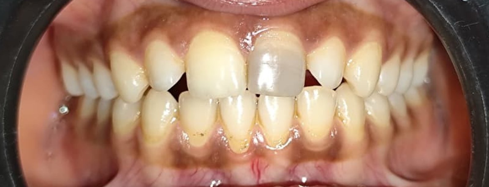
Clinical evaluation demonstrated uneventful healing in all cases. Patients reported minimal postoperative discomfort that subsided within a few days. No swelling, sinus tract formation, or periodontal complications were observed during follow-up.
Radiographic assessment at one week showed initial reduction in radiolucency and early signs of bone fill. At two weeks, lesion size reduction exceeded 50% in several cases, indicating rapid healing response. At 1 month, significant decrease in radiolucent area was observed, accompanied by improvement in PAI scores. By two months, near-complete radiographic healing was evident in most cases.
CBCT evaluation confirmed substantial increase in bone density within the defect area. Three-dimensional analysis revealed marked reduction in lesion volume and restoration of cortical integrity. Statistical analysis demonstrated significant reduction in lesion dimensions over time, confirming accelerated bone regeneration following simvastatin placement.
DISCUSSION
The regeneration of periapical bone following surgery is a complex biological process requiring coordinated cellular and molecular events. Simvastatin enhances this process through multiple mechanisms. By upregulating BMP-2 expression, it promotes differentiation of mesenchymal stem cells into osteoblasts, thereby accelerating new bone formation. Inhibition of the mevalonate pathway reduces osteoclast activity, minimizing bone resorption and creating a favorable environment for regeneration.
Simvastatin also exhibits anti-inflammatory properties by decreasing levels of tumor necrosis factor-alpha and interleukin-6, thereby reducing inflammatory tissue destruction. Enhanced angiogenesis mediated by improved endothelial function further contributes to nutrient supply and cellular recruitment necessary for bone repair.
The rapid reduction in lesion size observed within two weeks in this study suggests that local delivery of simvastatin provides a high concentration of drug at the defect site, maximizing its osteogenic potential. The gelatin sponge carrier likely facilitated controlled release and stabilization of the drug within the surgical cavity.
CBCT imaging proved valuable in assessing true three-dimensional healing, as conventional radiographs may underestimate lesion size due to superimposition of structures. The significant improvement in bone density and cortical repair observed on CBCT confirms the regenerative potential of simvastatin.
Although the findings are promising, limitations include limited sample size and relatively short follow-up duration. Long-term studies with histological analysis would provide stronger evidence of true bone regeneration.
CONCLUSION
Within the limitations of this study, surgical placement of simvastatin in periapical defects resulted in accelerated and enhanced bone regeneration. Significant reduction in lesion size, increased bone density, and favourable clinical outcomes were observed within two months. The osteo-inductive, anti-inflammatory, and angiogenic properties of simvastatin appear to contribute significantly to periapical healing. Simvastatin shows promising potential as a regenerative adjunct in endodontic surgery, particularly in large or persistent lesions. Further long-term clinical trials are recommended to establish standardized protocols and confirm its long-term efficacy.
Disclaimer from Guident
The views and opinions expressed in articles published in Guident are solely those of the authors and do not necessarily reflect the views of the editors, the editorial board or the publisher, Ivory India. Guident does not endorse any product, technique or treatment described. Clinical content is intended for qualified dental and medical professionals and is not a substitute for individual clinical judgement. Guident, its editors and the publisher accept no liability for any consequence arising from the use of information published in the journal.
How to cite this article
Dr. Sumaiya Khan, Dr. Anil Dhingra. Periapical Healing Using Simvastatin by Surgical Intervention: A Clinical, Radiographic and CBCT Evaluation: A Case Report. Guident. 2026 Mar;19(4). Available from: https://ivoryindia.in/articles/endodontics/periapical-healing-using-simvastatin-by-surgical-intervention-a-clinical-radiographic-and-cbct-evaluation-a.html