Magnification Error Assessment of Intraoral Periapical Radiograph
Department of Prosthodontics, Meenakshi Ammal Dental College, Chennai, TamilNadu, India, Tamil Nadu Government Dental College, Chennai, Tamil Nadu, India
ABSTRACT
Magnification error in the diagnostic intra oral periapical radiograph could mislead the diagnosis and treatment plan in dentistry. This article describes a simplest chair side procedure for preoperative assessment of the magnification error using gutterpercha points.
Keywords: Radiography, intra oral periapical radiograph, Cost-Benefit analysis, magnification error, Gutta-percha.
BACKGROUND
In our day-to-day clinical practice, intra oral periapical radiograph (IOPA) are primarily considered to visualise the periapical region of the target tooth. IOPA has the advantages of lesser radiation exposure, lesser distortion, with high reproducibility and cost effective compared to other imaging modalities.1,2 Parallelling angle technique is more preferred IOPA technique compared to bisecting angle technique due to its less magnification error.3 Use of positioning devices for the paralleling angle technique are restricted in cases of tori, shallow palate and shallow floor of mouth.4 In such cases, IOPA have to be made with bisecting angle technique but magnification error is its major drawback which could mislead its use in assessment of periodontal evaluation, implants, endodontic evaluation etc.5 To overcome this error, magnification factor of every radiograph needs to be assessed individually. This paper describes a simple procedure that enables the use of gutterpercha points to assess the magnification error of the intra oral periapical radiographs made with bisecting angle technique. To show the efficiency of this technique, IOPA was made for a patient with implants of 3.5mm x 13mm in maxillary right central incisor region and the length of the implants was assessed using values driven from IOPA made using the below mentioned technique.
TECHNIQUE
- Take of 40 size gutter percha point of measured length 8mm. (Figure 1)
- The area of target is thoroughly dried and cleaned to prevent the interference of the saliva.
- The measured 8mm gutterpercha point was held to the buccal surface of the clinical crown adjacent to the target area using carding wax. (Figure 2)
- Intra oral periapical radiograph films was placed on the lingual surface of the target area and held in position using the fingers of the patient.
- Radiograph was made using bisecting angle technique.
- As the image was developed and dried, the length of the gutterpercha was measured. (Figure 3)
- Asses the variation in the radiographic length of the gutterpercha point and its actual length which provides the magnification error factor for the particular image.
- Using the magnification error factor, the actual length of the target area can be assessed more accurately using the formula given below:-
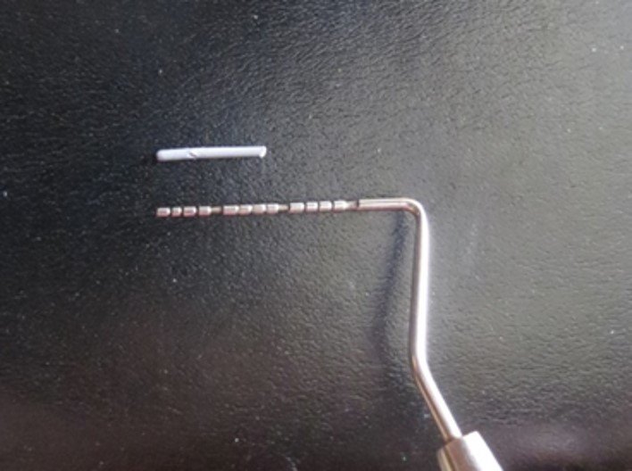
Fig 1- Guttapercha point of 8mm

Fig 2 - Using the carding wax, G.P.points are fixed
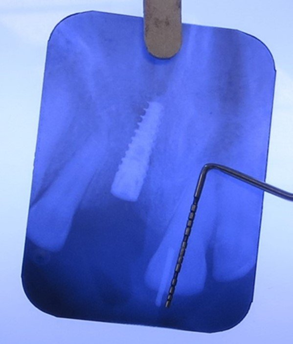
Fig 3 - Measurement of radiographic length of the G.P point (Radiographic length of G.P= 8mm)

The measured values of the maxillary right central incisor implant region are as follows:
Radiographic Guttapercha point length = 8mm
Radiographic implant length = 13mm

Hence the actual length of the implant is derived by knowing the magnification factor of the IOPA which justifies the efficiency of the technique.
DISCUSSION
Proper periapical radiographs with greater accuracy are needed to inform diagnosis, treatment planning, and prognosis evaluation. Greater accuracy of an image is obtained by assessing its actual magnification error. It can be assessed by measuring the magnification of the objects of known dimensions in the image and comparing it with the dimensions of the targeted area. Based on the previous literature, the various techniques for the magnification error assessment are implants, endodontic files during root canal treatment or by placing lead ball bearing stent. Each of the above said technique has their own drawbacks. implants and endodontic files limit its use in preoperative assessment. In lead ball bearing technique, stent fabrication postpones the assessment to the next appointment. In this technique, Guttapercha points was used to assess the magnification error as it offers excellent radiopacity, non toxic, high biocompatibility and structural stability. As they are easy to manipulate, a desired dimension were sectioned at ease. It was secured to the tooth with help of wax over the dry surface of the tooth. The magnification error of the radiographic image was assessed by comparing the radiographic image dimension of the gutta-percha point and its actual dimension. Based on the error value, the target area dimension was evaluated. To assess the efficiency of the technique, it was reported with an implant placed individual. Hence with use of this technique, we could evaluate the amount of radiographic parameters during diagnostic evaluation and post operative evaluation with better accuracy using intraoral periapical radiograph made with bisecting angle technique.
CONCLUSION
The above technique uses armamentarium such as gutterpercha points and carding wax which are readily available in our dental clinics. Identification of the magnification error helps in a precise evaluation and better treatment planning with the available IOPA irrespective of the radiographic technique. The data obtained from periapical radiograph using the above technique will be free of magnification error there by the need for the panoramic radiographs and cone beam CT scan can be minimized to some extent.
CLINICAL SIGNIFICANCE
As the technique is insensitive and cost effective, it should be implicated in our day-to-day practice to evaluate the magnification error of periapical radiograph for better diagnosis and clinical outcome. It can be implicated with digital radiography.
REFERENCES
- JacobsR, van Steenberghe D. Radiographicplanning and assessment of endosseous oral implants. Berlin: Springer-Verlag 1998.
- Beason RC, Brooks SL. Preoperative implant site assessment in southeast Michigan. J Dent Res 2001;80:136
- Gupta, A., Devi, P., Srivastava, R., Jyoti, B. 2014. Intra oral periapical radiography - basics yet intrigue: A review. Bangladesh Journal of Dental Research & Education 4(2): 83-7.
- Yen M, Yeung AWK. The Performance of Paralleling Technique and Bisecting Angle Technique for Taking Periapical Radiographs: A Systematic Review. Dent J (Basel). 2023 Jun 21;11(7):155.
- Stuart C. White DDS PhD, Michael J. Pharoah DDS . Oral Radiology: Principles and Interpretation- 6 edition; 76 -100
Disclaimer from Guident
The views and opinions expressed in articles published in Guident are solely those of the authors and do not necessarily reflect the views of the editors, the editorial board or the publisher, Ivory India. Guident does not endorse any product, technique or treatment described. Clinical content is intended for qualified dental and medical professionals and is not a substitute for individual clinical judgement. Guident, its editors and the publisher accept no liability for any consequence arising from the use of information published in the journal.
How to cite this article
Dr. A Mahalakshmi, Dr. A Selvam. Magnification Error Assessment of Intraoral Periapical Radiograph. Guident. 2026 Mar;19(4). Available from: https://ivoryindia.in/articles/periodontics/magnification-error-assessment-of-intraoral-periapical-radiograph.html