Surgical Management of Root Fracture
Department of Conservative Dentistry and Endodontics, Seema Dental College And Hospital, Rishikesh
INTRODUCTION
Traumatic injuries to a tooth can vary in severity from a simple enamel infraction to a complete ex-articulation of tooth (avulsion). Among these injuries, tooth fracture (crown fractures, crown-root fractures and root fractures) are considered to be the third most common cause of tooth loss.1 Of particular interest to clinicians (and clinically challenging) are the cases of root fractures as their management may involve an interdisciplinary/multidisciplinary treatment approach.2 Root fractures are defined as fractures involving the dentine, cementum and pulp.1 They comprise 0.5 to 7% of the injuries affecting the permanent dentition and commonly occur between the age group of 11 to 20 years.3,4,5 Root fractures can be broadly classified as:Horizontal (transverse); or Vertical.
A radicular fracture involves cementum, dentin and pulp and can be located in either the radicular apparatus only (root fracture) or the coronal portion of the tooth (coronal–root fracture). The prevalence of this sort of fracture ranges between 2% and 7%, depending on whether primary or permanent teeth are involved. The maxillary central incisors are most commonly involved, making up 80% of cases. The most affected age range is 3–4 years for primary tooth involvement and 11–20 years for permanent teeth involvement. Thus root frac-tures are rare in incompletely formed permanent teeth. Associated injuries (bone, dental, oral and peri-oral soft tissues) often complicate the clinical presentation of these traumas, making diagnosis and treat-ment a significant challenge
Horizontal root fractures are the most common type and occur mainly in the anterior region of the maxilla (maxillary central incisor region) in fully erupted teeth with complete root formation, owing to a frontal impact.1,3,6 They occur most commonly in the middle-third and rarely in the apical and coronal-third of the root.6,7 They show the highest chances of preservation of pulp vitality as compared to other luxation injuries.8 Another rare type of root fracture is a vertical root fracture that extends through the long axis of the root toward the apex. An interdisciplinary and/or multidisciplinary approach may be required for the functional and aesthetic rehabilitation of the tooth following such fractures.2,6,9
Horizontal/transverse root fractures are most commonly seen in young adults due to direct physical trauma in the anterior region.
In the case of apical-third fractures of the root, there is usually no mobility and the tooth may be asymptomatic. Also, it has been observed that the apical segment of a transversely fractured tooth remains vital in most of the cases. Thus no treatment is required and a watch and observe policy is advocated. If the pulp undergoes necrosis in the apical fragment, surgical removal of the apical fragment is indicated followed by root end preparation with retrograde root end filling. ZOE Cement IRM, Super EBA, cavit, zincpolycarboxylate, zinc phosphate and glass Ionomer cements, mineral trioxide aggregate, calcium phosphate cement and bone graft have also been employed for retro-fillings.6
The commonly used root end filling material is Bone Graft. Bone Formation following Periapical Surgery can be accelerated by placing bone graft in the bony defect. Various kinds of bone grafts available are autografts, allografts, xenografts and alloplasts.
Bone graft is biologically inert, non carcinogenic, easily maneuverable ans structurally stable. It serves as a base for new bone formation and solely resorbs to permit new bone formation.
CASE REPORT
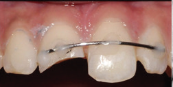
A 22-year-old male patient came to the Department of Conservative Dentistry and Endodontics at Seema Dental College and Hospital, Rishikesh with the chief complaint of pain in his upper left front teeth since last 3-4 months. Patient gave history of trauma few days back. Tooth was Root Canal initiated. On vitality tests tooth was unresponsive. On palpation vestibular tenderness was evident. Tooth was tender on percussion and grade I mobile. Radiographic examination revealed horizontal fracture of Apical third of the root. Apical segment was displaced from its anatomical position.
Root canal treatment was attempted and access cavity was restored with temporary restoration. Endodontic management of coronal segment followed by surgical removal of granulation tissue and displaced apical segment was planned. Informed consent was obtained. After removal of temporary restoration, working length is taken. After that master cone was selected and obturation with lateral compaction, using gutta-percha points of 20 0.6% taper(Dentsply Maillefer, Ballaigues, Switzerland) and Apexit plus (ivoclar vivadent) under rubber dam isolation, 1 day prior to the surgery.
After effective local anesthesia with 2% lignocaine, bleeding points were marked after that a Lubin oschbein flap was reflected. A small defect was visible in the cortical plate with the central incisor, which was enlarged to aid in complete curettage of the granulation tissue (which was sent for biopsy). This was followed by irrigation with betadine (Win Medicare; Win Medicare Dental Products, New Delhi, India) and sterile saline solution. Fractured root end is remove. After that Bone Graft was placed into the defect and completely used as root end filling material in teeth. Finally the flap was repositioned and closed with 4-0 silk suture.


Post operative radiograph
DISCUSSION
Root fractures are relatively uncommon clinical condition with a frequency of 0.5% to 7% in permanent teeth.1 Although the prognosis of root fractures is generally good2 histological reaction at fracture line can occur in one of the following 4 types: interposition of calcified tissue (calus formation), interposition of calcified and connective tissue, interposition of connective tissue and by interposition of granulation tissue, caused by an infected or necrotic pulp.7 Though most frequent tissue reaction at the fracture line is interposition of connective tissue, which occurs in almost 65% of cases 4. Interposition of granulation tissue occurs when the coronal segment loses its vitality and infective products in the coronal pulp cause an inflammatory response.
Tobón et al. concluded that the use of bone regeneration materials, such as non bioabsorbable membranes and resorbable hydroxylapatite improved the predictability of clinical, radiographic, and histologic healing. The use of biomaterials must be necessary when there is a large destruction of the bone circumjacent to the apex. However, a consensus has not yet been reached regarding whether the use of these biomaterials is indispensable for new bone formation and consequent remodeling.
Treatment approach for horizontal root fracture is complex and depends on degree of displacement of fracture fragment, patient’s age, stage of root growth, mobility of the coronal fragment, and diastasis of the fragments.11 Necrotic apical segment should be removed surgically if the remaining coronal root segment is long enough to provide adequate periodontal support. In the present case apical root segment along with granulation tissue was removed surgically followed by root end preparation and placement of Bone graft as root end filling material.
bone graft was shown to be helpful in other studies, especially when the new cementum formed over the dentinal surface and the connective insertion are considered
CONCLUSION
Management of intra alveolar root fracture with complications like calcific metamorphosis, periodontal communication and root resorption is challenging. A tooth with non healing horizontal root fracture with granulation tissue can be treated surgically by removal of fractured apical segment and granulation tissue followed by placement of bioactive materials like bone graft and techniques like retro filling, in its healthy state functionally and aesthetically.
REFERENCES
- Andreasen JO, Andreasen FM, Andersson L, editors. Textbook and Color Atlas of Traumatic Injuries to the Teeth. Munksgaard, Kopenhagen; 2007. Pp. 337-71.
- Andreasen JO, Hjørting-Hansen E: Intra-alveolar root fractures: radiographic and histologic study of 50 cases. J Oral Surg. 1967;25:414-26.
- Caliskan MK, Pehlivan Y. Prognosis of root-fractured permanent incisors. Endod Dent Traumatol. 1996;12:129-36.
- Jacobsen I, Zachrisson BU. Repair characteristics of root fracture-s in permanent anterior teeth. Scand J Dent Res. 1975;83(6):355-34.
- Andreasen JO, Andreasen FM, editors. Root fractures. In: Textbook and color atlas of traumatic injuries to the teeth, 3 rd ed. Copenhagen: Munksgaard; 1994. Pp. 79-314.
- Cvek M, Tsilingaridis G, Andreasen JO. Survival of 534 incisors after intra-alveolar root fracture in patients aged 7–17 years. Dental Traumatology. 2008; 24(4): 379–87.
- Majorana A, Bardellini E, Conti G, Keller E, Pasini S. Root resorption in dental trauma: 45 cases followed for 5 years. Dent Traumatol. 2003;19(5):262-65.
- McCabe PS, Dummer PMH. Pulp canal obliteration: an endodontic diagnosis and treatment challenge. International Endodontic Journal. 2012;45:177–97.
- Roberts HW, Toth JM, Berzins DW, Charlton DG. Mineral trioxide aggregate material used in endodontic treatment: a review of literature. Dent Mater. 2008;24(20):149-64.
- Agrawal Vineet S, K Sonali. Clinical management of severe external root resorption and immature open apex with MTA and calcium hydroxide- A case report. Endodontology. 2013;83-88.
Disclaimer from Guident
The views and opinions expressed in articles published in Guident are solely those of the authors and do not necessarily reflect the views of the editors, the editorial board or the publisher, Ivory India. Guident does not endorse any product, technique or treatment described. Clinical content is intended for qualified dental and medical professionals and is not a substitute for individual clinical judgement. Guident, its editors and the publisher accept no liability for any consequence arising from the use of information published in the journal.
How to cite this article
Dr. Annil Dhingra, Dr. Shalini Sharma. Surgical Management of Root Fracture. Guident. 2019 Dec;13(1). Available from: https://ivoryindia.in/articles/endodontics/surgical-management-of-root-fracture.html