
Dental Stem Cells: Miracle Cells of Dentistry
SIPS Hospital, Lucknow (U.P), India
ABSTRACT
Stem cells are the unique cells that have the potential to develop into different type of cells in the body. These are undifferentiated cells that can differentiate into specialized cells and can divide (through Mitosis) to produce more stem cells. They are found in multicellular organisms1.Stem cells have been isolated from many tissues and organs, including dental tissue. The tooth is a nature’s safe place for these precious stem cells and there are loads of these cells in wisdom teeth, permanent teeth and deciduous teeth. The stem cells contained within teeth are capable of reproducing themselves and can be voluntarily recovered at the time of an intended dental procedure. When stem cells divide, each new cell has the potential either to remain a stem cell or become another type of cell with a more specialized function, such as muscle cell, a red blood cell or a brain cell.
KEYWORDS- Stem cells, differentiation, regeneration, potency.
INTRODUCTION
Stem cells are biological cells that possess two properties: self-renewal, i.e. the ability to go through unlimited cycles of cell division for the purpose of replenishing the cell pool, and potency, i.e. the capacity to differentiate into other cell types .Stem cells are defined as clonogenic cells capable of both self-renewal and multilineage differentiation3.Due to their properties, stem cells have the potency to become an important tool of tissue engineering and regenerative medicine. In mammals, there are 2 broad types of cells-Embryonic stem cells, which are isolated from the inner cell mass of blastocysts, and adult stem cells which are found in various adult tissues including skin, adipose tissues, peripheral blood, bone marrow, pancreas, intestine, brain, hair follicles, as well as in the dental pulp cells. Dental stem cells can differentiate into odontoblasts, adipocytes, neuronal like cells, glial cells, osteoblasts, chondrocytes, melanocytes, myotubes and endothelial cells3.The stem cells used for tooth and periodontal regeneration are both dental and non-dental mesenchymal stem cells.
CHARECTERSTICS OF STEM CELLS
According to the ability and potency to differentiate into different cellular types, 4 types of Stem cells have been established1.
- TOTIPOTENCY-Produce all types of cells as well as germ cells.
- PLURIPOTENCY-Produce all types of cells apart from cells apart from cells of the embryonic membrane.
- MULTIPOTENCY-Distinguish into more than one adult cell.
- UNIPOTENCY-Produce one particular cell type.
Stem cells are distinguished from other cell types by two important characteristics. First, they are unspecialized cells capable of renewing themselves through cell division, sometimes after long periods of inactivity. Second, under certain physiologic or experimental conditions, they can be induced to become tissue-or organ-specific cells with special functions.
In medical therapy, stem cells have been used for engineering many tissues and organs. Stem cell therapy has also been used to treat diseases including Parkinson’s and Alzheimer’s diseases, stroke, burns, heart diseases, diabetes, osteoarthritis and rheumatoid arthritis.2
At present, teeth can only be replaced with prosthesis i.e. removable, fixed or implants with prior bone augmentation if necessary. Stem cell biology and tissue engineering may present new options for replacing damaged or lost tooth.2
TYPE OF STEM CELLS1
1) Embryonic stem cells-These are imitative from embryos that are 2-11 days old known as Blastocysts. These stem cells have the maximum potential to regenerate and repair unhealthy organs and tissues in the body.
2) Adult stem cells-These are undifferentiated cells that occur in a differentiated tissue. Sources of adult stem cells consists of bone marrow ,brain, blood, eye, skeletal muscle, lining of GIT, pancreas, dental pulp, skin, these are multipotent. Depending on their beginning and differential, they can be classified into-a) Hematopoietic stem cells b) Non-hematopoietic or Mesenchymal stem cells(MSC).
DENTAL STEM CELLS-Dental stem cells, along with other stem cells from the craniofacial region, develop from material that is created during the development of the nervous system and have the potential to differentiate into neural cell lines4.The finding of stem cells in natal teeth, supernumerary teeth and odontoma reinforces the concept that stem cells play a key role in the formation of every tooth.2 Two main cell types are involved in dental tissue formation: the ameloblasts, of epithelial origin, that form enamel and the odontoblasts of mesenchymal origin, that are responsible for dentin production. Uses of stem cells in dentistry are to regenerate the dental hard and soft tissues i.e. enamel, dentin, pulp and periodontal tissues.
Enamel is formed from ameloblasts that derive from epithelial stem cells. They are the only cells of ectodermal origin that play a role in tooth development. These cells and their ancestor are lost just after the eruption of the tooth and as a result they do not exist in permanent teeth and cannot therefore be stimulated in vivo, to produce enamel. Regeneration of enamel is dependent on ameloblasts, which are lost as soon as the tooth erupts in the mouth. One of the major problem researchers are facing is to produce sufficient number of enamel forming cells that have the capacity to produce enamel.
Advantages of dental stem cell-
The advantages of dental stem cells are that:
1.) These cells have high plasticity.
2).Dental stem cells are ideal for stem cell banking as they can be preserved for a longer period.
3).These cells have shown a favorable response with scaffold and growth factors.
SOURCES OF DENTAL STEM CELLS
According to the position of mesenchymal stem cells in the tooth, they can be grouped as:3
1).DPSC- Dental pulp stem cells.
2).SHED-Stem cells from exfoliated deciduous teeth.
3).SCAP-Stem cells from apical papilla.
4).PDLSC-Periodontal ligament stem cells.
5).DFPC-Dental follicle progenitor cells.
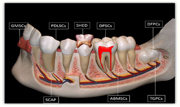
Figure 1.Schematic drawing illustrating sources of human dental tissue-derived MSCs.
ABMSCs: alveolar bone-derived mesenchymal stem cells; DFPCs: dental follicle progenitor cells; DPSCs: dental pulp stem cells; GMSCs: gingival mesenchymal stem cells; PDLSCs: periodontal ligament stem cells; SCAP: stem cells from the apical part of the human dental papilla;
SHED: stem cells from human exfoliated deciduous teeth; TGPCs:tooth germ progenitor cells.
Dental Pulp Stem Cells (DPSC)
The presence of stem cells in dental pulp was proposed by Fitzgerald et al. However, the identification and isolation of DPSC in the adult dental pulp was first reported by Gronthos and coworkers in 20003.Dental pulp tissue is extracted from the teeth recovered during routine dental procedure throughout the life and these teeth are the most convenient and valuable source of DPSCs9.These cells are the most attractive cells for periodontal tissue engineering based on their good growth and differentiation capacity5.Dental pulp stem cells can be found both in adults and children. The DPSC represents less than 1% of total cell population present in dental pulp3. Dental pulp was rich in different types of stem cells like adipocytes, chondrocytes, osteoblasts, and mesenchymal stem cells1.It is a non invasive process of extraction of MSC’s from dental pulp tissues. DPSC’s can be cryopreserved and retain their multipotential differentiation ability and revived whenever needed for future regenerative therapies3. Some of the diseases which are being cured by DPSCs include type 1 diabetes, neurological diseases, immunodeficiency diseases and diseases of bone and cartilages 9.
During the development of teeth, there is an interaction between epithelial cells of dental pulp which lead to the differentiation of ameloblasts and odontoblasts, resulting into deposition of specialized mineralized matrices, i.e., enamel and dentin respectively. The inner area of dental pulp chamber contains a highly proliferative stem/progenitor cells possessing a self-renewal and differentiation properties. It has been shown that after teeth eruption, there is an induction of reparatitive dentin formation which protects dental pulp from further degradation. MSCs constitutes a heterogeneous population of cells which are found first in BM and later in multiple tissues like adipose tissue, skin, cartilage, umbilical cord , placenta and now in dental pulp. As DPSCs have comparable therapeutic potential similar to BMMSCs, DPSCs is another alternative noninvasive source to be used for future regenerative therapies9.
It has been shown that DPSCs can be differentiated by modulation with growth factors, transcriptional factors, extracellular matrix proteins and receptor molecules into different cell types include odontoblast, osteoblast, chondrocyte, cardiomyocytes, neuron cells, adipocyte, corneal epithelial cell, melanoma cell and insulin secreting Beta cells. DPSCs usually remain quiescent when they are within the dental pulps, but respond quickly after injury. Theses DPSCs have a high proliferative capacity and immediately differentiate into odontoblasts, osteoblasts, and chondrocytes to produce dentin, bone, and cartilage tissues respectively for this repair process. It has been shown that the potentiality of DPSCs differentiation into odontoblastsis reduced after passage9 and they can only differentiate into osteoblast cells. DPSCs are mostly derived from cranial neural crest cells because they expressed GFAP, HNK-1, Nestin, P75 and S-100 which are neural crest-stem cell markers. Our recent studies on DPSCs have shown the differentiation of DPSCs into neuron like cells, cardiomyocytes, adipocytes and insulin secreting beta cells9.
STEM CELLS FROM EXFOLIATED DECIDUOUS TEETH (SHED)
Dental pulp from deciduous (baby) teeth, which are discarded after exfoliation, represents an advantageous source of young stem cells. SHED were isolated from dental pulp derived from exfoliated deciduous teeth. Compared to DPSC,SHED show a higher proliferation rate, increased cell-population doublings, sphere-like cell-cluster formation, osteoinductive capacity in vivo, and failure to reconstitute a complete dentin pulp–like complex. These cells have the ability to differentiate into odontoblast-like cells and the regenerated dentin shows immunoreactivity to dentin-specific dentin sialophosphoprotein (DSPP) antibody. Also, SHED is capable of inducing recipient murine cells to differentiate into bone-forming cells3.Due to their origin; DTSCs could be expected to be a good candidate for dental tissue regeneration. To demonstrate this capacity, SHED was seeded in biodegradable scaffolds prepared within human tooth slices and implanted subcutaneously into immunodeficient mice. Differentiation of multicellular SHED population into functional odontoblastos (which expressed dentin sialoprotein and generated tubular dentin) was observed.In the same study, SHED differentiation into endothelial-like cells was shown.This investigation confirmed that SHED has the capacity to generate dental pulp-like tissue in vivo, whose architecture and cellularity resemble that of the natural tooth.This finding suggests the potential use of DTSCs for dental pulp tissue reconstruction and biologically relevant tooth formation8.SHED, unlike DPSCs, cannot be differentiated into osteoblasts or osteocytes but are able to induce the host cells to undergo osteogenic differentiation. SHED possess a higher proliferation rate, as well as high odontogenic and osteogenic differentiation
potential, which make them distinct from the DPSC and represent the more immature form than DPSC11.These cells can repair calvarial defects in micedue to their ability to differentiate into osteoblasts. SHED secretes neurotropic factor for repair of motor neurons following dental injury and therefore it has proposed that SHED can be useful for the treatment of neurodegenerative diseases9.
Neural crest cells could give rise to dental pulp stem cells. Neural crests are multipotent cells with a high regenerative capacity (Rinon et al. 2011).They play a vital role in tooth development by inducing the growth of the mesenchymal component, including odontoblasts, dental pulp, apical vessels and periodontal ligaments (Dangaria et al. 2011). SHEDs have the same molecular properties as neural crests and stem cells. SHEDs are quickly transformed into nerve cells (Seo et al. 2004). Cells from human exfoliated deciduous teeth may differentiate in vitro into nerve cells through the expression of combined gene and protein sets. SHEDs may be used in autologous transplants in the treatment of various neurological diseases and neural traumas (Nourbakhsh et al. 2011)10.
STEM CELLS FROM APICAL PAPILLA (SCAP)
Stem cells from apical papilla (SCAP) are the cells which are found at the tooth root apex. In developing teeth, root formation starts as the epithelial cells from the cervical loop proliferate apically, and influence the differentiation of odontoblasts from undifferentiated mesenchymal cells and cementoblasts from follicle mesenchyme. It has been known that the dental papilla contributes to tooth formation and eventually converts to pulp tissue. As the root continues to develop after the bell stage, the location of the dental papilla becomes apical to the pulp tissue3.They havehigher proliferation rates as well as have a differentiation property in vitro similar to DPSCs. SCAP, similar to DPSC, are more committed to osteo/dentinogenicity. They are capable of differentiating into odontoblast cells and produce dentin in vivo. Due to their higher proliferative potential, SCAPs are also suitable for cell-based therapy for formation of apex roots9.White Mineral Trioxide Aggregate (WMTA) is a capping material has a property to differentiate SCAPs into osteoblast cells and thus SCAPs are involved in this regenerative process. WMTA significantly increase the proliferation of SCAPs within 1-5 days whereas, calcium enriched medium take 7 days to proliferate these cells9.
The discovery of SCAP may also explain a clinical phenomenon, apexogenesis that can occur in infected immature permanent teeth with periradicular periodontitis or abscess. It is likely that stem cells residing in the apical papilla survived the infection due to their proximity to the periapical tissues. Therefore, after endodontic disinfection, under the influence of the survived Hertwig’s epithelial root sheath (HERS), these cells give rise to primary odontoblasts to complete the root formation3.
PERIODONTAL LIGAMENT STEM CELLS (PDLSC)
The periodontal ligament (PDL) is a specialized connective tissue derived from the dental follicle and originates from neural crest cells. The PDL connects the cementum to the alveolar bone, and functions primarily to support the tooth in the alveolar socket. Human periodontal ligament stem cells (PDLSCs) can differentiate into cementoblast-like cells. They also have a capacity to form connective tissue which is riched in collagen I fiber. Human PDLSCs when seeded on 3D scaffolds such as fibrin sponge generate bone in vivo and retain stem cell properties and tissue regeneration capacity9. A recent report identified stem cells in human PDL and found that PDLSC implanted into nude mice generated cementum/PDL-like structures that resemble the native PDL as a thin layer of cementum that interfaced with dense collagen fibers, similar to Sharpey’s fibers. These cells can also differentiate into adipocytes, odontoblasts, myotubes, NFM-positive neuron-like cells, GFAP-positive astrocyte-like cells, and CNPase-positive oligodendrocyte-like cells 3.Periodontal ligament stem cells are responsible for the osseointegration of titanium implants. It has been demonstrated that stem cells proliferate more efficiently on rough surfaces (Heo et al. 2011)10.These cells showed a high rate of proliferation than DPSCs and demonstrated expression of STRO-1, CD146, and a tendon-specific transcription factor. It is thus obvious that PDL itself contains progenitors, which can be activated to self-renew and regenerate other tissues such as cementum and alveolar bone. PDLSCs possessed multilineage differentiation capabilities and were able to undergo osteogenic, adipogenic, and chondrogenic differentiations when cultured with the suitable inductive medium11.
DENTAL FOLLICLE PROGENITOR CELLS (DFPC)
Dental follicle precursor cells (DFPCs) are derived from dental follicle tissue which is a loose connectivetissue that surrounds the developing tooth. These cells have an ability to produce bone and cementum. Therefore, these stem cells have great potentiality to use in periodontal and bone regeneration therapies9.
DFPC are localized in the dental follicle, a mesenchymal tissue that surrounds the tooth germ and can be easily isolated after wisdom tooth extraction. However, these cells are available only from patients during wisdom tooth eruption, usually between 15 and 28 years of age.Human DFPC have the ability to differentiate toward alveolar osteoblasts, PDL fibroblasts, cementoblasts, adipocytes, and neuron-like cells3.DFPCs derived from human third molars teeth are rapidly attached to the culture plates and form a calcified nodule. They also exhibit a better plasticity than other dental stem cells. Dental pulp and dental follicle stem cells have similar mesenchymal stem cell characteristics, but DFPCs are easily accessible for cell culture and have a higher proliferation capacity than DPSCs. Therefore it appears that DFPCs might have more advantages as a stem cell resource for regenerative therapies in dental abnormalities9.
ISOLATION OF DPSCs
Several investigators have described various methods for isolation of stem cells from human dental pulp. Raoof et al have used three different methods for isolation of DPSCs from dental pulp tissue:1 Dental pulp tissue is digested with collagenase or dispase enzyme and isolated trypsinised cells are plated in culture dishes;2 They have explanted undigested dental pulp small tissue pieces directly to petridishes and3 Dental pulp tissues are initially trypsinised and then small tissue pieces are explanted to petridishes for their outgrowth. They have grown these cultures in Minimum Essential Medium (MEM) supplemented with 20% FBS at 37 ℃with 5% CO2 and 90% humidity in CO2 incubator. They have recommended the third method for isolation of DPSCs from dental pulp. This method gives better cell outgrowth with achieving confluency at 60% within 2 d of culture. They have further checked the pluripotency of these cells by studying the expression of Nanog, OCT-4, and Nucleotoxin markers by RT/PCR analysis. Thus, this study proposes the third method for obtaining better DPSCs with high efficacy in a short time9.
STEM CELL HANDLING AND CRYOPRESERVATION
Stem cells are discharge from minute amounts of tissues, in the case of stem cells from dental pulp. The tissue is positioned in an enzyme solution so as to release the stem cells, which are then cultured to increase. This can be completed using serum-free medium, eliminating the need for use of animal serum. Differentiation then arises and the cells are transplanted – either single or with a scaffold or other biomaterials, depending on the purpose.
Extracted permanent and primary (including exfoliating) teeth can be conserved for future use with cryopreservation. The cells are quickly cooled to sub-zero temperature as low as -196° C, impeding any cellular or biochemical action. Rapid freezing is essential to prevent ice from forming around or within the cells and to avoid dehydration, as these would origin cell harm and demise. Research has confirmed that stem cells obtained from the dental pulp of extracted third molars hold on to the ability to differentiate into numerous cell types following defrosting after cryopreservation using liquid nitrogen. Stem cells removed from the periodontal ligament are workable following cryopreservation. After two years of cryopreservation, stem cells have been capable to distinguish and to multiply, and it has been concluded that dental stem cells can go through long-term cryopreservation1.
DERIVATIVES OF DENTAL STEM CELLS

Figure 2.The figure enumerates various cell types derived from dental stem cells: (a) cementoblast, (b) adipocyte, (c) odontoblasts, (d) neuronal cells, (e) myoblast, (f) chondrocyte, (g) pulp cells, (h) hepatocyte, (i) endothelial cell, (j) osteoblast, and (k)melanocyte.
THE DENTIN–PULP REGENERATION
Dental pulp is a complex organized tissue with various types of cells and structures, providing nutrition, sensation, and defense against the various pathogens; additionally, it produces dentin and maintains the biological and physiological vitality of the dentin. Pulpitis , which is one of the most common diseases related to pulp, usually caused by dental trauma and caries. Due to the complex structure, small volume, and insufficient blood supply of dental pulp, it is difficult to eradicate the infection and initiate self-healing in pulpitis. The traditional therapy for pulpitis is root canal therapy, by removing the inflamed pulp and replacing it with inorganic material. However, this treatment usually leads to tooth fragility and fracture (Ricketts, 2001). As a result, keeping or regenerating vital pulps is a better choice. There are primarily two approaches for dental pulp regeneration: pulp revascularization; and scaffold and/or stem cell-based pulp regeneration. Dental pulp revascularization usually depends on inducing host cells from the apical region to migrate into the root canal and differentiate into a vascularized pulp tissue. The root canal space should be disinfected, and the resulting blood clots induce cell migration and initiate pulp tissue formation. Another approach is scaffold and/or stem cell-based pulp regeneration. Since Mooney and colleagues’ first study of pulp tissue engineering (Mooney, Powell, Piana, & Rutherford, 1996), numerous studies have been made in a variety of animal models to achieve complete pulp regeneration7.
PERIODONTAL REGENERATION
Periodontitis is a chronic infectious disease of the soft and hard tissues supporting the teeth and is a major cause of tooth loss in adults (Pihlstrom, Michalowicz, & Johnson, 2005). Mesenchymal stem cells derived from inflamed periodontal ligaments exhibit impaired immunomodulation (Liu et al., 2012). Conventional periodontal therapy involves debridement of the root surface to induce healing, guided tissue regeneration (Needleman et al., 2006), and bone graft placement. Indeed, the traditional treatment for periodontitis is associated with a relatively high degree of variability in clinical outcome (Aichelmann-Reidy & Reynolds, 2008), and the curative effect remains unsatisfactory. The advances in regenerative medicine have made it possible for periodontal regeneration based on MSC-mediated tissue engineering (Bartold, McCulloch, Narayanan, & Pitaru, 2000). Among all the mesenchymal stem cells, PDLSCs are the main candidate stem cells in periodontal regeneration. Transplanting PDLSCs directly into periodontal defect areas resulted in periodontal regeneration (Bartold, Shi, & Gronthos, 2006). When transplanted into surgically created periodontal defect areas in miniature pigs, autologous and allogeneic PDLSCs also were capable of regenerating periodontal tissues (Ding, Liu, et al., 2010; Ding, Wang, et al., 2010; Liu et al., 2008), indicating
PDLSC-mediated tissue engineering could be a useful treatment for periodontitis. The potential mechanism has also been clarified and is largely dependent on the immunomodulatory properties of PDLSCs7.
For regenerating periodontal tissue, the use of growth and differentiation factors is one of the most popular tissue engineering approaches. Till date, several growth factors including transforming growth factors-b (TGF-b) superfamily members, such as, BMP-2, BMP-6, BMP-7, BMP-12, TGF-b, basic fibroblast growth factors (bFGF), and platelet-derived growth factors(PDGF) have been used. These factors have been used as a protein-based approach for periodontium regeneration.
Nagatomo et al. in their experimental studies found that periodontium can be regenerated by using PDL cells that have stem cell properties.
Iwata et al. did a similar in vitro study wherein PDL cells were harvested and expanded. He also made transplantable constructs of primary canine containing PGA scaffold and PDL cell sheets. Regeneration of periodontal structures that included cementum, alveolar bone, and periodontal fibers was induced by the transplanting constructs in combination with porous bTCP (b-tricalcium phosphate)2.
Previous studies mainly combined the stem cells with a scaffoldfor periodontal regeneration, for example, collagen, fibrin, hydrogel, and gelatin. However, major concerns are the complicated transplantation process and the potential for host rejection. Recently, non-scaffold tissue engineering has been of increasing interest to researchers. There are primarily two non-scaffolds strategies-cell injection and cell sheets. Cell injection has been the common treatment in stem cell-based tissue engineering (Kinnaird et al., 2004). Local injection of DPSCs or PDLSCs has also been shown effective for treating periodontal diseases (Baik, Park, Lee, & Chung, 2014). Periodontal regeneration was observed in a rat periodontitis model after local injection of a BMMSC suspension (Du, Shan, Ma, Wang, & Fan, 2014). Cell sheet engineering has been developed as a unique, scaffold-free method of cell processing by culturing in ascorbic acid (Wei et al., 2012; Du et al., 2014) or utilizing temperature-responsive cell culture vessels (Owaki, Shimizu, Yamato, & Okano, 2014). Cell sheets can maintain extracellular matrix and cell–cell junctions, which would be degraded by proteolytic enzymes such as trypsin and/or dispase, and has shown positive outcomes in treating many diseases (Nishida et al., 2004; Ohashi et al., 2007; Owaki et al., 2014).Besides PDLSCs (Ding, Liu, et al., 2010; Ding, Wang, et al., 2010; Liu et al., 2008), DPSCs (Hu et al., 2016; Khorsand et al., 2013), SHEDs (Fu, Jin, Ma, Fan, &Wang, 2014), BMMSCs (Du et al., 2014), ADSCs also have the potential for periodontal regeneration (Tobita& Mizuno, 2013)7.

FIGURE-3 Regenerative strategy of periodontal tissue.
TOOTH REGENERATION:
The regeneration of adult teeth will be possible in future with the help of tissue engineering and newer expansion in stem cell therapy. Regenerative procedures would be improved fitting and substitutes in place of dental implants. Experimental studies with animal models have exposed that the tooth crown formation can be regenerated using tissue engineering techniques that merge stem cells and recyclable scaffolds. Epithelial mesenchymal exchanges are mandatory in tooth development. “These exchanges are considered by the reciprocal exchange of signals between these two native germ layer tissues and outcome in the emergence of inimitable terminal phenotypes with their supporting cells”.
Three key elements are involved in tooth regeneration which includes:
- Inductive morphogenes
- Stem cells
- Scaffold
Following steps are involved in regeneration of tooth:
1. Harvesting and spreading out of adult stem cells.
2. seeding the stem cells into scaffold which provides optimize environment.
3. Cells are instructed with targeted soluble molecular signals spatially.
4. The gene expression profile is confirming by the cells for next phase in odontogenesis.
Duailibi et al., in their studies were able to form tooth from single cell suspensions of cultured rat tooth bud cells. They confirmed bioengineered rat teeth grown in 12 weeks with PLGA and PGA scaffold. Honda et al. developed tissue engineered teeth, when implanted into omentum of rat utilizing porcine tooth bud cells and PGA fiber engage scaffold that reminds of the model of odontogenesis. Young et al., using porcine tooth bud cells, PLGA and PGA scaffolds produced a crossbreed tooth bone for the cure of tooth loss beside with alveolar bone resorption1.
THE BIO ROOT REGENERATION
Although great progress has been made in whole tooth regeneration, there are still some obstacles, such as uncontrolled morphology and tooth eruption. The tooth root provides a stable anchor for a natural or post supported crown. Compared to whole tooth regeneration, regenerating the root may be more feasible in the near future. A bioroot was regenerated by implanting preshaped root-like scaffolds combined with mesenchymal stem cells into the alveolar bone to form a functional root, having root-likestructure, similar biomechanical properties and elements to natural teeth, periodontal ligament-like tissue and dentin-like matrix structure, and is capable of supporting post crown prostheses.Compared to a dental implant, a biorootis superior in biomechanical properties, such as compressive strength, modulus of elasticity, and torsional force, that is, all more similar to the properties of the natural tooth root (Gao et al., 2016). In addition, treated dentin matrix (TDM) could be a biological scaffold for dentinogenesis and tooth root construction (Luo et al., 2015; Yang et al., 2012)7.

FIGURE-2 Regenerative strategy of whole tooth and bioroot.
Deciduous tooth stem cells (DTSCs) Versus Dental pulp stem cells(DPSCs)
DPs from deciduous and permanent teeth differ both anatomically and functionally and hence could differentially determine stem cell fate. The gene expression profile determines tissue and stem cell capacity to differentiate toward a specific lineage. This analysis also allows a composition of the properties of different stem cell populations isolated from the same or different sources. DNA microarray analysis revealed 4386 genes with a differential expression between DPSCs and DTSCs (SHED) by 2.0-fold or more. In particular, higher expression in DTSCs was observed for the genes related to pluripotency (OCT4, SOX2, NANOG, and REX-1), cell proliferation, and extracellular matrix, including several cytokines such as fibroblast growth factor and tumor growth factor b. The most prominent up regulation was observed in expression of collagens (Col I, III, VII, and XIII) and proteoglycans (glypican and versican). Both of these studies infer that DTSCs retained, whereas DPSCs lost, their plasticity through passaging. Taken together these results provide an additional evidence of the divergence in stem cell properties between DTSCs (deciduous teeth) and DPSCs (adult teeth) and indicated that DTSCs could be useful for cell-based regeneration therapy not only for oral disease but also for systemic diseases8.
NONDENTAL STEM CELLS FOR DENTAL APPLICATIONS
Just as dental stem cells have the potential for therapeutic use throughout the body; stem cells from outside the oral cavity may be used for regenerating tissue in the mouth. Ivan Ho, DDS, is the founder of Platinum Dental in Rancho Santa Margarita, Calif. In March 2009, he performed an outpatient procedure called Bone Marrow Aspirate Concentration (BMAC) to strengthen the insufficiently dense mandible of a patient in preparation for a permanent dental implant.
BMAC has been used for many years by neurosurgeons and orthopedic surgeons to generate bone for procedures such as spinal and tibia fusions. It is a minimally invasive, relatively painless outpatient procedure that takes between one and three hours to perform, providing faster healing, less pain and better results than other implant procedures.
“This minimally invasive procedure is an innovative, advanced long-term alternative to existing tooth replacement options such as dentures, bridges and even traditional bone-grafting procedures,” Ho explained. “In addition to providing better and faster healing, the transplantation of the patient’s own stem cells enables the body to increase bone growth in the jaw through angiogenesis to permanently support the dental implant. Because the use of dentures and bridges carries a high risk of problems—in many cases resulting in gum disease, tooth decay and the loss of viable teeth—additional costly surgical procedures are necessary. Dental implants are the most natural solution and provide the best long-term results.”
Ho harvested his patient’s own bone marrow mesenchymal stem cells through a small incision in the hip bone the morning of the procedure. Using a centrifuge, he isolated and concentrated the stem cells from the plasma, transplanted them into the patient’s jaw and added an allograft. The stem cells then began to work with the surrounding tissue to eventually generate healthy, dense bone tissue in preparation for the implant4.
STEM CELL BANKS-
“Dental stem cells are a valuable source of stem cells and are found in teeth with healthy pulp,” StemSave’s Chotkowski explained. “These teeth could be deciduous teeth, wisdom teeth and other permanent teeth. Presently, these teeth are being discarded as medical waste. A stem cell bank allows an individual the opportunity to preserve their biomaterial for future regenerative therapies.”
Chotkowski cited the banking of umbilical cord blood as a source of stem cells, which has been advocated for the past 15 years to parents of newborns. “To date, there have been many lives saved through the transplantation of these stem cells,” he said. “Cryopreservation or banking of stem cells maintains the viability of these cells indefinitely.
“During cryopreservation, the cells are put to sleep through a process called vitrification, in which the tissue is placed in liquid nitrogen at a temperature of –196 degrees Celsius. The cryopreservation process stops all cellular metabolism involving both cell growth and cell death. The cells preserved today can be applied to future regenerative therapies. There are many parents who did not have the opportunity to bank their children’s cord blood. StemSave allows these parents another chance to bank their children’s accessible and valuable stem cells when they are undergoing a routine dental procedure4.”
Compared to other stem cells’ banking, tooth stem cell banking is justat the initial stage, but is catching up quickly. Cryopreservation does not affect the biological and immunological properties of SCAPs, supporting the feasibility of SCAP cryopreservation in nitrogen (Ding, Liu, et al., 2010; Ding, Wang, et al., 2010). In the USA, there are several companies or institutions (BioEden, StemSave, The Tooth Bank, National Dental Pulp Laboratory, and Store-A- Tooth) involved in banking tooth stem cells. In Japan, Hiroshima University established a toothbank named “Three Brackets,” and Nagoya University also developed a tooth bank in 20077.
THE FUTURE OF DENTAL STEM CELL RESEARCH
Chotkowski said that this is an exciting time for the dental professional. “We will continue to see the advancement of dental stem cell research and dental stem cells applied to clinical therapies,” he explained, noting that several clinical trials utilizing dental stem cells have been proposed outside the United States. “In Japan, they have been able to form a whole tooth from stem cells in mice. Stem cells are being applied to dental implants and they have been shown to form a ligamentous attachment between the implant and bone.
“Regeneration or regrowing the pulp of a tooth is right around the corner, and it will change the specialty of endodontic within the next five years. On a global scale, there is a tremendous investment from both the public and private sectors in stem cell research. The Obama administration has made stem cell research one of the pillars of his health program. The U.S. Army is investing over $250 million in stem cell research to treat injuredsoldiers in a project called AFIRM (Armed Forces Institute for Regenerative Medicine).”
“Dental stem cells may be able to repair cardiac muscle that becomes damaged during a heart attack or from ischemic heart disease,” Spolarich said. “Also, laboratory research has shown that roots of teeth can be regrown in animal models, suggesting that in the future, people may be able to regrow their own tooth roots, replacing the use of traditional metal implants4.”
CONCLUSION
Stem cell therapy has a promising future in tissue regeneration and the management of disease. It allows the repair of defective tissues or functions through the transplantation of autologous cells. Several kinds of stem cells have been isolated from human adult teeth and have been used for tooth and periodontal regeneration. Stem cell therapy has got a cardinal role as a forthcoming treatment modality in dentistry. The ultimate goal of tooth regeneration is to replace the lost teeth by a natural tooth. Stem cell-based tooth engineering is conceived as an upcoming approach in the making of a biological tooth (biotooth)2. Stem cell-based therapies represent the most promising alternative for successful regeneration of damaged or pathological dental tissues or even the entire tooth following tooth loss. The identification of a good stem cell source is a major key for a success in tissue regeneration. It is therefore fundamental to understand the exact mechanisms underlying the potential of the various dental stem cell populations as well as their behavior after transplantation in ectopic sites.
REFERENCES
1). Harendersingh, D.J.Bhasker, Rahilarehman, Chatainya Dev Jain, Mohsin Khan-“Stem cells: An emerging future in dentistry”.
2). Rani Somani, Shipra Jaidka, Neeti Bajaj, Sameksha Arora-“Miracle cells for natural dentistry – A review”.
3). Vladimir Petrovic and Vladisav Stefanovic University of Nis School of Medicine, 18000 Nis, Serbia-“Dental Tissue – New Source for Stem Cells”.
4).Jean Majeski-“Dental Stem Cells in Research and Practice”.
5). Nahid Y. Ashri, BDS, MSc, Sumaiah A. Ajlan, BDS, MSc, Abdullah M. Aldahmash, MSc, PhD.-“Dental pulp stem cells-Biology and use for periodontal tissue engineering”.
6). Shayee Miran, Thimios A. Mitsiadis, and Pier Francesco Pagella-“Innovative Dental Stem Cell-Based Research Approaches:The Future of Dentistry”.
7). L Hu,Y Liu,S Wang-“Stem cell-based tooth and periodontal regeneration”.
8). Irina Kerkis, and Arnold I Caplan-“Stem Cells in Dental Pulp of Deciduous Teeth”.
9)Pravin D Potdar, Yogita D Jethmalani-“Human dental pulp stem cells: Applications in futureregenerative medicine”.
10). P. DziubiÅ„ska, M. Jaskólska, P. Przyborowska, Z. Adamiak-“Stem cells in dentistry – Review of literature”.
11).Elna Paul Chalisserry,Seung Yun Nam Sang Hyug Park and Sukumaran Anil-“Therapeutic potential of dental stem cells”.
12. Murray PE, Garcia-Godoy F, Hargreaves KM. Regenerative endodontics: a review of current status and a call for action. J Endod.2007;33:377–390.
13. Bluteau G, Luder HU, De Bari C, Mitsiadis TA. Stem cells for tooth engineering. Eur Cell Mater.2008;16:1–9.
14. Jiang Y, Jahagirdar BN, Reinhardt RL, et al. Pluripotency of mesenchymal stem cells derived from adult marrow. J DentRes. 2002;418:41–49.
15. Bonassar LJ, Vacanti CA. Tissue engineering: the first decade and beyond. J Cell Biochem Suppl. 1998;30(31):297–303.
16. Shaikh R. Therapeutic potential of stem cells in regenerative dentistry; a review of literature. Int J Stud Res. 2013;1(May (4):22–30.
17. Jamal M, Chogle S, Goodis H, Karam SM. Dental stem cells and thier potential role in regenerative medicine. J Med Sci. 2011;4:53–61.
18. Honda MJ, Imaizumi M, Tsuchiya S, Morsczeck C. Dental follicle stem cells and tissue engineering. J Oral Sci. 2010;52(4):541–552.
19. Estrela C, Alencar AH, Kitten GT, Vencio EF, Gava E. Mesenchymal stem cells in the dental tissues: perspectives for tissue regeneration. Braz Dent J. 2011;22(2):91–98.
20. Weissman, I.L. (2000) Stem cells: units of development, units of regeneration, and units in evolution. Cell 100, 157–168.
21. Wagers, A.J. and Weissman, I.L. (2004) Plasticity of adult stem cells. Cell 116, 639–648.
22. Gronthos, S., Brahim, J., Li, W., Fisher, L.W., Cherman, N., Boyde, A., DenBesten, P., GehronRobey, P., and Shi, S. (2002) Stem cell properties of human dental pulp stem cells. J. Dent. Res. 81, 531–535.
23. Gronthos, S., Mankani, M., Brahim, J., Robey, P.G., and Shi, S. (2000) Postnatal human dental pulp stem cells(DPSC) in vitro and in vivo. Proc. Natl. Acad. Sci. U. S. A. 97, 13625–13630.
24. Miura, M., Gronthos, S., Zhao, M., Lu, B., Fisher, L.W., Robey, P.G., and Shi, S. (2003) SHED: stem cells from human exfoliated deciduous teeth. Proc. Natl. Acad. Sci. U. S. A. 100, 5807-5812.
25. Stem cell - Wikipedia, the free encyclopedia. En.wikipedia. org/wiki/ Stem cell.
26. Sloan AJ and Waddington RJ. Dental pulp stem cells: what, where, how? Int J Pediatric Dent 2009; 19: 61–70.
27.Abdul R and Shaikh R. Therapeutic Potential Of Stem Cells In Regenerative Dentistry; A Review Of Literature. International Dental Journal Of Student’s Research. 2013; 14: 22-30.
28. Desai VD, Varma b, Maheshwari S, Bumb D. Tooth Regeneration By Stem Cells -An Innovative Approach. Asian J. Pharm. Hea. Sci. 2012; 23:433-7.
29. Saraswathi K Gopal Lankupalli AR, Stem Cell Therapy. J Clin Diagn Res. 2012 61: 142-4.
30.Kompalli PV, Stem Cells - A Review. Webmedcentral Biomedical Engineering 2013; 42:Wmc004046.
31.Buch A & Choksi D. Clinical Applications Of Stem Cells In Dentistry. Dharmsinh Desai University, Faculty od Dental Science, Journal od Dental Sciences. 2010; 11: 26-31
32.Masthan KMK, Babu NA, Sankari SL, Krisnan TG And Elumalai M. Life Saving Is Cost Effective - Dental Pulp Stem Cells. Int J Pharm Bio Sci 2013; 42: 361–5.
Disclaimer from Guident
The views and opinions expressed in articles published in Guident are solely those of the authors and do not necessarily reflect the views of the editors, the editorial board or the publisher, Ivory India. Guident does not endorse any product, technique or treatment described. Clinical content is intended for qualified dental and medical professionals and is not a substitute for individual clinical judgement. Guident, its editors and the publisher accept no liability for any consequence arising from the use of information published in the journal.
How to cite this article
Dr. Vikram Ahuja Dr. Abhishek Kr. Pandey Department of Dentistry. Dental Stem Cells: Miracle Cells of Dentistry. Guident. 2018 Mar;11(4). Available from: https://ivoryindia.in/articles/general/dental-stem-cells-miracle-cells-of-dentistry.html


