Management of Pregnant Patient in Dental Practice
Department of Oral and Maxillofacial Surgery Jodhpur Dental College Jodhpur National University, Boranada, Jodhpur
INTRODUCTION
Pregnancy is a process comprised of complex physical and psychological changes that profoundly affect even healthy women. The changes that occur are the result of increasing maternal and fetal requirements for the growth of the fetus and the preparation of the mother for delivery. Agents such as pharmacologic, radiologic, and environmental influences all need to be evaluated when preventive dentistry during pregnancy is considered.. At this point, the dentist can render treatment which alleviates the dental complaint without creating undue risks for either the mother or the fetus
ORAL AND FACIAL CHANGES
Oral changes seen in pregnancy include gingivitis,gingival hyperplasia, pyogenic granuloma, and salivary changes.2Increased facial pigmentation is also seen. Elevated circulating estrogen, which causes increased capillary permeability, predisposes pregnant women to gingivitis and gingival hyperplasia3,4.Pregnancy gingivitis usually affects marginal and interdental papilla 5 and is related to preexisting gingivitis. Good oral hygiene can help to prevent or reduce the severity of the hormone-mediated inflammatory oral changes. Pregnancy does not cause periodontal disease but doesworsen an existing condition6.Pyogenic granuloma (pregnancy tumor) occurs in about 1% to 5% of pregnant women.7 Increased angiogenesis, due to sex
hormones coupled with gingival irritation by local factors such as plaque, is believed to cause pyogenic granuloma.8 It occurs mainly on the labial aspect of the interdental papilla.
The main salivary changes in pregnancy involve its flow, composition, pH, and hormone levels. The change in composition of the saliva includes a decrease in sodium and pH, and an increase in potassium, protein, and estrogen levels.7,8 Salivary estrogen level has been suggested as a screening test to detect the risk potential for preterm labor. Salivary estrogen levels are higher in the women destined to have preterm babies than in women having normal term deliveries.9,10
Salivary estrogen also increases the proliferation and desquamation of the oral mucosa and an increase in subgingival crevicular fluid levels. The desquamating cells provide a suitable environment for bacterial growth by providing nutrition predisposing the pregnant woman to dental caries.7 There is an increase in facial pigmentation called melasma or the ‘‘mask of pregnancy,’’ appearing as bilateral brown patches in the mid-face.11 These facial changes begin during the first trimester12 and are seen in up to 73% of pregnant women.11 The etiology of this condition is unknown, but it might be related to the increase in serum estrogen and progesterone 13Melasma usually resolves after parturition.5
OPTIMAL TRIMESTER OF PREGNANCY FOR DENTAL TREATMENT
Any dental treatment of an urgent nature can be provided at any time during pregnancy 14 .Traditionally, dental treatment of any kind has been avoided during the first trimester of pregnancy, so as not to harm the fetus during the stage of organogenesis. However, there is not enough evidence to forbid dental treatment even during the first trimester of pregnancy. The ideal period for a complete dental treatment of a pregnant woman is the beginning of the second trimester (14-20 weeks of pregnancy) 15.At this stage, there is no risk of teratogenesis, nausea and vomiting have subsided, and the uterus is not yet large enough to cause discomfort. It is essential to treat all dental problems during the second trimester, not only because the
woman should have a healthy mouth, but also because during labour she might need to be intubated, in which case teeth with a bad prognosis (e.g.,tooth mobility due to periodontal disease) may be damaged or lost 16
POSITIONING OF PREGNANT WOMEN INTHE DENTAL CHAIR
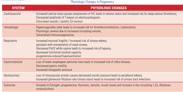
During the third trimester of pregnancy, when the woman is supine, the uterus may press on the inferior vena cava and impede venous return to the heart, which may lead to hypotensive syndrome (occurring at a rate of 15- 20%) and loss of consciousness 17.In order to prevent this from happening during dental treatment, when seated in the dentist’s chair the pregnant woman’s head should always be higher than her feet, and, if necessary, a small pillow (or a folded blanket) should be placed under her right hip (“leftuterine displacement”) so that the uterus is moved away from the inferior vena cava18, 19
RADIOGRAPHS IN PREGNANCY
Dental radiographs may be prescribed during pregnancy, because radiation exposure to the fetus in utero is negligible.20The dose to the fetus is about 1/50 000 of the direct exposure to the head.20
Teratogenecity of radiation depends on fetal age and the dose ofradiation.21 The greatest risk to the fetus for teratogenecity and death isduring the first 10 days after conception. The most critical period of fetal development is between 4 and 18 weeks after conception. The chance offetal teratogenecity with an exposure of 0.01Gy is about 0.1% and radiationdoses of up to 0.05 Gy or less are not associated with significant increase intetratogenecity.22The National Commission for Radiation Protection (NCRP) recommends that thecumulative fetal dose should not exceed 0.005 Gy.23Fetal exposure to radiation of more than 0.20 Gy will cause microcephalyand mental retardation.21 Radiographs employed in dentistry such as the panoramic and full mouth intraoral series are generally safe during pregnancy. The average radiation doses absorbed by the fetus in panoramicand full mouth radiographs are 1.53104 Gy and 105 Gy respectively . The dental radiation received is also 40-fold less than the naturally occurring background radiation.22, 23Although the risk of teratogenecity is exceedinglylow with dental radiographs, the amount of radiation exposure to thepregnant mother and fetus can and must be minimized even further byusing bitewing radiographs instead of panoramic radiographs, usinghigh-speed films (E speed), the use of rectangular collimation instead ofcircular collimation, properly collimated beam, and lead aprons over theabdomen.24,25 Following all the above-mentioned precautions will furtherreduce the patient’s radiation exposure.25Estimated radiation exposure to the human body in Gray (Gy) 22,25,26

DRUGS USED IN PREGNANCY
The Swedish system for the classification of fetal risk of drugs was the firstof its kind and was implemented in 1978. Drugs for use in pregnant women are classified in 4 general categories — A to D.27

The US Food and Drug Administration (FDA) introduced a system in 1979 also using the letters A to D, together with an X category27 Category X—Risk of use in pregnant women clearly outweighs possible benefits


Small number but a wide variety of drugs that are teratogens (ie, drugs that can cause either structural or functional birth defects.28

DENTAL MANAGEMENT GUIDELINES29
First trimester (conception to 14th week)
The most critical and rapid cell division and active organogenesis occur between the second and the eighth
week of postconception. Therefore, the greater risk of susceptibility to stress and teratogens occurs during this time and 50% to 75% of all spontaneous abortions occur during this period.30
The recommendations are:
1. Educate the patient about maternal oral changes during pregnancy.
2. Emphasize strict oral hygiene instructions and thereby plaquecontrol.
3. Limit dental treatment to periodontal prophylaxis and emergency treatments only.
4. Avoid routine radiographs. Use selectively and when needed.
Second trimester (14th to 28th week)
Organogenesis is completed and therefore the risk to the fetus is low. This is the safest period for providing dental care during pregnancy
The recommendations are:
1. Oral hygiene, instruction, and plaque control.
2. Scaling, polishing, and curettage may be performed if necessary.
3. Control of active oral diseases, if any.
4. Elective dental care is safe.
5. Avoid routine radiographs. Use selectively and when needed
Third trimester (29th week until childbirth)
Although there is no risk to the fetus during this trimester, the pregnant mother may experience an Increasing level of discomfort. Short dental appointments should be scheduled with appropriate positioning while in the chair to prevent supine hypotension. It is safe to perform routine dental treatment in the early part of the third trimester, but from the middle of the
third trimester routine dental treatment should be avoided.
The recommendations are:
1. Oral hygiene, instruction, and plaque control.
2. Scaling, polishing, and curettage may be performed if necessary.
3. Avoid elective dental care during the second half of the third trimester.
4. Avoid routine radiographs. Use selectively and when needed
CONCLUSION
Good oral health during pregnancy is important because the condition of a pregnant woman’s mouth can affect her health and that of the fetus. It is vital that health professionals collaborate to ensure pregnant women receive proper oral assessment and intervention as well as oral health education. Prescribing of systemic drugs of any kind ideally should be performed after consultation with the general medical practitioner or obstetrician. Oral health assessment should be part of comprehensive prenatal care for all women and every general medical practitioner and obstetrician should consider referral of a newly pregnant woman to a dentist as routine.
REFERENCES
- Management of Pregnant Patient in Dentistry Sophia Kurien, Vivekanand S Kattimani, Roopa Rani Sriram etal Journal of International Oral Health. Jan-Feb 2013; 5(1):88-97
- Lakshmanan Suresh, BDS,and Lida Radfar, DDS, MS,b Buffalo, NY. Pregnancy and lactation (Oral Surg Oral Med Oral Pathol Oral Radiol Endod 2004;97:672-82
- Soory M. Hormonal factors in periodontal disease. Dent Update 2000;27:380-3.
- Hugoson A. Gingivitis in pregnant women. A longitudinal clinical study. Odontol Revy 1971;22:65-84
- Neville BW, Damm DD, Allen CM, Bouquot JE. Oral & Maxillofacial Pathology. 3rd ed. Philadelphia: W B Saunders; 2002. p. 329-30, 447-9.
- Tilakaratne A, Soory M, Ranasinghe AW, Corea SM,etal Periodontal disease status during pregnancy and 3months post-partum in rural population of Sri-Lankan women.
Disclaimer from Guident
The views and opinions expressed in articles published in Guident are solely those of the authors and do not necessarily reflect the views of the editors, the editorial board or the publisher, Ivory India. Guident does not endorse any product, technique or treatment described. Clinical content is intended for qualified dental and medical professionals and is not a substitute for individual clinical judgement. Guident, its editors and the publisher accept no liability for any consequence arising from the use of information published in the journal.
How to cite this article
Dr. Ashish Vyas Assistant. Management of Pregnant Patient in Dental Practice. Guident. 2014 Mar;7(4). Available from: https://ivoryindia.in/articles/general/management-of-pregnant-patient-in-dental-practice.html