Rationale for a Bi-Jaw Surgery in an Asymmetric Class III Mandible
Department of Orthodontics, Manipal College of Dental Sciences, Manipal University, Manipal, Karnataka, Manipal Melaka College of Dental Sciences, Manipal University, Manipal, Karnataka, Department of Oral Biology, Department of Orthodontics, Manipal College of Dental Sciences, Manipal University, Manipal, Karnataka
Abstract
A 22 yr-old male patient from Malaysia reported with a chief complaint of large lower jaw in July 2011. His problem list included mandibular prognathism, an asymmetrical jaw deviating 6mm to the left, and a skeletal posterior crossbite on the left side. His molar relationship was class-III. He had an openbite of 1.5mm and an overjet of -7mm; the maxillary dental midline was deviated 2mm to the right, and the mandibular midline 6mm to the left. The patient had 0.5 mm of lower anterior crowding. Orthognathic surgery involved a LeFort I maxillary superior impaction and advancement osteotomy and a mandibular Bilateral Sagital Split Osteotomy (BSSO) with rotation for aligning midlines, to position the maxilla 3mm anteriorly into a 2mm overbite-overjet relationship and to correct the crossbite. The segments were stabilized using rigid internal fixation. The entire treatment took two years and four months, partly because waiting for authorization and scheduling the surgery took an additional eight months. Detailed finishing of the occlusion was accomplished with fixed appliances during the four months after surgery.
Keywords: mandible, prognathism, crossbite, Class III molar relationship, BSSO.
INTRODUCTION
<5/p>
John Hunter defined mandibular prognathism as: “The lower jaw projecting too far forward so that the foreteeth pass before those of the upper jaw, therefore disfigurement and malocclusion are two of the main facial characteristics.” Forty percent of facial asymmetrie shave Class III malocclusion compared to 28% of asymmetric Class II patients.1 These Class III patients are also associated with a deviated mandibular symphysis(chin). Total 80% of those deviations are to the left side.1Laterality is more common in males (41%) than females (40%). In addition, orbital dystopia, nasal tip deviation, maxillary occlusal cant, and zygomatic arch discrepancy are seen in 25% of Class III patients.
Intra-orally, a Class III molar relation with an anterior cross bite and a dental compensation, (consisting of labial tipping of the maxillary anterior teeth with lingual tipping of the mandibular anterior teeth) is a common feature. Other distinguishing features are incomplete closure of lip, deviation of the midline, and decrease of mento-labial fold.
The etiology of facial asymmetry is categorized into genetic and environmental origins. In one study of Class III children, it was reported that one-third had a parent with the same skeletal pattern and one-sixth had a Class III sibling.2 We can also cite the historic Hapsburg family, as an example of a Class III pattern. Finally, the association of the class IIIproblem (especially maxillary deficiency) with syndromes such as Crouzon, Pffeifer and achondroplastic dwarfism supports the genetic etiology. The postnatal factors included trauma (mandibular condyle fractures in children) and infection or inflammation in the TMJ. Condylar trauma and ankylosis may cause abnormality in condylar head size on the affected side leading to facial asymmetry.
We have described a case that explains the surgical options and rationale behind choosing a bi-maxillary (two-jaw) surgical approach for an asymmetric class III mandible.The outcome of any orthognathic procedure (that results in facial harmony and functional occlusion), depends on an accurate diagnosis of the skeletal malocclusion and inter-disciplinary pre-surgical planning. The purpose of pre-surgical orthodontics was (1) to decompensate the anterior teeth (remove the masking effect of teeth) and expose skeletal deformity to maximum and (2) orthognathic surgery included the pre- and postoperative orthodontic treatment, cephalometric prediction tracing (CPT), model surgery for splint preparation.
DIAGNOSIS AND ETIOLOGY:
A 22 yr-old male patient from Malaysia reported with a chief complaint of large lower jaw in July 2011. His problem list included Class III mandibular prognathism, an asymmetrical jaw deviating 6mm to the left, and a skeletal posterior crossbite on the left side. He had an openbite of 1.5mm and an overjet of -7mm; the maxillary dental midline was deviated 2mm to the right, and the mandibular midline 6mm to the left. The patient had 0.5 mm of lower anterior crowding (Figure 1).
Cephalometric measurements showed a skeletal class III (Wits appraisal= -10mm and N-A-Pg =-14°),with a retrognathic maxilla (Nasion Perpendicular to A= -4.7mm), prognathic mandible (Nasion Perpendicular to B=+6.8mm) and a normo-divergent face (MP-HP=29.9°). The maxillary incisors were proclined, with the upper incisor at 6mm and 32° to NA.The lower incisors were retroclined, with the lower incisor at –5mm, and 10° to NB and IMPA of 89°(Figure 2). Panoramic evaluation revealed presence of all 3rd molars and patient’s periodontal status was healthy (Figure 3), with no bleeding on probing.
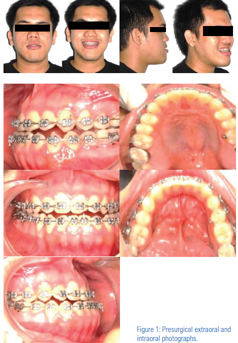


Figure 3: Presurgical panoramic radiographs.
TREATMENT OBJECTIVES
The treatment goals for this patient were 1) to establish a Class I skeletal relationship with a Class I molar relationship, 2) correct the mandibular asymmetry and the left posterior crossbite, 3) align the mid- lines, and 4) finish with about 2mm of overbite and 2mm of overjet.
TREATMENT PROGRESS
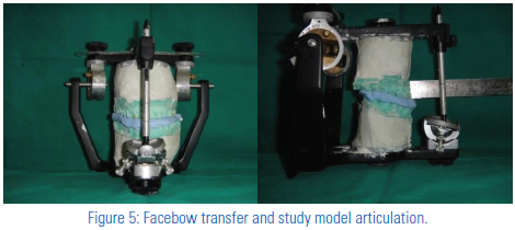
The treatment plan was non-extraction with pre-adjusted appliance (022” Roth prescription, 3MUnitek, Monrov,CA) was started in July 2011. Presurgical orthodontics included decompensation of anteriors by proclining the lower anteriors and controlling torque of upper anteriors. Levelling and aligning was complete in 4 months with wires in the following sequence:016”niti, 016x022” niti, 016x022”SS and 017x025”SS. All the third molars were extracted 6 months prior to surgery in-order to achieve good bone contact during rigid fixation. Surgical Prediction using manual tracing and Dolphin software prediction called for 1) 2mm maxillary advancement, 3mm maxillary impaction and rotation of 2mm to the left,2) a 4mm mandibular setback and rotation of 4mm to the right, 3) Vertical reduction genioplasty of 5mm (Figure 4). A facebow transfer and model articulation in a semi-adjustable articulator (Figure 5) helped perform a model surgery to prepare intermediate and a final surgical splint(to ensure functional occlusion of dental arches).

Figure 4: Surgical Prediction using Dolphin software prediction.
TREATMENT RESULTS:
Orthognathic surgery involved a LeFort I maxillary superior impaction and advancement osteotomy, with rotation for aligning midlines, to position the maxilla 3mm anteriorly into a 2mm overbite-overjetrelationship and to correct the crossbite (Figure 6). The segments were stabilized using rigid internal fixation and confirmed with a lateral cephalogram and orthopantomogram,1- week after surgery. OPG also confirmed root parallelism (Figure 7). Post-Treatment lateral cephalometric analysis revealed a class I skeletal base (Wits Appraisal =+2mm) with upright lower anteriors (IMPA = 92º, U1-NA= 32º, L1-NB= 25º) and a normo-diververgent facial pattern (MPHP= 28º).
Maxillary and Mandibular superimpositions show maxillary advancement of 3mm, impaction of 3mm and mandibular retraction of 7mm. Soft tissuesuperimpositions indicated an improvement in the profile with upper lipprotraction of 2mm and lower lip retraction of 5mm.(Figure10)(black lineindicates pre-treatment tracing and red line indicates post – treatment tracing).
DISCUSSION:
The clinical presentation of a true skeletal Class III may be:1) mandibular prognathism, 2) maxillary deficiency, or 3) a combination of both. True mandibular prognathism is evaluated by comparing the neck form and throat length. These patients frequently present with a decreased mentolabial fold and a thin lower lip.3 Other features include flared incisors, moderate generalized dental spacing and an open bite tendency. A large anteriorly-

positioned tongue is often present with scalloped indentations on the lateral margin from the mandibular teeth.
Maxillary deficiency patients often have an increased neck-chin angle and submental area with a sunken mid-face region and thin upper lip.3Our patient with a maxillary deficiency showed infraorbital, malar andparanasal deficiencies. Maxillary incisor display at rest and his upper lip length were increased. He presented an obtuse nasiolabial angle,a narrowed alar base, and a more horizontally oriented columella. As a consequence of infraorbital deficiency, an area of sclera is seen beneath the pupils is a classical feature. Due to the small dimension of the maxilla in all three planes of space, maxillary deficient patients can present with severe maxillary crowding and malformed or missing maxillary lateral incisors. They have a bilateral posterior cross bite causing buccal tipping of the maxillary molars to compensate for transverse deficiency.3The role of cephalometric analysis in surgical planning is providing theskeletal dimensions and dental inclinations, thus facilitating a more precisediagnosis. Several parameters are available,of which the relationships of the maxilla and mandible to cranial base are vital. Two classical findingsin Class III are; 1) a decreased cranial base length(S-N) 4 and 2) an acutecranial base angle (N-S-Ba).This results in a more forwardly positionedglenoid fossa that positions the mandible more anteriorly. Also, lengthof the maxilla and mandible (N-A and N-B) will determine if the Class III malocclusion is caused by maxillary deficiency, mandibular prognathism,or a combination of both. This will help finalize whether BSSO alone or a bi-jaw surgical plan is needed.
For example, in cases of mandibular setback surgery alone, facial profilechanges include increase in the 1) facial convexity, 2) upper lip length and 3)reduction of lower lip protrusion.5 However, to reduce vertical and horizontalexcess, the recommended surgery included a maxillary superior impactionand mandibular setback to improve the profile and the lip posture.6,7The softtissue responses to a two-jaw surgery vs mandibular setback surgery aloneare similar, except for changes in the nasal tip and the upper lip.8-10 Profile changes in our patient(with an asymmetric mandible) were desirablenot only in the sagittal but also in the vertical plane, hence a verticalreduction genioplasty of 5mm was added to the surgical plan. Although studies on the soft tissue response to genioplasty are diverse, the ratios of soft-tissue to hard-tissue changes are quite predictable.11-12

Figure 7: Post-surgical lateral cephalogram and OPG.

Figure 8: Profile change and facial Symmetry assessed with PA and lateral cephalogram.
The mandibular body is shaped like a triangle, with occlusal and mandibular planes intersecting toward the posterior portion of the face. An asymmetric prognathic mandible in our patient required a rotational bilateral sagittalsplit osteotomy (BSSO) to correct lower midline and set back the mandible. Only a BSSO would have lengthened the posterior components of the mandibular body at the osteotomy site. For our patient with an anterior open bite tendency, it was crucial to pivot the distal portion of the bony segments at the osteotomy site, else it would have worsened the openbite. This procedure also inadvertently increases the burden on the masseter, medial pterygoid, and pterygo-masseteric sling,13 risking potential relapse.14
So, reduction of the total facial length through a Le Fort I maxillaryosteotomy in conjunction with superior impaction was necessary to fit the facial complex within normal physiological function. Our patient with mandibular prognathism presented with a 5mm gummy smile characterized by a long incision-stomion (i.e., incisor display with lips in repose), requiring the need for maxillary superior impaction to minimize display of the teeth.The entire treatment took two years and four months, partly because of waiting for parental/patient authorization and scheduling the surgery took an additional eight months. The archwires were cut to allow settling and detailed finishing of the occlusion was accomplished with 3/16” long class III elastics during the four months after surgery. Radiographic comparison of pre-surgical and post-surgical symmetry, lower facial height and overjet are shown in Figure 8. Improvement in Facial profile and symmetry is illustrated in Figure 9. Table 1 presents the comparison of pre and post-surgical Grummon’s and COGS analysis.

CONCLUSION
:
A combination of LeFort I Maxillary impaction with advancement and BSSO setback did not lengthen the facial complex due to autorotation of themandible. This was the rationale in adopting bi-maxillary surgery in our patient with mandibular prognathism. This resulted in a favourable anterior and posterior lower facial height in this patient. Normal physiological function was not violated by bimaxillary surgical correction in our patient, ensuring a stable surgical correction.

ACKNOWLEDGEMENTS:
We appreciate the contribution of Dr. AbhayTaranathKamath (Prof. andHOD,Oral Surgery), Dr. Manish Kumar Bhagnania(Associate Professor, Oral Surgery) Dr. Amit (Post graduate resident,Oral Surgery), Dr. Prem(Post graduate resident,Oral Surgery), during surgical procedure and Mr. SharathShetty (Orthodontic lab technician) during splint preparation. We are also thankful to Dr. NirmalaRao (Professor and Dean of Manipal College of Dental Sciences, Karnataka, India) for always being a source of inspiration and guidance during our research work. We also are grateful to management of Manipal University, Karnataka, India for providing us the scientific journals for our research.
REFERENCES
- Severt T. R., Proffit W. R. The prevalence of facial asymmetry in the dentofacial deformities population at the University of North Carolina. Int J Adult Orthodon Orthognath Surg 1997. 12:171–176.
- Litton SF, Ackerman LV, Isaacson RJ, Shapiro B. A genetic study of Class III malocclusion. Am J Orthod 1970;58:565-577.
- Sinclair PM, Proffit WR. Class III problems: Mandibular excess/ maxillary deficiency. In Proffit WR, White RP Jr. (eds): Surgical- Orthodontic Treatment. St. Louis, CV Mosby Co., 1991: pp. 428-482.
Disclaimer from Guident
The views and opinions expressed in articles published in Guident are solely those of the authors and do not necessarily reflect the views of the editors, the editorial board or the publisher, Ivory India. Guident does not endorse any product, technique or treatment described. Clinical content is intended for qualified dental and medical professionals and is not a substitute for individual clinical judgement. Guident, its editors and the publisher accept no liability for any consequence arising from the use of information published in the journal.
How to cite this article
Dr. Madhumitha, Dr. Bharath Rao, Dr. Parul Dasson. Rationale for a Bi-Jaw Surgery in an Asymmetric Class III Mandible. Guident. 2014 Mar;7(4). Available from: https://ivoryindia.in/articles/orthodontics/rationale-for-a-bi-jaw-surgery-in-an-asymmetric-class-iii-mandible.html