Enhancing Esthetics in Craniofacial Deformities by Orthodontics
Department of Orthodontics and Dentofacial Orthopedics, Rishi Raj Dental College and Hospital, Bhopal, Department of Anatomy, L.N. Medical College and Research Centre. Bhopal, Department of Orthodontics and Dentofacial Orthopedics, Darshan Dental College and Hospital, Udaipur
ABSTRACT
The comprehensive care of craniofacial patients requires the efforts of a multidisciplinary craniofacial team, including the pediatric neurosurgeon, craniofacial surgeon, geneticist, speech pathologist, nutritionist, orthodontist, oral surgeon, pediatrician, ophthalmologist and pulmonologist, in order to accurately diag nose, evaluate and treat their challenging problems. A careful history (including family, pregnancy and birth) and systemic evaluation, as well as a functional and aesthetic assessment, are vital to a successful outcome. Key physical- examination findings include height, weight, head circumference, head shape, head positioning, muscle tone, movements, airway noises, intraoral findings and facial morphology. Imaging studies are an invaluable aid in evaluating patients with syndromic craniosynostosis. A skull series can help evaluate cranial sutures for signs of fusion; however, CT is more diagnostic in assessing the cranial vault as well as the brain.
Keywords- Syndrome. Craniofacial, Deformity, Orthodontics
INTRODUCTION
A Craniofacial malformation is an anomaly of embryonic development that results in a serious impairment of the normal anatomy of the skull, the jaws and the adjacent soft tissues. Three-quarters of the malformations diagnosed at birth fall in the category “craniofacial”. The most common facial malformations are cleft lip and cleft palate. Less frequent are the syndromes of the I and II branchial arches, malformations that predominantly affect the mandible and the adjacent soft tissues, symmetrically or asymmetrically, such as hemifacial microsomia (Oto-mandibular Syndrome) or Franceschetti syndrome (or Treacher Collins). Even more rare are the forms more accurately called” craniofacial”, that primarily involve the midface and the skull, called cranio-facial-synostosis, such as Apert Syndrome (1 born in 100,000) or Crouzon syndrome (1 in 25,000). Then there are even more rare syndromes, such as Beckwith Wiedemann syndrome, Achondroplasia, Oro-facialdigital syndrome, which the orthodontist is required to know in depth, as involving the jaws, they have a strong involvement in orthodontics1-3.
The birth of a child with a facial deformity is a devastating event for most parents, who often have never seen or heard about similar malformations. For some parents it is very difficult to accept the deformity of the child because it involves the face making the child appear completely different from all the others. Feelings of rejection, guilt, and anxiety gradually develop. Therefore, the geneticist, the surgeon, the neonatologist, the pediatrician, the neurosurgeon, the ENT, the orthodontist, the ophthalmologist, the speech therapist and speech pathologist, and many other specialists who will take care of these patients should all have a very specific expertise in the field and a habit of close collaboration, because the problems of these patients often differ substantially from those of the so-called normal patients. Through a multidisciplinary treatment by these different specialists, a child affected by a craniofacial malformation can, nowadays, achieve results which could not have been hoped for, only a few years ago4-5.
BRIEF OUTLINE OF FEW IMPORTANT SYNDROMES
STICKLER SYNDROME
Stickler syndrome is an autosomal dominant disease characterized by midfacial flattening and variable disorders of vision, hearing and articulation. There are three types of the syndrome caused by mutations in different genes (type 1, COL2A1; type 2, COL11A1; and type 3, COL11A2). Most of the stickler syndrome patients exhibited pretreatment characteristics of small SNA and SNB angles, steep mandibular planes, enlarged gonial angles, and retroclined incisors of both arches, demonstrating the characteristic skeletal and occlusal features of this syndrome6.
OCULO-FACIO-CARDIO-DENTAL (OFCD) SYNDROME
Oculo-facio-cardio-dental (OFCD) syndrome is a rare syndrome characterized by ocular, facial, cardiac, and dental disorders. The most prominent of the various features of this syndrome is canine radiculomegaly. Other features include a long and narrow face, a high nasal bridge, a broad and pointed nose, a bifid nose, ear deformity, cleft palate or submucous cleft palate, maxillary growth retardation, a large gonial angle, open apices, delayed eruption, persistent deciduous teeth, extreme overbite, and constricted maxilla1-2.
APERT SYNDROME
Apert syndrome, is a rare congenital anomaly, appearing with a frequency of 1 in 55,000 to 90,909 live births is one of the five craniosynostosis syndromes caused by allelic mutations of the fibroblast growth-factor receptor 2 (FGFR2) . Externally, this progessive disease of growth is characterized by acrocephaly, syndactyly, and typical features in the orofacial region. The lips are crossbow-shaped or trapezoidal, and the lower lip may seem to protrude. The midface is retruded, which is already marked at birth. The Class III discrepancy is progressive , the maxilla is hypoplastic in all three dimensions, and anteriorly there is edge-to-edge contact or an open bite. Maxillary height is extremely short, and upper facial height markedly reduced, whereas total facial height is increased as compared with the norm. The morphological defects may induce functional defects, such as sleep apnea, disturbances of breathing, feeding, speech and lip closure7-8.
CROUZON’S SYNDROME
Crouzon’s Syndrome occurs in 1/25,000 births and is inherited as an autosomal dominant trait with variable expressivity. Four major physical findings that are characteristic of the syndrome: exorbitism, retromaxillism, inframaxillism, and paradoxic retrognathia. The craniostenosis of the most commonly involved coronal suture leads to brachycephaly. The deformity results in a foreshortened anterior cranial base and a retro-positioned frontal bone. There is an underdeveloped midface, and receded cheekbones. They exhibit an Angle Class III malocclusion due to the midface deficiency, while the mandibular growth potential is normal. Because of the underdeveloped midface and high arched palate, nasal airway obstruction may occur. Other features commonly encountered are visual disturbances due to eye muscle imbalance and frequent hearing loss due to recurrent ear infections8-9.
MARFAN SYNDROME
Marfan syndrome is a heritable disorder of connective tissue that can affect the heart, bloodvessels, lungs, eyes, bones, and ligaments. It is characterized by tall stature, elongated extremities, scoliosis, and a protruded or caved-in breastbone. Patients typically have a long, narrow face. A high-arched palate produced by a narrow maxilla and skeletal Class II malocclusion due to mandibular retrognathia are other common features. For a patient with no family history of the disorder, at least three body systems must be affected before a diagnosis can be made. Oral manifestations of Marfan syndrome, although not speciï¬c, are identiï¬able during a routine intraoral examination. Dental caries and periodontal disease have not been reported to occur more frequently in patients with Marfan, although certain orofacial defects are more prevalent. Constriction of the maxilla and a high-arched palate are important from an orthodontic standpoint, as are concomitant crowding and posterior cross-bite. A dolichofacial face type and skeletal Class II malocclusion are commonly noted10-11.
PIERRE ROBIN SYNDROME
Pierre Robin syndrome (abbreviated to PRS, and also known as Pierre Robin malformation, Pierre Robin sequence, Pierre Robin anomalyor Pierre Robin anomalad), is a congenital condition of facial abnormalities in humans. The 3 main features are cleft palate, micrognathia (a small jaw) and glossoptosis (airway obstruction caused by backwards displacement of the tongue base). A genetic cause to PRS was recently identified. Pierre Robin sequence may be caused by genetic anomalies at chromosomes 2, 11, or 17. PRS is characterized by an unusually small mandible (micrognathia),[1] posterior displacement or retraction of the tongue (glossoptosis), and upper airway obstruction. Incomplete closure of theroof of the mouth (cleft palate) is present in the majority of patients, and is commonly U-shaped12.
DOWN SYNDROME
Down syndrome is the commonest autosomal chromosomal anomaly with an incidence of 1 in 600 to 1000 live births in all races and economic groups. In this condition extra genetic material causes delays in the way a child develops, both mentally and physically. The primary skeletal anomaly affecting the orofacial structures in Down syndrome is hypoplasia of the mid-facial region, with the bridge of the nose, bones of the mid-face and maxilla being relatively small in size. Many children with Down syndrome children exhibit a narrow palate, probably due to the sides of the palate being abnormally thick. This narrowness may sometimes give the impression of a high palatal vault; however the height is generally not significantly greater than normal. Cleft lip and/or palate are reported in 3%-5% of Down syndrome patients. Hypoplasia of the mid-facial region is common in Down syndrome children. In many instances this causes a relative prognathic Class III occlusal relationship which may contribute to an open bite. Other common orthodontic features of Down syndrome include irregular alignment of the teeth, posterior crossbite, anterior open bite, crowding of teeth, and widely spaced teeth. Down syndrome patients tend to have teeth with small conical roots. This is an important factor when considering orthodontic tooth movement and also contributes to early tooth loss in periodontal disease13-14.
GOLDENHAR SYNDROME
Goldenhar syndrome (hemifacial microsomia) is a rare hereditary condition characterized by numerous anomalies affecting the first and second branchial arches of the first pharyngeal pouch, the first branchial cleft, and the primordia of the temporal bone. Goldenhar syndrome is characterized by abnormalities of the face (hemifacial microsomia, unilateral facial hypoplasia, and lateral facial cleft), eyes (epibulbar dermoid or lipodermoid [mostly bilateral]; colobomas of the upper eyelid, iris, choroidea, and retina; and other eye anomalies), and ears (microtia, anotia, preauricular skin tags or blind fistulas, and other external ear malformations). The clinical characteristics of Goldenhar syndrome vary from minor facial asymmetry to severe underdevelopment of one half of the face, with orbital deformation and microtia, or sometimes total absence of the ear . Goldenhar syndrome consists of the complete triad of epibulbar dermoids, accessory auricular appendages, and pretragal fistulae15-17 .
ORTHODONTIC CONSIDERATION
The orthodontist is only one of the many specialists within the craniofacial team, playing a role, albeit not of primary importance, which should necessarily be coordinated with the surgical treatment, speech therapy and other treatments needed by the child. Only in this manner we are able to provide a real service to the patient and the burden of care of these children can be significantly reduced. Malocclusion is a common occurrence in Syndrome patients and is principally related to the dentoalveolar imbalance resulting from the craniofacial abnormality. Since surgery primarily addresses skeletal disharmonies, so the key to successful treatment of these patients is interdisciplinary coordination and sequencing. Frequently, when the patient is first seen by the orthodontist after eruption of the permanent dentition, the most common approach is to prepare the patient for the surgical procedure by aligning and leveling the dental arches to remove dental compensations that have developed as a result of the skeletal imbalance1-3.
If the midface dysplasia is severe, extractions and additional tooth movement will be performed prior to surgery to exaggerate the dental sagittal discrepancy and allow for maximal midfacial advancement and improved facial esthetics. Following surgical correction, the patient will complete orthodontic treatment with final alignment and detailing of the occlusion. If a coordinated approach between the specialties is not implemented, a compromised result with neither the malocclusion nor the facial dysplasia fully corrected may occur. The patient then faces additional surgical and orthodontic care to reverse previous treatment that could have been easily avoided with a coordinated approach6-9.
The therapeutic sequence may be:
• orthodontics or surgery alone
• orthodontics-surgery-orthodontics
• surgery-orthodontics
• surgery-orthodontics-surgery-orthodontics.
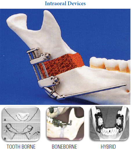
The correction of deformities of patients’ with syndromes is complex because of the esthetic and functional difficulties associated with these disorders. Depending on the specific requirements of each case, a coordinated interdisciplinary approach is essential to the achievement of an optimal result. For example, there are several methods of surgical treatment in Goldenhar syndrome, such as conventional surgical procedures (costochondral rib graft and classical osteotomy) and the distraction technique. Using the distraction technique, it is possible to lengthen the jaw and the ramus of the mandible to the desired size; however, this technique does not result in normal growth and function of the temporomandibular joint. In addition, there is a risk of mild infection during the period of lengthening. Treatment of deformities requires multiple procedures performed by a multidisciplinary team, and long-term regular follow-up is important to monitor the growth and development of patients18. Whereas connective tissue disorders often are associated with extensive periodontal tissue breakdown. In this context, a definite correlation between Marfan syndrome and an increased susceptibility to periodontal disease is uncertain. Early diagnosis and treatment are important factors that play a major role in delaying or altogether avoiding the onset of severe symptoms later in life. Patients with Marfan syndrome can undergo orthodontic treatment, just as their healthy counterparts do. Surgical procedures for patients with Marfan syndrome can be undertaken as long as appropriate precautions are taken19-20.
CONCLUSION
The major advances in diagnostics, materials and techniques have provided the tools that allow the various specialties to significantly improve the quality of life for these individuals who have frequently been ostracized from normal societal interactions. The outcomes achieved not
only benefit the patients we serve but provide enormous satisfaction to all those involved in their care.
BIBLIOGRAPHY
- Regezi S. Oral pathology. Clinical pathologic correlations, 1st ed. Philadelphia: Saunders, 1989: 450-51.
- Muller RFO, Young IDO, Emery’s elements of medical genetics. 11th ed. Churchill Livingstone; 2001.pp-52.
- Vogel F, Motulsky AGO. Human genetics, problems and approaches. 2nd ed. Springer-Verlag; 1996 pp-59.
- Smith WB. Recognizable pattern of human malformations, 4th edition. Jones LK, ed. Philadelphia: Saunders; 1988: 10-12.
- Sassouni V, Forrest E. Dentofacial pathology related to malocclusions. Orthodontics in Dental Practice. St. Louis: Mosby, 1971: 169-97.
- Parke DW. Stickler syndrome: clinical care and molecular genetic. Am J Ophthalmol 2002; 134 (5): 746–8.
- Kaplan, L C. Clinical assessment and multispecialty management of Apert syndrome. Clinics in plastic surgery 1991; 18 (2): 217–25.
- Moss ML: The pathogenesis of premature cranial synostosis in man. Acta Anat. 1959;37;351.
- Moss ML: The primacy of functional matrices in orofacial growth. Dent Pract Rec. 1968;19:65.
- von Kodolitsch Y, Robinson PN. Marfan syndrome: an update of genetics, medical and surgical management. Heart. 2007;93:755–760.
- Crosher R, Holmes A. Marfan syndrome: dental problems and management. Dent Update. 1988;15:120–122.
- Jakobsen LP, Knudsen MA, Lespinasse J, et al . "The genetic basis of the Pierre Robin Sequence". Cleft Palate Craniofac. J 2006; 43 (2): 155–9.
- Sterling ES. Oral and dental considerations in Down syndrome. Down syndrome Advances in Medical Care. In Lott I, McCoy E, editors. New York: Wiley-Liss; 1992: 135-45.
- Sassouni V, Forrest E. Dentofacial pathology related to malocclusions. Orthodontics in Dental Practice. St. Louis: Mosby, 1971: 169-97
- Kokavec R (2006) Goldenhar syndrome with various clinical manifestations. Cleft Palate Craniofac J 2006;43: 628-634.
- Touliatou V, Fryssira H, Mavrou A, Kanavakis E, Kitsiou-Tzeli S. Clinical manifestations in 17 Greek patients with Goldenhar syndrome. Genet. Couns2006; 17 (3): 359–70.
- Vinay C, Reddy RS, Uloopi KS, Madhuri V, Sekhar RC. Craniofacial features in Goldenhar syndrome. J Indian Soc Pedod Prev Dent 2009; 27:121-124.
- Bielicka B, Necka A, Andrych M. Interdisciplinary treatment of patients with Goldenhar syndrome – clinical reports. Dent Med Probl 2006;43: 458-462.
- Nualart Grollmus ZC, Morales Chavez MC, Silvestre Donat FJ. Periodontal disease associated to systemic genetic disorders. Med Oral Patol Oral Cir Bucal 2007;12:E211–E215.
- Straub AM, Grahame R, Scully C, Tonetti MS. Severe periodontitis in Marfan’s syndrome: a case report. J Periodontol 2002;73:823–826
Disclaimer from Guident
The views and opinions expressed in articles published in Guident are solely those of the authors and do not necessarily reflect the views of the editors, the editorial board or the publisher, Ivory India. Guident does not endorse any product, technique or treatment described. Clinical content is intended for qualified dental and medical professionals and is not a substitute for individual clinical judgement. Guident, its editors and the publisher accept no liability for any consequence arising from the use of information published in the journal.
How to cite this article
Dr. Amit Prakash, Dr. Shanker Dayal Gupta, Dr. Adit Arora. Enhancing Esthetics in Craniofacial Deformities by Orthodontics. Guident. 2014 Feb;7(3). Available from: https://ivoryindia.in/articles/orthodontics/enhancing-esthetics-in-craniofacial-deformities-by-orthodontics.html