Expanding Boundaries - Distraction Osteogenesis
Department of Orthodontics and Dentofacial Orthopaedics A. B. Shetty Memorial Institute of Dental Sciences, Derlakatte, Mangalore
INTRODUCTION
Distraction Osteogenesis(callus distraction, callotasis, and osteodistraction) is a biologic process that leads to bone formation between two bony segments that are mechanically separated at a constant rate.
New bone is generated in an osteotomy gap in response to tension stresses placed across the bone gap.
It enables the clinician to lengthen and widen bone and fill in gaps between bones without the need for bone or soft tissue grafts.
The simultaneous expansion of the soft tissues, including muscles, ligaments , fat and skin produces excellent aesthetic and functional results and minimizes the skeletal relapse.
HISTORY
1905 - First bone distraction was performed by Codivilla -for the treatment of shortened femur
1927 - Lengthening of tibia – Abbott
1954 - Ilizarov began his work on the lower extremity
1972 – Snyder et al – used external fixator to lengthen a canine mandible.
1989 –McCarthy et al - First human mandibular distraction
1997 –Polley & Figueroa – Discussed the management of severe maxillary deficiency in childhood & adolescence performing DO with an external adjustable,rigid distraction device.
INDICATIONS
Cranio-facial microsomia ( unilateral / bilateral)
Nager’s syndrome
Treacher collins syndrome
Pierre robin syndrome
Cleft lip and palate
TMJ ankylosis
Post traumatic growth disturbances
Regeneration of mandibular condyle
Distraction of mandibular symphysis to correct anterior crowding.
Maxillary deficiency
ADVANTAGES
There is no need of autogenous bone grafting
Gradual distraction of not only the hard tissues but of the soft tissues also
Multi-directional expansion of the skeleton
Minimal evidence of relapse
Ease to open & close the mouth & ease of mastication
Reduced length of hospitalization & operating time
Applied at an younger age group
DISADVANTAGES
Cutaneous scarring resulting from transcutaneous fixation pins
CLASSIFICATION
Distraction devices can be classified into
Intraoral devices
Extraoral devices
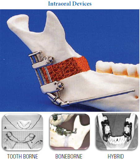

DEVICE PLACEMENT
Vertical device placement causes an increase in the vertical dimension of the mandible. Horizontal device placement causes sagittal advancement of the mandible. Oblique device placement results in an increase in both horizontal & vertical dimensions of the ramus & body
MAXILLARY DISTRACTION
Maxillary advancement by Distraction has many advantages over conventional orthognathic procedures :
- Can be done at young age
- Direction of distraction can easily be controlled by the RED device
- Minimal morbidity & blood loss
- No need for bone grafts
- Minimal relapse
- Can be easily removed without anaesthesia after distraction
The only limitation of this technique is in patients who lack teeth or adequate bone in the cranial vault .Complication includes velopharyngeal incompetency
Transverse,vertical and horizontal maxillary deficiency needing an advancement
- greater than 6 mm to 8 mm
- palatal clefts with severe scarring
- normal mandibular morphology and position
- normal neck/chin angle
- patients in the full primary dentition or older
- patients with an intact cranium
PRINCIPLES OF DISTRACTION OSTEOGENESIS (ILIZAROV PRINCIPLES)
- BONE CUT: It is important to preserve the osseous blood supply. CORTICOTOMY AND PRESERVATION OF PERIOSTEAL ENVELOPE.
- LATENCY : After bone cut is performed,a latency period of 5 to 7 days is observed before device activation.This allows for the formation of an adequate fibrovascular bridge between the bone Latency period may be shortened (1 to 2 days) if the patient is young.
- RATE: A regenerate can best be generated when the tensile stress is applied and bone edges separated 1.0 mm per day. For young child, the rate may be increased upto 1.5 to 2 mm per day .Advancing the bone segments more than 2 mm per day may exceed the limit of vascular supply of the overlying soft tissue.
- RHYTHM : Continuous application of distraction force is ideal. Clinically, application of the distraction is best performed by activating the device twice a day(0.5 mm twice a day).If the patient experiences discomfort ,then the rhythm should be altered to allow for a smaller incremental application(0.25 mm for four times a day)
- CONSOLIDATION : Once the regenerate has been created, the distraction device is held in neutral fixation allowing the neomandible to ossify. The timing of the ossification process is similar to that of fracture healing(6 to 8 weeks).For younger children ,ossification can occur quicker.It is best to observe a cortical outline on the radiograph of the regenerate before device removal.
CASE REPORT

A female patient age 12 years reported to our department with the chief complaint of the face being shifted to one side. She presented with a history of a repaired facial cleft extending from corner of the mouth to the tragus on the right side and scar tissue visible on her face. On extra - oral examination we found that there was a gross facial asymmetry due to flattening of the face on the affected side which was because of the scar tissue of the repaired facial cleft leading to mandibular hypoplasia on that side.
Pre Treatment – Intra Oral


Pre Treatment – Occlusal


Distraction was indicated for the mandibular hypoplasia on the affected site.
Distractor Being Placed


After the distraction was done the patient was strapped up with 0.22 Roth prescription
Post Surgical Intraoral

Present Status Intra Oral


Case II
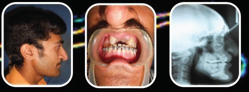
RAPID MAXILLARY EXPANSION APPLIANCE USED FOR ANTERIOR MAXILLARY DISTRACTION
Pre Treatment Photos
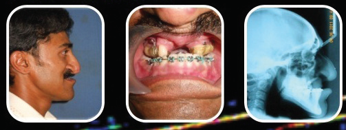
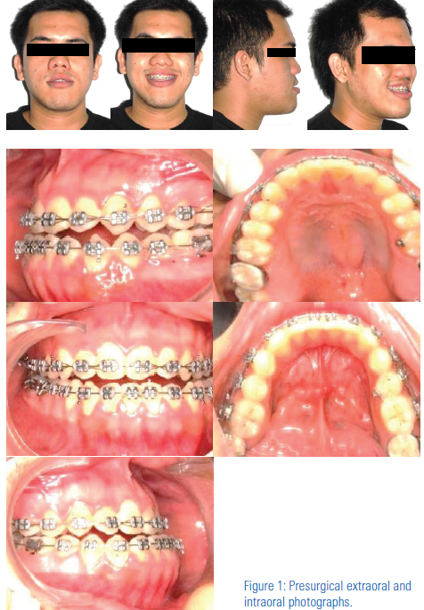
A male patient named rakesh reported to our department with a chief complaint of forwardely placed lower jaw. Cephalomteric findings revealed that he had a retrusive maxilla with a normal mandible which lead to the prognathic lower jaw appearance.
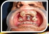
The treatment plan was to give two OSTEOTOMY bone cuts in the premolar region followed by the use of a RME appliance placed in the antero – postero being used as an intraoral distractor as shown in the intro oral picture. By turning the screw two quarter turns daily sufficient protaction of the maxilla was achieved in about 2 weeks.
Distractor in Place
Post Treatment Photos.

CONCLUSION
Osteodisraction provides a means whereby bone can be remodelled into different shapes to more adequately address the nature of skeletal deformities & asymmetries
Many of the congenital deformities that require extensive musculoskeletal movements may be addressed with fewer procedures eventually achieving the same structural,functional & esthetic results commonly seen with modern orthognathic procedures
Future may witness the use of the concepts of distraction osteogenesis to achieve better,faster & more efficient tooth movement
REFRENCES
- Wolford LM, Wardrop RW, Hartog JM. Coralline porous hydroxylapatite as a bone graft substitute in orthognathic surgery. J Oral Maxillofac Surg 1987;45:1034-42.
- Trauner R, Obwegeser H. The surgical correction of mandibular prognathism and retrognathia with consideration of genioplasty: part I, surgical procedures to correctmandibular prognathism and reshaping of the chin. Oral Surg Oral Med Oral Path 1957;10:677-89.
- Caldwell JB, Amaral WJ. Mandibular micrognathia corrected by vertical osteotomy in the rami and iliac bone graft. J Oral Surg 1960;18:3-15.
- Schendel SA, Epker BN. Results after mandibular advancement surgery: an analysis of 87 cases. J Oral Surg 1980;38:265-82.
- Converse JM, Horowitz SL. The surgical-orthodontic approach to the treatment of dentofacial deformities. Am J Orthod Dentofacial Orthop 1969;55:217-43.
- Cassidy DW, Herbosa EG, Rotskoff KS, Johnston LE Jr. A comparison of surgery and orthodontics in “borderline” adults with Class II, Division 1 malocclusions. Am J Orthod Dentofacial Orthop 1993;104:455-70.
- Blair VP. Operations on the jaw-bone and face. Surg Gyn Obstet 1907;4:67-78.
- Babcock WW. The surgical treatment of certain deformities of the jaw associated with malocclusion of the teeth. J Am Med Assoc 1909;53:833-9.
- Ellis E, III, Carlson DS. Stability two years after mandibular advancement with and without suprahyoid myotomy: an experimental study. J Oral Maxillofac Surg 1983;41:426-37.
- Caldwell JB, Hayward JR, Lister RL. Correction of mandibular retrognathia by vertical L osteotomy: a new technic. J Oral Surg 1968;26:259-64.
- Longaker MT, Siebert JW. Microsurgical correction of facial contour in congenital craniofacial malformations: the marriage of hard and soft tissue. Plast Reconstr Surg 1996;98:942-50.
- Vargervik K, Ousterhout DK, Farias M. Factors affecting long-term results in hemifacial microsomia. Cleft Palate Craniofac J 1986;23(Suppl 1):53-68.
- Ilizarov GA. The tension-stress effect on the genesis and growth of tissues: part II, the influence of the rate and frequency of distraction. Clin Orthop Rel Res 1989;239:263-85.
- lizarov GA. The tension-stress effect on the genesis and growth of tissues: part I, the influence of stability of fixation and soft tissue preservation. Clin Orthop Rel Res 1989;238:249-81.
- Ilizarov GA. Clinical application of the tension-stress effect for limb lengthening. ClinOrthop Rel Res 1990;250:8-26.
- Samchukov ML, Cherkashin AM, Cope JB. Distraction osteogenesis: origins and evolution. In: McNamara JA Jr, Trotman CA, eds. Advances in Craniofacial Orthopedics. Volume 34. Craniofacial Growth Series. Ann Arbor: Center for Human Growth and Development, University of Michigan; 1998:1-35.
Disclaimer from Guident
The views and opinions expressed in articles published in Guident are solely those of the authors and do not necessarily reflect the views of the editors, the editorial board or the publisher, Ivory India. Guident does not endorse any product, technique or treatment described. Clinical content is intended for qualified dental and medical professionals and is not a substitute for individual clinical judgement. Guident, its editors and the publisher accept no liability for any consequence arising from the use of information published in the journal.
How to cite this article
Dr. U.S.Krishna Nayak, Dr. K. Saidath )Reader), Dr. Neelen Shah. Expanding Boundaries - Distraction Osteogenesis. Guident. 2014 Feb;7(3). Available from: https://ivoryindia.in/articles/orthodontics/expanding-boundaries-distraction-osteogenesis.html