Failures and Complications in Implants
Department of Prosthodontics, Crown, Bridge and Implantology, AME’s Dental college and Hospital, Raichur
ABSTRACT
Implant therapy is, today a routinely used treatment option reducing the limitations of the conventional fixed partial dentures. However, it is a well-established fact that failure of the implant therapy can happen even under best care. Yet, with proper diagnosis and observation at the right time ,the failures can be avoided and the finest management can be done to a great extent. This review article provides a checklist of the main causes associated with the complications as well as of the implant failures, the knowledge of which is essential for a successful implant placement.
Keywords: Implant failure, Complications, causes
INTRODUCTION
The concept “osseointegration” as a direct anchorage of the implant fixture to surrounding host bone is apparently the most important feature to affirm the reported long-term clinical success of dental implants. However, in spite of the high success rate, implant failure has been reported.1 Implant failure is a static outcome situation that requires removal of a failed implant. It may be referred to as the position status of the implant that when using some quantitative measurements falls below an acceptable level. This definition includes clinical situations, ranging from all symptomatic mobile implants to implants that show more than 0.2 mm of crestal bone loss after the first year of loading.2
SUCCESS CRITERIA FOR DENTAL IMPLANTS
According to Alberktson, Zarb, Washington, and Erickson3:- Individual unattached implant that is immobile when tested clinically
- Radiograph that does not demonstrate evidence of peri implant radiolucency.
- Bone loss that is less than 0.2mm annually after the implant’s first year of service.
- Individual implant performance that is characterized by an absence of persistent and/or irreversible signs and symptoms of pain, infections, neuropathies, paresthesia or violation of the mandibular canal.
In content of criteria mentioned, a success rate of 85% at the end of a 5 –year observation period and 80% at the end of 10-year observation is a minimum criterion for success.
COMPLICATIONS IN IMPLANTS
Dental implants may fail for different reasons, with a range that differentiates between a failure and a complication. An Unmanageable degree of complication is considered as a failure.
Example, fracture of the connecting screw or the prosthetic part a complication and not a failure because it can be managed in most cases.
IMPLANT COMPLICATIONS can be classified as follows (Kelly and Wang)10:-

Table 1

Fig. 1: Anatomy related complications

Fig. 2: Treatment Plan Related Complications

Fig. 3: Procedure Related Complications
IMPLANT FAILURE
Dental implants may fail for different reasons, with a range that Differentiates between a failure and a complication.
Esposito et al definition on implant failures includes biologic failures (related to biologic processes) and mechanical failure of the component (including fractures of implants, coatings, connecting screws, and prosthesis, fractures of the implant connecting screws and prosthesis.
Since the introduction of the concept of osseointegration, the success of dental implants has increased dramatically’ because of better understanding of bone response and the improvement in bone loading concepts. Improper patient selection, accumulation of bacterial plaque because of poor oral hygiene, traumatic occlusion, debris retention resulting from improper prosthetic restoration, and bone prep- aration without the use of internally cooled, high-torque, slow-speed handpieces, have been the factors contributing to the breakdown of otherwise successful implants Identification of the cause and type of implant failure alone will help in the major management of the case.
CLASSIFICATION OF IMPLANT FAILURES
Implant failures can be categorized into Seven categories based on the different aspects of their failures:-CATEGORY A: - ETIOLOGICAL FACTORS
CATEGORY B: - TIMING OF FAILURE
CATEGORY C: - ORIGIN OF INFECTION
CATEGORY D: - CONDITION OF FAILURE
CATEGORY E: - RESPONSIBLE PERSONNEL
CATEGORY F: - FAILURE MODE
CATEGORY G: - SUPPORTING TISSUE TYPE
CATEGORY A - ETIOLOGIC FACTORS
a. Host factors- Medical status - Osteoporosis and other bone diseases; uncontrolled diabetes.
- Habits - smoking, para-functional habits.
- Oral status - poor home care, juvenile, and rapidly progressive periodontitis, irradiation therapy.

Fig. 4: Implant failure of a 60 yr old patient due to poor oral hygiene
b. Surgical placement
- Off axis placement (severe angulation): Improper implant placement can result in a framework design that compromises esthetics and distribution of force on implants.

Fig. 5: Off Axis placement of Implants
- Lack of initial stabilization: Primary stability has been described as the absence of mobility in the bone bed immediately after implant placement, which is achieved through mechanical engagement between the implant surface and surrounding bone. Primary stability is influenced by multiple factors such as materials used, the local bone tissue characteristics, the surgical/placement technique.11
- Impaired healing and infection because of improper flap design or others.
- Overheating the bone and exerting too much pressure12: Implant drilling procedures could cause a temperature rise in the surrounding hard tissue.

Fig. 6: Thermally induced bone necrosis during implant surgery
- Minimal space between implant: Most implant manufacturers recommend a space of 4 mm to 7 mm between the neighboring implants to allow sufficient biologic space to avoid the necrosis that could happen because of blood supply impairment.6

Fig. 7: Improper spacing between implants
- Placing the implant in immature bone grafted sites: Placing an implant in immature grafted bone will not provide the implantologist with intimate implant-to-bone contact, which is essential for the implant to resist applied torque. Placing the implant in the mature fresh bone, the maximum implant-to-bone contact will be obtained.6
- Placement of the implant in an infected socket or a pathologic lesion

Fig. 8: Peri implant lesion caused by periapical pathology of adjacent infected teeth
- Contamination of the implant body before insertion: Contaminated handling of the implant is poor protocol and may alter the surface chemistry. The implant may be contaminated because of manufacturer errors, by the operator, from nontitanium instruments, or by bacteria (oral cavity).
c. Implant selection:
- Improper implant type in improper bone type.
- Length of the implant (too short, crown-implant ratio unfavorable)
- Diameter of the implant.
d. Restorative problems:
- Excessive cantilever, pier abutments, no passive fit, improper fit of the abutment, improper prosthetic design, improper occlusal scheme, bending moments, connecting implants to natural dentition, premature loading, excessive torquing.
2. CATEGORY B - Timing of occurrence.
- Before stage II (after surgery): Traditionally, before placing dental implants, the compromised teeth are removed and the extraction sockets are left to heal for several months. To preserve the alveolar bone level from the collapse caused by healing and to reduce treatment time in situations in which tooth extraction precedes implant placement, some clinicians began to install the implant immediately into the postextraction socket without waiting for the site to heal.13

Fig. 9: Immediate placement of implants after extraction
- At stage II (With healing head and or abutment insertion): Dental implants may fail at a certain stage of treatment that does not fall in either of the two categories of early and late failure. It can fail at the second stage of surgery, during healing or head placement, at abutment connection , and before prosthetic placement. This could be due to excessive torquing during abutment connection when inserted into grafted or D4 bone or possibly because of poor surface treatment of the fixture.Recently various surface coating treatments are being proven to increase the antibacterial efficacy at the fixture as well as improved osseointegration.14
- After restoration: Occlusal overload has been regarded as a major cause of biomechanical complications, including screw loosening, prosthesis failure, and the fracture of screws, veneering material, or the implant.15

Fig. 10: Abutments of an FPD debonded due to the improper axial convergence

Fig. 11: Veneer ceramic of an implant supported bilayer zirconia single crown replacing tooth no14 fractured due to the premature contact occurred in lateral movement

Fig. 12: An abutment fractured under overload and/or excessive tightening with its intact original
3. CATEGORY C - Origin
- Peri-implantitis (infective process, bacterial origin).
CLINICAL SIGNS OF PERIIMPLANTITIS:- The clinical signs of14
- inflammation,
- bleeding, and purulence,
- in addition to increased mobility,
- peri- implant radiolucency, and
- probing depths greater than 6 mm, are associated with failing implants

Fig. 13: Radiographic representation of an implant failed due to the periimplantitis and/or overload.
Retrograde peri-implantitis (traumatic occlusion origin, non-infective, forces off the long axis, premature, or excessive loading): It is defined as a clinically symptomatic periapical lesion (diagnosed as a radiolucency) that develops shortly after implant insertion while the coronal portion of the implant achieves a normal bone to implant interface.16

Fig. 14a: Clinical exam revealing probing depths around all implants ranging from 2 to 4 mm with no bleeding on probing and no mobility and the presence of a fistula apical to implant #8 (black arrow) & Fig. 14b: A periapical radiograph revealed the presence of radiolucencies associated with the apices of implants #8 and #9 and marginal discrepancy of the implant-supported crowns
4. CATEGORY D - Condition of failure. 17,14
a) Ailingb) Failing
c) Failed implants by radiographic analysis.
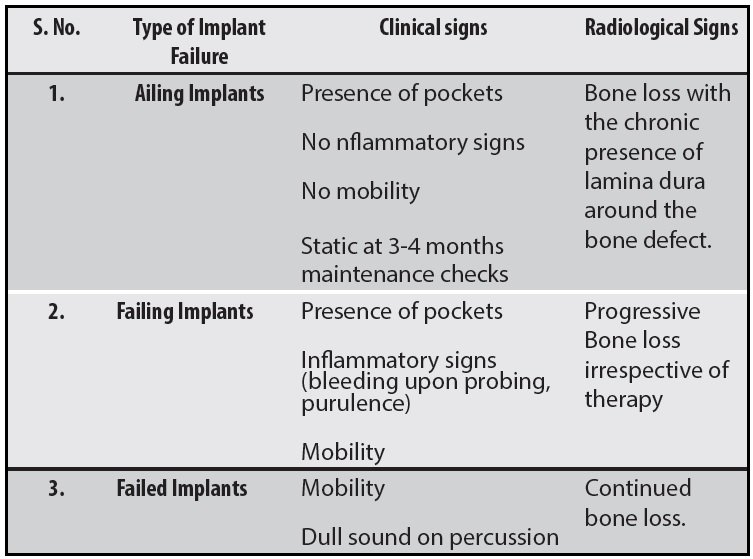
The identification of the above-mentioned implants clinically and radiographically are as follows:
5. CATEGORY E -Responsible personnel include 14
- Dentist (oral surgeon, prosthodontist, periodontist)
- Dental hygienist
- Laboratory technician
- Patient.
The success and integrity of the dental implant rely on cooperation among a dental team that consists of the general dentist, surgeon, prosthodontist, periodontist, dental hygienist, laboratory technician, and even the patient. Each member has his or her own role for certain stages of treatment. Therefore, each individual on the dental team, including the patient, shares a part of the responsibility. This must be verified in the implant failure checklist to identify the personnel responsible for a failure and to deal with the medico legal problems that could result.
6. CATEGORY F - Failure mode. 6, 14
- Lack of osseointegration (usually mobility)
- Unacceptable esthetics
- Functional problems
- Psychological problems.
7. CATEGORY F –SUPPORTING TISSUES involved. 6, 14
- Soft tissue problems (lack of keratinized tissues, inflammation, etc.
- Bone loss (Radiographic changes, etc.
- Both soft tissue and bone loss.
CLINICAL AND RADIOGRAPHIC CONDITION OF A FAILED IMPLANT 17,14,6
Meffert proposed a classification of failure including
- Ailing,
- Failing, and
- Failed implants.
1. Ailing implants are those showing radiographic bone loss without inflammatory signs or mobility. Such implants do not pose any indication of failure, but with the progression of bone loss, they could be at a higher risk of failure.

Fig. 15a: Ailing implants secondary to calculus

Fig. 15b: Ailing implants with soft tissue involvement only
2. Failing implants are characterized by progressive bone loss, signs of inflammation, and no mobility. These implants are usually in a reversible state (i.e. the condition can be treated). It is therefore necessary to determine and eliminate the etiological factor(s) causing such a condition.

Fig. 16: Failing implant
3. Failed implants are those with progressive bone loss with clinical mobility and that are not functioning in the intended sense. Failed implants are usually encapsulated in a fibrous capsule. Radiographically, failed implants are characterized by diffuse radiolucency.

Fig. 17: Failed implants
DIAGNOSIS OF FAILED IMPLANTS 9
The most common diagnostic criteria employed for the evaluation of established implant failures (failed implants) are as follows:
1. Clinical signs of early infection
During the healing period (3-9 months) complications such as swelling, fistulas, suppuration, early/late mucosal dehiscences, and oseteomyelitis, can occasionally be present and may indicate implant failure. Signs of infection occurring during at an early stage of healing is more critical than if they occur at a later stage. This is because; the infection occurring at an early stage will lead to disturbance in the osseointegration of the implant to the surrounding bone.
2. Pain or sensitivity
Pain or discomfort is often associated with mobility and could be one of the first signs which indicate an implant failure.
3. Clinical discernible mobility
Mobility is always a clear sign of failure. Once the clinician has distinguished between the mobility of a poorly connected abutment and the mobility of the underlying implant, the implant must be suspected to be surrounded by a fibrous tissue capsule.
Several different types of mobility have been recognized as follows:8
- Rotation mobility
- Lateral or horizontal mobility
- Axial or vertical mobility.
- Occasionally, clinically discernible mobility can be present without distinct radiographic bone changes. Therefore, mobility is the cardinal sign of implant failure.
4. Radiographic signs of failure
There can be two well-distinct radiographic pictures: a thin peri-fixtural radiolucency surrounding the entire implant, suggesting the absence of a direct bone-implant contact and possibly a loss of stability, and an increased marginal bone loss.
Since the distinction between the two radiographic pictures is not always clear, when a suspected peri-fixtural radiolucency or excessive marginal bone loss is observed, it is recommended to remove the prosthetic construction and cheek the implants for stability. Clinically discernible mobility after bridge removal can confirm the presumptive radiographic diagnosis of implant failure.
5. Dull sound at percussion
It has been suggested that a subdued sound upon percussion is indicative of soft tissue encapsulation, whereas a clear crystallization sound indicates successful osseointegration.7
Although it is a rather subjective test without a solid scientific background, it can provide a useful indication to the examiner.
MANAGEMENT OF FAILED IMPLANTS 18
The management of failed implants can be categorized into 2 ways -
REMOVAL OF FAILED IMPLANT
A mobile implant is extracted easily with a forceps, and the adjacent granulomatous tissue should be removed. However, a late implant failure due to a fracture or peri-implantitis probably will require additional efforts. It should be noted that implant removal is quite different from extracting a tooth that has a periodontal ligament. Great caution has to be exercised not to remove a buccal or lingual plate during explantation, because an implant is circumferentially osseointegrated.18
METHODS OF EXTRACTING DENTAL IMPLANTS
Froum and colleagues reviewed different methods regarding extraction of dental implants These techniques include use of a counter-torque ratchet, Piezo tips, high-speed burs, elevators, forceps, reverse screw and trephine burs and combinations of these tools. Use of the counter-torque ratchet is the least invasive technique for removing an implant. It is screwed into a failed implant and reverse torqued.
DENTAL IMPLANT REIMPLANTATIONS
After diagnosing an implant as failed, the clinician needs to formulate a treatment plan with respect to replacing or not replacing a lost fixture. In that regard, the clinician needs to consider the extent of osseous destruction, which will influence the timing of placing another implant.18
IMMEDIATE VERSUS DELAYED REIMPLANTATION AFTER LOSS OF AN IMPLANT.
With respect to the timing of reimplantations, Kim and colleagues18 reported that failure rates after implant removal were not significantly different between delayed and immediate reimplantations. They indicated that replaced implants were larger in length and diameter than the implants placed first. It can be surmised that if there is adequate bone to achieve primary stability, delayed placement does not appear to provide any advantages.
CONCLUSION
Success in implants is achieved if due respects are given to function, esthetics and hygiene and it is the role of the clinician to identify the possible complications at the right time to avoid failures. Long term acceptable criteria should be established and limiting factors should be identified before prosthetic reconstruction to minimize occurrence of complications related to maintenance and patient management. So with proper patient selection and treatment planning, using dental implants to support restorations replacing missing teeth can provide long lasting functional and aesthetic restorations.
As with any Prosthodontic restoration, meticulous attention must be given to treatment planning. Subsequent to the customary medical and dental history analysis, intraoral and extra oral examination, and radiographic analysis, diagnostic casts should be made. These casts, along with a face bow transfer and occlusal registration, are essential for treatment planning and restoration design. Also, properly oriented diagnostic casts are needed to evaluate the remaining dentition, residual bone, and maxillomandibular relationships.
REFERENCES
- Sakka S, Baroudi K, Nassani MZ. Factors associated with early and late failure of dental implants. Journal of investigative and clinical dentistry. 2012 Nov;3(4):258-61.
- Albrektsson T, Zarb G, Worthington P, Eriksson AR. The long-term efficacy of currently used dental implants: a review and proposed criteria of success. Int j oral maxillofac implants. 1986 Jan 1;1(1):11-25.
- Smith DE, Zarb GA. Criteria for success of osseointegrated endosseous implants. The Journal of prosthetic dentistry. 1989 Nov 1;62(5):567-72.
- Misch CE, Perel ML, Wang HL, Sammartino G, Galindo-Moreno P, Trisi P, Steigmann M, Rebaudi A, Palti A, Pikos MA, Schwartz-Arad D. Implant success, survival, and failure: the International Congress of Oral Implantologists (ICOI) pisa consensus conference. Implant dentistry. 2008 Mar 1;17(1):5-15.
- Sones AD. Complications with osseointegrated implants. The Journal of prosthetic dentistry. 1989 Nov 1;62(5):581-5.
- El Askary AS, Meffert RM, Griffin T. Why do dental implants fail? Part I. Implant dentistry. 1999 Jan 1;8(2):173-85.
- Esposito M, Hirsch JM, Lekholm U, Thomsen P. Biological factors contributing to failures of osseointegrated oral implants.(II). Etiopathogenesis. European journal of oral sciences. 1998 Jun 1;106(3):721-64.
- Hämmerle CH, Schmid J, Olah AJ, Lang NP. Influence of initial implant mobility on the integration of titanium implants. An experimental study in rabbits. Clinical oral implants research. 1996 Jun;7(2):120-7.
- Prashanti E, Sajjan S, Reddy JM. Failures in implants. Indian Journal of Dental Research. 2011 May 1;22(3):446.
- Misch K, Wang HL. Implant surgery complications: etiology and treatment. Implant dentistry. 2008 Jun 1;17(2):159-68.
- Manzano-Moreno FJ, Herrera-Briones FJ, Bassam T, Vallecillo-Capilla MF, Reyes-Botella C. Factors affecting dental implant stability measured using the ostell mentor device: A systematic review. Implant dentistry. 2015 Oct 1;24(5):565-77.
- Manzano-Moreno FJ, Herrera-Briones FJ, Bassam T, Vallecillo-Capilla MF, Reyes-Botella C. Factors affecting dental implant stability measured using the ostell mentor device: A systematic review. Implant dentistry. 2015 Oct 1;24(5):565-77.
- Chrcanovic BR, Martins MD, Wennerberg A. Immediate placement of implants into infected sites: a systematic review. Clinical implant dentistry and related research. 2015 Jan;17:e1-6.
- El Askary AS, Meffert RM, Griffin T. Why do dental implants fail? Part II. Implant dentistry. 1999 Jan 1;8(3):265-77
- Sheridan RA, Decker AM, Plonka AB, Wang HL. The role of occlusion in implant therapy: a comprehensive updated review. Implant dentistry. 2016 Dec 1;25(6):829-38
- Quirynen M, Vogels R, Alsaadi G, Naert I, Jacobs R, Steenberghe DV. Predisposing conditions for retrograde peri?implantitis, and treatment suggestions. Clinical oral implants research. 2005 Oct;16(5):599-608.
- Meffert RM. How to treat ailing and failing implants. Implant Dentistry. 1992 Jan 1;1(1):25-33.
- Greenstein G, Cavallaro J. Failed dental implants: diagnosis, removal and survival of reimplantations. The Journal of the American Dental Association. 2014 Aug 1;145(8):835-41.
- Kim YK, Park JY, Kim SG, Lee HJ. Prognosis of the implants replaced after removal of failed dental implants. Oral Surgery, Oral Medicine, Oral Pathology, Oral Radiology, and Endodontology. 2010 Sep 1;110(3):281-6.
Disclaimer from Guident
The views and opinions expressed in articles published in Guident are solely those of the authors and do not necessarily reflect the views of the editors, the editorial board or the publisher, Ivory India. Guident does not endorse any product, technique or treatment described. Clinical content is intended for qualified dental and medical professionals and is not a substitute for individual clinical judgement. Guident, its editors and the publisher accept no liability for any consequence arising from the use of information published in the journal.
How to cite this article
Dr. Amritha Chandran, Dr. Sunil Dhaded. Failures and Complications in Implants. Guident. 2022 Aug;15(9-10). Available from: https://ivoryindia.in/articles/implantology/failures-and-complications-in-implants.html