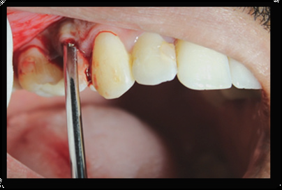
Full Mouth Rehabilitation of an Ectodermal Dysplasia Patient
Jaipur Dental College, Jaipur, India, Wisdom Dental Clinics, Jaipur, India
ABSTRACT
Ectodermal dysplasia (ED) is a hereditary disorder associated with abnormal development of embryonic ectodermally-derived organs including teeth, nails, hair and sweat glands. Hypodontia of the primary and permanent dentition is the most common oral finding.Full mouth rehabilitation in these patients is difficult to manage, especially because the afflicted individuals are quite young when they are evaluated for treatment; therefore, esthetics is an important concern. This clinical report describes the rehabilitation of a18-year-old girl diagnosed with ectodermal dysplasia. Nine implants were placed in the maxilla and mandible and cement retained crowns were delivered on all these implants, along with PFM crown units and bridges. This treatment plan seems to be favorable for ED patients.
Key Words: Ectodermal Dysplasia, Dental Implants, Fixed Partial Dentures, Rehabilitation
INTRODUCTION
Ectodermal dysplasia (ED) is described as an inherited disorder characterized by the abnormality of at least two structures of ectodermal origin.1-3 The different forms of EDof ectodermal tissues, such as skin, hair, nails, sweat glands and the teeth ,leads to dysplasia or aplasia.4 This condition classified into two types: (1) Hidrotic, which is an autosomal triad with normal sweat glands; and (2) Anhidrotic (hypohidrotic), which is a X-linked recessive triad with the absence of, or considerable decrease in the sweat glands.5-7
ED significantly involves more males because X-linked is the most prevalent form and the affected men often have extensive dental anomalies, such as missing most of their deciduous and permanent teeth or sometimes multiple retained teeth.8-10
The most common oral manifestations are hypodontia, anodontia, tapered or malformed teeth, delayed dental eruption and because there is a deficiency in the alveolar process, it often leads to the loss of vertical dimension.11-15
External characteristics are an indistinct vermilion border, protuberant lips, a prominent forehead and a depressed nasal bridge.14-16
Even literature supports the placement of osseo-integrated implants when patients are in their late teens .1-3,7
Finally, for every patient a treatment plan should be personalized depending on a significant factors, such as the patient's age, medical history, the number of present teeth, alveolar process, esthetics and the patient's attitudes.
The present study describes using osseointegrated dental implants to full mouth rehabilitate an ED patient with maxillary and mandibular multiple retained teeth.
CASE REPORT
An 18 year old girl reported to our clinic with the chief complain of missing teeth and small sized existing teeth. (Figure. 1) On general examination, she had thin hair on extremities. The palms and soles showed marked hyperkeratosis. There was lack of sweating and complaints of hyperthermia in warm conditions.
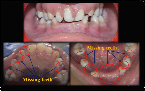
Figure.1 - Pre-operative view of the patient
On extraoral examination, lips appeared protuberant and lower facial height was reduced, giving the patient a concave facial form and an overaged look. On intraoral examination, multiple retained teeth with hypodontia was observed. Teeth were spaced and conical shaped with deep overbite.
Patient was advised for OPG. It revealed presence of retained deciduous teeth .Roots were conical shaped with compromised periodontal support. (Figure.2) After history and examination, patient was given a detailed overview of the condition and all treatment options were discussed. Finally, full mouth rehabilitation was finalized with multiple implants in the edentulous areas along with possibility of 3 unit bridge, depending on space availability and other essential parameters.

Figure.2 - OPG of the patient
Firstly retained deciduous teeth were extracted from both the arches under LA. After that fixed orthodontic therapy was started to align and upright all the teeth, correction of midline and other refinements to create proper space for restoration of missing teeth. After 6 months Denta-scan was done to assess the bone for placement of implants. (Figure. 3)

Figure. 3 - Extraction of retained deciduous teeth and alignment of teeth
Based on Denta-scan evaluation, 9 Noble biocare active implants were placed with free hands in 15,13,23,25,33,31,41,42 and 47 positions. (Figure.4a, b, c)

(a)

(b)

(c)
After 4 months of period of osteointegration and space stabilization, brackets were debonded and intentional endodontic therapy was done irt 44 and 46 to give 3 unit PFM bridge. Meanwhile, implant level impressions were made for both the arches along with crown preparations. (Figure.5)

(Figure.5 - Implant level impression)
Full mouth rehabilitation was done after establishing adequate occlusion stability and function, jaw relation records and esthetics. Cement retained crowns were delivered on all the implants, along with PFM crown units and bridges.(Figure.6a,b)

Figure.6a, b - Crown preparation and PFM crown placed
Patient was instructed about maintenance and oral flosser was given to maintain better hygiene (Figure.7a,b).Regular follow-ups were planned after one week, one month and then every 6 months.

(a)

(b)
Figure.7 a, b - Final prosthesis
After 3 year of follow up, patient is satisfied and comfortable with her aesthetics. She has adult looking appearance and can chew properly with his prostheses.(Figure. 8)

Figure.8 - Follow up after 3 years
DISCUSSION
Oral rehabilitation of the ectodermal dysplasia patient is necessary to improve both the sagittal and vertical skeletal relationship as well as to improve the esthetics, speech, and masticatory efficiency.18 The most common treatment method is removable prostheses but nowadays dental implants are considered better option.
For ED patients it requires teamwork endeavors which is a controversial issue. In young adult patients osseo-integrated implants often provide more retention and esthetics. Patients who are in their growing period lack sufficient bone in the jaws, dental implants should be used carefully.19
In contrast, according to Celar et al20, since implant placement can improve denture stability, esthetics and function, growing patients can benefit from it. In addition, Escobar and Epker21supported the theory that implant insertion in ED children can stimulate alveolar development. Hence, for best result, the treatment plan should be individualized for every patient with ED.
Advantages of dental prostheses are that they improve the tone of the muscles of mastication and may also compensate for the reduced vertical dimension.22 Major problem arising from loss of teeth is difficulty with mastication.23 As seen in the present case, the facial profile and expression improved significantly in addition, mastication and dietary patterns also improved.
CONCLUSION
The aim of the treatment should be realistic without overburdening the patient. Special dental care is essential to ensure retention of the reduced number of teeth. Optimization of the spaces orthodontically combined with composite additions, resin retained bridges, veneers, PFM crown with 3 bridge unit and implant retained prosthesis contribute to an improvement in aesthetics and functions.
REFERENCES
- Kearns G, Sharma A, Perrott D, Schmidt B, Kaban L, Vargervik K. Placement of endosseous implants in children and adolescents with hereditary ectodermal dysplasia. Oral Surg Oral Med Oral Pathol Oral RadiolEndod. 1999 Jul;88(1):5–10. [PubMed]
- Smith RA, Vargervik K, Kearns G, Bosch C, Koumjian J. Placement of an endosseous implant in a growing child with ectodermal dysplasia. Oral Surg Oral Med Oral Pathol. 1993 Jun;75(6):669–73.[PubMed]
- Sweeney IP, Ferguson JW, Heggie AA, Lucas JO. Treatment outcomes for adolescent ectodermal dysplasia patients treated with dental implants. Int J Paediatr Dent. 2005 Jul;15(4):241–8. [PubMed]
- Neville BW, Douglas DD, Allen CM, Bouquot E. Oral and maxillofacial pathology. 1st ed. . Philadelphia: Saunders; 1995. pp. 541–3.
- Rimoin DL, Connor JM, Pyeritz RE. 3rd ed. . London: Churchill Livingstone; 1996. Emery and Rimoin’s principles and practice of medical genetics; pp. 1279–83.
- Abadi B, Herren C. Clinical treatment of ectodermal dysplasia: a case report. Quintessence Int. 2001 Oct;32(9):743–5. [PubMed]
- Guckes AD, Scurria MS, King TS, McCarthy GR, Brahim JS. Prospective clinical trial of dental implants in persons with ectodermal dysplasia. J Prosthet Dent. 2002 Jul;88(1):21–5. [PubMed]
- Nakata M, Koshiba H, Eto K, Nance WE. A genetic study of anodontia in X-linked hypohydrotic ectodermal dysplasia. Am J Hum Genet. 1980 Nov;32(6):908–19. [PMC free article] [PubMed]
- Crawford PJ, Aldred MJ, Clarke A. Clinical and radiographic dental findings in X linked hypohidrotic ectodermal dysplasia. J Med Genet. 1991 Mar;28(3):181–5. [PMC free article] [PubMed]
- Imirzalioglu P, Uckan S, Haydar SG. Surgical and prosthodontic treatment alternatives for children and adolescents with ectodermal dysplasia: a clinical report. J Prosthet Dent. 2002 Dec;88(6):569–72.[PubMed]
- Bondarets N, McDonald F. Analysis of the vertical facial form in patients with severe hypodontia. Am J Phys Anthropol. 2000 Feb;111(2):177–84. [PubMed]
- Hummel P, Guddack S. Psychosocial stress and adaptive functioning in children and adolescents suffering from hypohidrotic ectodermal dysplasia. Pediatr Dermatol. 1997 May;14(3):180–5. [PubMed]
- Ohno K, Ohmori I. Anodontia with hypohydrotic ectodermal dysplasia in a young female: a case report. Pediatr Dent. 2000 Jan-Feb;22(1):49–52. [PubMed]
- Rashedi B. Prosthodontic treatment with implant fixed prosthesis for a patient with ectodermal dysplasia: a clinical report. J Prosthodont. 2003 Sep;12(3):198–201. [PubMed]
- Ramos V, Giebink DL, Fisher JG, Christensen LC. Complete dentures for a child with hypohidrotic ectodermal dysplasia: a clinical report. J Prosthet Dent. 1995 Oct;74(4):329–31. [PubMed]
- Saksena SS, Bixler D. Facial morphometrics in the identification of gene carriers of X-linked hypohidrotic ectodermal dysplasia. Am J Med Genet. 1990 Jan;35(1):105–14. [PubMed]
- Bryant SR. The effects of age, jaw site, and bone condition on oral implant outcomes. Int J Prosthodont. 1998 Sep-Oct;11(5):470–90. [PubMed]
- Tarjan I, Gabris K, Rozsa N. Early prosthetic treatment of patients with ectodermal dysplasia: a clinical report. J Prosthet Dent. 2005;93:419–424. [PubMed]
- Rehabilitation of an adolescent with ectodermal dysplasia. Two-stage orthognathic, graft, and implant surgery: case report. Implant Dent. 2009 Aug;18(4):311–5. [PubMed]
- Celar AG, Durstberger G, Zauza K. Use of an individual traction prosthesis and distraction osteogenesis to reposition osseointegrated implants in a juvenile with ectodermal dysplasia: a clinical report. J Prosthet Dent. 2002 Feb;87(2):145–8. [PubMed]
- Escobar V, Epker BN. Alveolar bone growth in response to endosteal implants in two patients with ectodermal dysplasia. Int J Oral Maxillofac Surg. 1998 Dec;27(6):445–7. [PubMed]
- Itthagarun A, King NM. Ectodermal dysplasia: a review and case report. Quintessence Int. 1997;28:595–602. [PubMed]
- Vieira KA, Teixeira MS, Guirado CG, Gaviao MB. Prosthodontic treatment of hypohidrotic ectodermal dysplasia with complete anodontia: case report. Quintessence Int. 2007;38:75–80. [PubMed]
Disclaimer from Guident
The views and opinions expressed in articles published in Guident are solely those of the authors and do not necessarily reflect the views of the editors, the editorial board or the publisher, Ivory India. Guident does not endorse any product, technique or treatment described. Clinical content is intended for qualified dental and medical professionals and is not a substitute for individual clinical judgement. Guident, its editors and the publisher accept no liability for any consequence arising from the use of information published in the journal.
How to cite this article
Dr. Gaurav Gupta, Dr. D. K. Gupta, Dr. Neelja Gupta Consultant. Full Mouth Rehabilitation of an Ectodermal Dysplasia Patient. Guident. 2019 Feb;12(3). Available from: https://ivoryindia.in/articles/implantology/full-mouth-rehabilitation-of-an-ectodermal-dysplasia-patient.html