Full Rehabilitation with Metal-Ceramic Bridge Prostheses
National Medical University, Ukraine
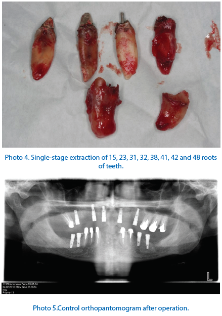
A 36-year-old patient turned to the clinic with the complaints of absence of teeth and impaired chewing function. Diagnosis: root caries of 15, 23, 38, 48, 31, 32, 41 and 42 teeth. Adentia of 12, 14, 16, 22, 25, 36, 37, 46 and 47 teeth (Photos 1 and 2). Plan of treatment: Extraction of roots of 15, 23, 38, 48, 31, 32, 41 and 42 teeth with placement of Alpha Deny Implants in the area of 12, 14, 16, 22, 25, 36, 37, 46 and 47 teeth.

Photo 1. Initial clinical situation.
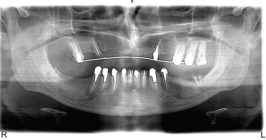
Photo 2. Patient’s orthopantomogram at the stage of planning.
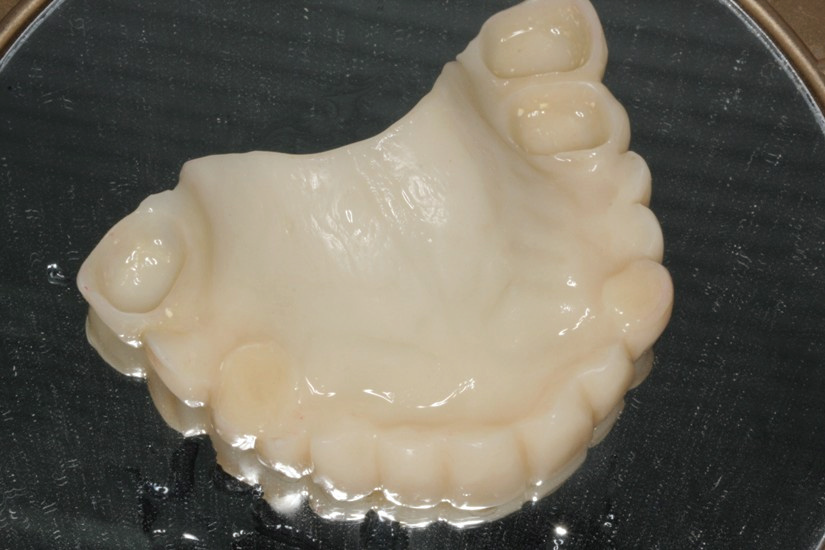
Photo 3. Temporary removable dental prosthesis after extraction of teeth.
A temporary removable dental prosthesis was made at the stage of preparation for the operation (Photo 3).
COURSE OF OPERATION
Removal of 15, 23, 31, 32, 38, 41, 42, 48 teeth and curettage of alveolarsockets were performed under infiltration anesthesia. The mucous membrane was cut along the alveolar crest in the area of 16, 14, 12, 22, 25, 36, 37, 46 and 47 teeth. Exfoliation of periosteal mucosal graft. Formation of bone bed. Placement of Alpha Dent Implants in the area of 12, 14, 16, 22, 25, 36, 37, 46 and 47 teeth (Photo 5). The implants are covered with screw-type plug. Suture of mucous membrane. Adjustment of temporary removable dental prosthesis. Administrations: antibiotic therapy, removal of stitches in seven days.
The doctors set to orthopedic part of treatment in six months. Opening of implants and placement of the gingiva formers were followed by correction and insertion of immediate prosthesis being already available. The individual abutments were made (Photos 6 and 7). The imprints were taken with individual spoons (Photos 8 and 9). Then removable orthopedic construction was replaced by fixed one – temporary plastic bridge prosthesis was made (Photos 11, 12 and 13).

Photo 6. Individual mandibular abutments.


Photo 9. Individual maxillary spoon.


Photo 12. Temporary plastic mandibular bridge.

Photo 13. Temporary plastic prostheses after placement of implants.
This was done in order to eliminate gingival reduction around the implants as well as to determine the real height of bite, horizontal line of cutting edge, location of central incisor midline and their compliance with the implants. Then the metal-ceramic bridge prostheses were made (Photo 14).
RESULT
There was complete and total rehabilitation attained with insertion of maxillary and mandibular metal-ceramic bridge prostheses. The chewing and aesthetic functions were restored (Photo 15).

Disclaimer from Guident
The views and opinions expressed in articles published in Guident are solely those of the authors and do not necessarily reflect the views of the editors, the editorial board or the publisher, Ivory India. Guident does not endorse any product, technique or treatment described. Clinical content is intended for qualified dental and medical professionals and is not a substitute for individual clinical judgement. Guident, its editors and the publisher accept no liability for any consequence arising from the use of information published in the journal.
How to cite this article
Dr. Kuts Pavel Department of Prosthetic Dentistry. Full Rehabilitation with Metal-Ceramic Bridge Prostheses. Guident. 2015 Apr;8(5). Available from: https://ivoryindia.in/articles/implantology/full-rehabilitation-with-metal-ceramic-bridge-prostheses.html