
Healing Abutment Versus Cover Screw After Implant Placement
Jaipur Dental College, Jaipur, India, Wisdom Dental Clinics, Jaipur, India, Wisdom Dental Clinics, Jaipur, India, Department of Pedodontics and Preventive Dentistry
ABSTRACT
This article is based on the most debated topic in the implantlogy i.e, healing abutment versus cover screw after implant placement. So this article emphasis on these two points. First, if there is a strong clinical rationale for immediate healing abutment placement (HAP) and Second, the risks and benefits of delayed implant abutment placement versus HAP. Hence in this article we discuss two cases proving thatif there is no intention to augment the zone of keratinized tissue, healing abutments should be placed at the time of implant placement to avoid unnecessary surgical re-entry.
Keywords - Implant, Healing abutment, Cover screw
INTRODUCTION
Historically, a dental implant was placed into an edentulous site, at which time a cover screw was affixed. Ideally thedepth of implant platform is about one to 2 mm subcrestally so as to be “fully embedded”— thus, the soft-tissue flap could be replaced over the surgical site after placement of the cover screw and would be able to rest on the jaw between fixtures and not directly on the cover screws.1
According to original Brånemark protocols and depending on the site, after waiting anywhere from 4 to 6 months for osseointegration, an abutment was placed during a second-stage surgical entry as part of what is termed a 2-stage procedure—this can also be referred to as delayed abutment placement (DAP).
Thirty years later, some would argue that due to both our more advanced understanding of osseointegration and our remarkably overwhelming emphasis on soft-tissue aesthetics of the final restoration, we witness the placement of healing abutments often at the same time as implant placement in what is termed a single-stage procedure—this can also be referred to as immediate healing abutment placement (HAP). But others might counter that healing abutments are placed with increasing frequency at the time of implant placement simply because it’s more convenient. The paucity of formal investigation on the relative risks and benefits between HAP and DAP unfortunately precludes a rigorous evaluation of evidence-based best practice. Instead, this article presents an informal review of the risks and benefits of the 2 procedures as currently understood.
THE ROLE OF THE HEALING ABUTMENT
Early descriptions in the literature involving the role of the healing abutment are difficult to locate. Some texts refer to Stage II (initial fixture uncovering after healing) by discussing the placement of a final abutment. Throughout time, a number of objectives have been developed for the second-stage surgery. It is important to note, however, that they are really objectives of abutment placement, notwithstanding the timing of this placement, and it is only because DAP was universally employed in all cases that the 2 became synonymous. Now that HAP is a clinical reality, placement of the healing abutment and second-stage surgery should be stated as distinct entities. In order to recognize HAP as an acceptable alternative to DAP, it must be established that HAP can be reasonably expected to meet or exceed the stated goals of DAP (and in reality, of abutment placement as a whole) without contributing unnecessary compromise from surgical, restorative, functional, and aesthetic perspectives.2
THE OBJECTIVES OF DAP AT SECOND-STAGE SURGERY ARE LISTED HERE 3,4 -
1.Firstlyto uncover the submerged implant without damaging the surrounding bone
Conclusion:Peri-implant bone preservation associated with HAP appears to be at least as good as, if not superior to, that associated with DAP.
2. To control the thickness of the soft tissue that is surrounding the implant
Conclusion: Soft-tissue thickness around an implant can be reduced with HAP in a similar fashion as it can be with DAP. Thickening soft tissue via soft-tissue grafting can also be done simultaneously with HAP, or can be attempted during Stage I while performing DAP.
3. To preserve or create attached keratinized tissue which is present around the implant
Conclusion: In a situation where keratinized tissue is lacking, DAP may be more appropriate, unless grafting procedures can be implemented simultaneously with HAP.
4. To ensure proper abutment seating
Conclusion: Full surgical and visual access to the implant platform is ideal when placing healing abutments. HAP affords this opportunity without additional compromise to the peri-implant bone (see objective 1).
5. To develop appropriate soft-tissue contours
Conclusion: HAP, and perhaps specifically with customized or specially designed abutments at immediate extraction sites, appears to favor preservation of the immediate socket soft-tissue profile more than DAP, so the final implant restoration may best mimic the tooth it is meant to replace.
OTHER CONSIDERATIONS
1. Time Management
The most obvious and objective gain of HAP is avoiding the need for a second surgery. The benefit of simplified patient management should not be understated. It is not a secret that some patients fear the dentist in general, and perhaps the underlying issue can be tied to a more specific fear of administration of local anesthesia and surgical intervention, no matter how seemingly insignificant. Furthermore, distinct financial reimbursement for DAP is often nonexistent; therefore subsuming the abutment placement procedure into the same visit as the implant placement procedure is both time and cost efficient. The fewer the number of procedures, the more superior the care, assuming no significant compromises are introduced as a result.5
2. Risk of Occlusal Loading
Occlusal loading may be the greatest concern related to HAP. Initially, it was thought that it was necessary to allow implant fixtures time to integrate prior to exposing them to the oral cavity with transmucosal attachments.1,4,6 Current information shows that this is not the case. If HAP is performed, some worry that forces applied to the exposed abutment during healing can result in failure to integrate successfully. In contrast to this, a remarkable volume of literature has been published endorsing HAP on the grounds that it carries many potential benefits (as discussed above) while not contributing to diminished survival.7-12 The literature even demonstrates that, immediate loading can be a predictably successful option when ample primary stability is achieved.13-16In contrast, early force application via an exposed healing fixture may interfere with osseointegration if an implant does not possess sufficient primary stability.
3.Bone Grafting at the Time of Implant Placement
Introducing bone graft material at an implant site during initial placement may make it more difficult to perform HAP. In such things as planning the incision design, use of membranes, flap advancement procedures, desire for primary closure, ease of suturing, and flap securement , DAP may be advantageous because presence of a healing abutment may interfere in it.3
CASE -1
Healing abtument is placed immediately after implant placement resulting in good emergence and contour of the crowns.(Figure.1a,b,c,d)
(Figure. 1 a, b, c, d)
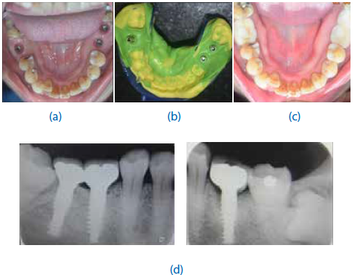
CASE - 2
Healing abtument is placed immediately after implant placement resulting into skipping 2nd stage surgery & directly impression making before crowns placement. (Figure. 2 a, b, c, d)

(Figure. 2 a, b, c, d)
CONCLUSION
Clinical Recommendations HAP appears to be equivalent to DAP in all objectives except when there is either a deficiency in primary stability, or softtissue thickness or keratinization. Notwithstanding, some investigators have developed methods of soft-tissue augmentation that have been shown to be predictable even when done at the same time as placement of a healing abutment.
Based on the available data and the clinical experience of those who have been performing HAP with regularity for a considerable amount of time, the following factors ought to be taken into account when considering the timing of abutment placement:
- HAP has been shown to meet or exceed the demands of healing abutment placement in many situations.
- HAP reduces both chair time and the number of surgical procedures. IAP also reduces the number of times a patient needs to be anesthetized, a procedure that most patients would prefer to avoid.
- When sufficient primary stability has been achieved, the concern of early overloading may be overemphasized. At this time, however, the characteristics of sufficiency of stability (ie, degree of stability and extent of fixture surface area engaging bone) have not been well defined. Two currently utilized parameters of stability, final surgical torque value and implant stability quotient, are only modestly informative, as they do not take primary stability into account as a function of the surface area, which may be a critical component that is overlooked. Further study is required in this area.
- Healing abutments should not extend more than 2 mm coronally from the gingival margin to limit force transmission during mastication. It should be out of occlusion in all excursions and protrusive. Care should be taken in patients with a history of parafunctional habits, and clinicians should confirm that healing abutments exhibit clearance not only from opposing teeth, but also from any removable appliances or devices, such as athletic guards or nightguards.
- If there is no intention to augment the zone of keratinized tissue, healing abutments should be placed at the time of implant placement to avoid unnecessary surgical re-entry. Even if keratinized tissue is deemed deficient, techniques do exist for augmenting soft tissue following fixation of transmucosal components.
REFERENCES
- Adell R, Lekholm U, Brånemark PI. Surgical procedures. In: Brånemark PI, Zarb GA, Albrektsson T, eds. Tissue-Integrated Prostheses: Osseointegration in Clinical Dentistry. Chicago, IL: Quintessence Publishing; 1985:221.
- Lekholm U. The surgical site. In: Lindhe J, Karring T, Lang NP, eds. Clinical Periodontology and Implant Dentistry. 4th ed. Oxford, England: Blackwell Munksgaard; 2003:863.
- Han TJ, Park KB, Klokkevold PR. Standard implant surgical procedures. In: Newman MG, Takei HH, Klokkevold PR, et al, eds. Carranza’s Clinical Periodontology. 10th ed. St. Louis, MO: Saunders Elsevier; 2006:1120-1132.
- McGlumphy EA. Implant-supported fixed prostheses. In: Rosenstiel SF, Land MF, Fujimoto J, eds. Contemporary Fixed Prosthodontics. 3rd ed. St. Louis, MO: Mosby; 2001:315,326,329.
- Buser D, Wittneben J, Bornstein MM, et al. Stability of contour augmentation and esthetic outcomes of implant-supported single crowns in the esthetic zone: 3-year results of a prospective study with early implant placement postextraction. J Periodontol. 2011;82:342-349.
- Block MS. Anesthesia, incision design, surgical principles and exposure techniques. In: Block MS, Kent JN, Guerra LR, eds. Implants in Dentistry: Essentials of Endosseous Implants for Maxillofacial Reconstruction. Philadelphia, PA: Saunders; 1997:153.
- De Rouck T, Collys K, Wyn I, et al. Instant provisionalization of immediate single-tooth implants is essential to optimize esthetic treatment outcome. Clin Oral Implants Res. 2009;20:566-570.
- Block MS, Mercante DE, Lirette D, et al. Prospective evaluation of immediate and delayed provisional single tooth restorations. J Oral Maxillofac Surg. 2009;67(suppl 11):89-107.
- Cooper LF, Raes F, Reside GJ, et al. Comparison of radiographic and clinical outcomes following immediate provisionalization of single-tooth dental implants placed in healed alveolar ridges and extraction sockets. Int J Oral Maxillofac Implants. 2010;25:1222-1232.
- Chen ST, Darby IB, Reynolds EC. A prospective clinical study of non-submerged immediate implants: clinical outcomes and esthetic results. Clin Oral Implants Res. 2007;18:552-562.
- Cordaro L, Torsello F, Roccuzzo M. Clinical outcome of submerged vs. non-submerged implants placed in fresh extraction sockets. Clin Oral Implants Res. 2009;20:1307-1313.
- Hämmerle CH, Lang NP. Single stage surgery combining transmucosal implant placement with guided bone regeneration and bioresorbable materials. Clin Oral Implants Res. 2001;12:9-18.
- Cooper LF, Scurria MS, Lang LA, et al. Treatment of edentulism using Astra Tech implants and ball abutments to retain mandibular overdentures. Int J Oral Maxillofac Implants. 1999;14:646-653.
- Chiapasco M, Gatti C. Implant-retained mandibular overdentures with immediate loading: a 3- to 8-year prospective study on 328 implants. Clin Implant Dent Relat Res. 2003;5:29-38.
- Raghoebar GM, Friberg B, Grunert I, et al. 3-year prospective multicenter study on one-stage implant surgery and early loading in the edentulous mandible. Clin Implant Dent Relat Res. 2003;5:39-46.
- Crespi R, Capparé P, Gherlone E, et al. Immediate versus delayed loading of dental implants placed in fresh extraction sockets in the maxillary esthetic zone: a clinical comparative study. Int J Oral Maxillofac Implants. 2008;23:753-758
Disclaimer from Guident
The views and opinions expressed in articles published in Guident are solely those of the authors and do not necessarily reflect the views of the editors, the editorial board or the publisher, Ivory India. Guident does not endorse any product, technique or treatment described. Clinical content is intended for qualified dental and medical professionals and is not a substitute for individual clinical judgement. Guident, its editors and the publisher accept no liability for any consequence arising from the use of information published in the journal.
How to cite this article
Dr. Gaurav Gupta, Dr. D. K. Gupta, Dr. Neelja Gupta. Healing Abutment Versus Cover Screw After Implant Placement. Guident. 2019 Apr;12(5). Available from: https://ivoryindia.in/articles/implantology/healing-abutment-versus-cover-screw-after-implant-placement.html


