Maintenance of Dental Implants
Department of Orthodontics and Dentofacial Orthopedics, manipal college of Dental Science Manipal, Karnatka, India
Abstract
This article reviews various implant maintenance procedures to increase the longevity of implants.
INTRODUCTION
An implant is not a tooth as it does not decay, has no periodontal membrane and a vital pulp as an early indicator of any disease. The strong biological bond formed by the dental implant enables the entire tooth replacement structure to remain firmly ‘rooted’ in the jaw bone and function like a natural tooth.1,2
The following are various implant maintenance procedures which are useful in the clinical practice.
Role of clinician2
- Identification of potential implant patients.
- Demonstrating oral hygiene options during each phase of treatment.
- Thoroughly educating the patients about the implant therapy and what is to be expected during each phase.
- Should assess the patients’ home care routine.
- Informing about the financial, time and maintenance appointment which is very crucial for the long term success of implant therapy.
- Use of instruments that are effective at removing biofilms and accretions.
- Should avoid damage to all portions of the implant, abutment restorations and tissues.
- Establishment and maintenance of soft tissue around the trans-mucosal portion of the implant which enhances the success of implant.
PLAQUE BIOFILM AND IMPLANT
Biofilms are primary causative factor of periodontal disease. If left undisturbed, it will lead to mature plaque on teeth. From teeth bacteria migrate to implant àimplants.
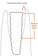
Similar to teeth, failing implants present inflammation, pockets and progressive bone loss.
Another similarity is between periodontitis microbiota and peri-implantitis.
Studies have found that patients with history of periodontitis had a greater impact on peri-implant microbiota than implant loading time. Thus, it is very important to educate patients about their responsibility to decrease plaque effectively especially with history of periodontitis.
Natural dentition with dental implants appears to increase the risk for implant infections compared to completely edentulous patients. This suggests that natural teeth may serve as reservoir for periodontal pathogen that may extend their growth to contiguous implants in the same oral cavity.
Listgarten et al3 reported proportions of coccoid forms (65.8%), motile rods (2.3%) and spirochetes (2.1%) in implant pocket area whereas in natural teeth they were 55.6%, 4.9% and 3.6% respectively. In fully edentulous dentition the same was found to be 71.3%, 0.4% and no spirochetes. Thus, they concluded that microflora in partially edentulous is more pathogenic than fully edentulous patients.
Peri-implant mucositis is similar to gingivitis as there is no bone loss. Therefore, similar to gingivitis, peri-implant mucositis is also reversible, once etiologic agent and plaque biofilm is removed. If undisturbed will lead to peri-implantitis and includes loss of Osseo-integration similar to loss of attachment in periodontitis.
PATIENT ORAL HYGIENE
Patients rely on clinicians to suggest or recommend products for oral hygiene procedures. With most of the patients “Tell, show, do” technique of home care instruction is very helpful. Clinician instructions and patient’s compliance is necessary to evaluate the long term success for each patient.
The clinician should take into consideration the location, length, angulation of abutments, super structure design, anatomical limitations, patient’s habits, motivation and manual dexterity of each patient while recommending oral hygiene methods.
Oral hygiene instructions should be simple to keep patient motivated. Final prosthesis should allow for access to the patient and clinician to keep area clean.
Clinician should instruct the patient the use of tooth brushes (manual and automatic), floss, tufted brushes, interdental brushes, oral irrigators etc.
Patient instructions may include the use of anti-microbial such as cetyl pyridinium chloride or chlorhexidine gluconate. It can be used as a rinse or applied to the area with brushes or cotton swabs. Oral irrigation if used should be properly instructed.
INSTRUMENT SELECTION
Maintaining a smooth surface of titanium without pits and scratches can prevent plaque accumulation.Instrument selection should depend upon tip design that is not bulky (to avoid unnecessary tissue manipulation and design should facilitate manipulation by clinician.
Evaluation of prosthesis design, location of deposits, and tenacity of calculus also help select appropriate instruments.
Metallic ultrasonic and sonic scalers have been reported to gouge titanium. A plastic or rubber sleeve over an ultrasonic scaler appears not to alter titanium.Conventional ultrasonic scalers with nonmetallic tip also are suitable for implant maintenance.2
Air polishers are effective and safe for maintenance procedures around implants.
Stainless steel tipped instruments have been found to be detrimental to a smooth titanium surface.This is important when sharpening implant hygiene instruments to ensure a sharpening stone free of steel scrapings. A variety of nonmetallic, plastic, graphite, nylon or Teflon coated instruments are available and have been proven to be safe to use on titanium implant surfaces.
A titanium curette and rubber cup with flow of pumice is suitable for cleaning implant surfaces. After removal of calculus, polishing with a rubber cup and tooth paste, fine prophy paste, commercial implant polishing pastes and tin oxide have been shown to be safe for titanium surfaces.
Ramaglia et al4 demonstrated that a plastic curette and air powder with water spray did not alter the implant surface but may leave deposits which should be removed by irrigation to avoid any adverse tissue healing.
IMPLANT MAINTENANCE PROCEDURES
Frequent recall visits after implant placement and restorations are necessary for evaluation and establishment of good oral hygiene after treatment.
Titanium usually does not have calculus embedded into it. Therefore adequate oral hygiene should prevent the accumulation of heavy accretions.
Healthy implant tissues may have tight seal and create challenges while scaling. Since, the peri-mucosal seal is more fragile than a normal tooth sulcus, it is important to use short exploratory working strokes with light pressure.
Depending on the location of calculus a horizontal, vertical or oblique strokes may need to be used while avoiding tissue trauma.
When an instrument must be used sub-gingivally to remove calculus or excess cement, insertion and instrumentation should be gentle and light strokes should be used in semi-circular pattern.
Attention to placing the blade carefully under the deposit and calculus or cement with compressed air may make detection and removal easier and more comfortable for the patient.
Surgical correction may be required sometimes to reduce chronic inflammation and infection.
Periodic occlusion monitoring may detect discrepancies to indicate occlusal changes needed.
CHEMOTHERAPEUTIC AGENTS
Chlorhexidine gluconate has been shown to reduce inflammation in the oral cavity and around dental implants.
Long term use of antimicrobials such as chlorhexidine gluconate, cetyl pyridinium chloride or phenolic compounds may be used along with brushes and floss to minimize staining.
If sub-gingival irrigation is used, cannula should be inserted carefully in peri-implant tissue to avoid gouging the surface.
Also, care should be taken to avoid gouging the surface. Care should also be taken to avoid inserting the cannula to the base of implant sulcus to prevent fluid distention into surrounding tissues.
IMPLANT QUALITY AND HEALTH SCALE
Implant quality scale has been established as an aid to diagnosis and treatment that also proposes management approaches according to the signs and symptoms. This scale based on clinical evaluation was agreed upon by the International congress of Oral Implantologists2 in 2007. The prognosis and appropriate treatment is related to the quality scale.
Table
|
Group |
Clinical conditions |
|
No pain or tenderness on function |
|
No pain |
|
No pain |
|
Any of the following: |
REPAIR OF AILING, FAILING DENTAL IMPLANTS
1. If an active infection (purulence, swelling) is present with radiographically visible bone loss and the disease process continuing, following steps should be implemented.
- Reflect the tissue and de-granulate the defect (metallic currettes are acceptable).
- If the implant is hydroxyapatite (HA) coated and HA is undergoing resorption and has changed color texture ïƒ requires removal of all the HA until the metallic surface is visible. Use of ultrasonic such as cavitron is best. Use of air abrasion is dangerous because of danger of air emboli in marrow spaces.
- Detoxify the dental implant with citric acid (40% ph-1) 30 seconds per surface is sufficient.
- Graft with freeze dried bone or alloplast if completely detoxified, or with alloplast such as HA or bio-glass if not completely detoxified.
- Protect the graft with a membrane for guided bone regeneration if needed.
- Leave the repaired implant out of function and covered for 10-12 weeks.
Note: If the surface of implant is metallic (Ti, Ti-6Al-4V, Ti plasma spray), go from step A to step C.
2. If no active infection is present and if HA coated implant is in place, HA looks intact without ongoing resorption (bone loss from traumatic occlusion, overloading, off axis loading) follow the steps
- Reflect the tissue and degranulate the defect with metallic currettes.
- Detoxify the HA surface with citric acid (40% ph-1) 30 seconds per surface. Flush and irrigate with sterile water or sterile saline to stop demineralization process of citric acid.
- Continuing with grafting and GBR material.
Important: Do not use Tetracycline on intact HA because it changes the Ca/P ratio of HA. Do not leave citric acid on HA surface for more than 1 minute, it continues to “remove”.
Absolute implant failure consists of implant surgically removed or exfoliated or no longer in the mouth.
The remaining edentulous area often is treated with autogenous or synthetic bone graft procedures which are performed to replace the missing bone. Once favorable bony conditions are restored, implants may be inserted with good prognosis.
IMPLANT- CROWN ESTHETIC INDEX
This was developed by Henny et al5 as an objective tool in rating esthetics of implant supported single crowns and adjacent soft tissue.
Esthetics can be rated by
- Subjective - use of Questionnaire completed by patient.
- Objective - by professional observer.
An objective rating score with a division in different items provides an insight into the esthetic results as a function of time to analyze the stability of treatment procedures.
9 selected items were as follows. These items should be in harmony with adjacent teeth and contralateral tooth.
Above 3 based on 5 point rating scale – Grossly under contoured
Slightly under contoured
No deviation
Slightly over contoured
Grossly over contoured
Above 2 based on 3 point rating scale – Gross mismatch
Slight mismatch
No mismatch
Above 2 based on 3 point rating scale – Deviation of 1.5 mm/ more
Deviation of less than 1.5 mm
No deviation
Based on 5 point scale - Grossly under contoured
Slightly under contoured
No deviation
Slightly over contoured
Grossly over contoured
Based on 3 point rating scale – Gross mismatch
Slight mismatch
No mismatch
- Mesio-distal dimension of crown.
- Position of incisal edge of crown.
- Labial convexity of crown.
- Color and translucency of crown.
- Surface of crown.
- Position of the labial margin of the peri-implant mucosa.
- Position of the mucosa in the approximal embrasures.
- Contour of labial surface of mucosa
- Color and surface of labial mucosa
Grading
0 points – excellent esthetics
1-2 points – satisfactory esthetics
3-4 points – moderate
5 or more – poor
CONCLUSIONS
Implant maintenance plays a key role in the success of dental implants through regular 3 month assessment and maintenance procedures.
REFERENCES
- Anjali mehta, Siddharth mehta. Tooth or Implant: whats the difference. A review. Guident 2014;7(12):48-50.
- Misch CE. Contemporary implant dentistry. Mosby III edition.
- Listgarten, M.A.,Hellden, L. Relative distribution of bacteria at clinically and periodontally diseased sites in humans, J ClinPeriodontol 1978;5:115-32.
- Ramaglia, L, di Lauro, A.E., Morgese, F., Squillace, A. Profilometric and standard error of the mean analysis of rough implant surfaces treated with different instrumentations. Implant Dentistry 2006;15:77–82.
- Henny J. A. Meijer,KeesStellingsma,Leo Meijndert,Gerry M. Raghoebar.A new index for rating aesthetics of implant-supported single crowns and adjacent soft tissues – the Implant Crown Aesthetic Index.Clinical Oral Implants Research 2006; 16(6):645-9.
Disclaimer from Guident
The views and opinions expressed in articles published in Guident are solely those of the authors and do not necessarily reflect the views of the editors, the editorial board or the publisher, Ivory India. Guident does not endorse any product, technique or treatment described. Clinical content is intended for qualified dental and medical professionals and is not a substitute for individual clinical judgement. Guident, its editors and the publisher accept no liability for any consequence arising from the use of information published in the journal.
How to cite this article
Dr. Siddharth Mehta, Dr. Anjali Mehta. Maintenance of Dental Implants. Guident. 2016 Mar;9(4). Available from: https://ivoryindia.in/articles/implantology/maintenance-of-dental-implants.html