Papilla Preservation- a Journey to Pink Health
Department of Prosthodontics K.D Dental College & Hospital, Mathura, (U.P), India
Abstract
Presented are the main stages of the origin and application of Plasmolifting technology, which is an injectable form of platelet-rich auto plasma; theoretical and pathogenetic mechanisms, acquired clinical, indications and recommendations for the technology application in dermatocosmetology, counter indications, method of the procedure fulfillment.
INTRODUCTION
Aesthetic implantology seeks harmony in all details to simulate the appearance of natural teeth to achieve the overall desired beauty. Under the scope of aesthetics natural teeth take on more from being merely white physical structures and assume social attributes integral to a person’s self-image, interaction and physical attractiveness.
Research shows besides most patients appreciating the functional improvement experienced by prosthodontic implant rehabilitation also note a marked upsurge of social and spiritual wellbeing as a result of the changes in appearance.
Morphology of Interproximal Papillae
Morphological configuration of interdental papilla depends on the following factors:
- Volume of underlying osseous support
- Proximal surface topography of adjacent teeth
- Assistance of sharpey’s fibres
The two crests of osseous bone are connected by gingival col which maybe
- Convex
- Concave
- Sawtoothed.(Holmes 1965, Cohen 1959).
The collagenous fibres run in buccolingually through the lamina propria (Stahl 1963, Melcher 1962, Hagerman 1965). (Type V collagen, more resistant to collagenase, is predominant around the implants.) This keeps marginal gingival closely adherend to the neck of the tooth.Thus these fibres are responsible for maintaining the shape and position of the papilla
Loss of Interproximal Papilla
The sequential loss of the interproximal papillae starts immediately after extraction. The various reasons for this can be listed as
- Thin crestal alveolar bone
- Reduced blood supply
- Possible direct contamination by oral bacteria
- Absence of Sharpey’s fibres in the peri-implant tissue
Classification Of Conditions Of Interproximal Papillae
- Tarnow et al 1992
- Salama et al 1998
- Gastaldo et al 2004
- Norland and Tarnow
- Furhausens Pink Esthetic Score
TARNOW ET AL 1992

This classification is considered to be the ultimate clinical parameter in predicting the presence of papilla around implants.

PAPILLA PRESERVATION TECHNIQUES
Correct treatment planning: Prior to even the extraction of teeth, thorough treatment planning for implants must be performed.
- Deficiencies in hard and soft tissues which preclude the esthetically optimal placement of an implant must be addressed and systematically corrected.
- It is only after the ridge has been restored to close to its original dimensions that soft tissue augmentation is considered.
BIO-COL ALVEOLAR RIDGE PRESERVATION TECHNIQUE
- Combines surgical and prosthetic protocols to preserve hard and soft tissue ridge anatomy in preparation for implant placement in aesthetic areas.Rationale is prevention of soft tissue collapse that occurs following tooth extraction, by immediately supporting these tissues with anatomically correct provisional restorations that replicate natural tooth contours

Immediate implants: Placement of immediate implants reduces the alveolar resorption which helps maintain osseous support for the interpapillary tissues thereby preserving the interproximal papillae.

Optimal implant position:

Implant position especially axial position determines the distance of the contact point to the osseous crest.
Tissue biotypes:

- thick flat tissue biotypes are more predictable.
- thin scalloped tissue biotypes
- insufficient volume
- liable to slough
- Recession
- In case of non-availability of the right biotype,
tissue augmentation procedures
orthodontic extrusion
FLAP DESIGN
Cosmetic incisions, innovative flap design and conservative exposure techniques aim at preserving soft tissue circulation and minimising shrinkage in aesthetically critical areas.

Flapless placement of implants:
- Reduced post operative bleeding
- less post operative discomfort to the patient
- minimally invasive nature of the procedure
- Preserves excess soft tissue volume on the facial aspect.
- Available in a variety of diameters

PROSTHETIC GUIDED SOFT TISSUE HEALING
Provisionalisation (Kan and Rungcharassaeg, 2003) has been introduced as the use of flared provisional crowns which put pressure onto the interdental papilla to maintain its shape.

Orthodontic solutions: Teeth are moved in an incisal direction, the new bone deposits above the root apex and an increase level of attached mucosa and proximal papillae is observed at cervical level. Requirements:
- Rate of extrusion should not exceed rate of bone deposition
- Tooth must be moved only in axial direction, tipping may cause penetration of labial plate.
- Light eruptive forces of 25-30g result in coronal migration of entire attachment apparatus
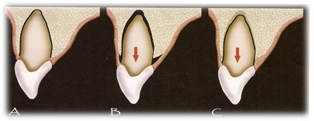
- be a highly predictable treatment modality for preserving interproximal papilla.
- prolongs the duration of treatment
- additional costs
- limited to immediate implant protocols.
Scalloped implant design: Scalloped designs can be helpful to preserve interproximal tissues.
Papilla Regeneration
Factors influencing treatment outcome (prognosis) of papilla reconstruction surgery:
Blood supply: blood supply maintained in the flap design, especially in complex grafting procedures involving both hard and soft tissues, is a key factor in a positive treatment outcome.
Crown shape
- Triangular crowns negatively influence the volume and height of gingival embrasure, thus affecting overall size of interimplant papilla.
- Square crown shapes show better prognosis

Use of wider implants induces more soft tissue loss.
Height of osseous crest: determines the predictability of interimplant papilla. In case of severe resorption, papillary morphology and soft tissue is lost. A staged approach wherein stable osseous topography is achieved then the implant is placed.

Platform switching: switching the platform to a smaller diameter at the interface level favors biologic width development in horizontal direction to compensate for vertical thereby minimizing post operative bone resorption and helping to maintain stable margins. Narrow and constricted sub-gingival components lead to better soft tissue marginal results

PROCEDURES FOR PAPILLA REGENERATION :

Soft Tissue Procedures
Due to the fragile nature of peri-implant soft tissues, decreased blood supply at soft tissue ends and post operative soft tissue remodeling factor, most soft tissue methods have attained neither great long term prognostic success, nor persistent clinical results. Several soft tissue surgical procedures have been introduced in an attempt to reconstruct the missing papillary tissues:
- Han and Takei 1996:
- Coronally positioned flaps with connective tissue grafts.
- Tinti and Parma-Benfenati 1995:
- Coronally positioned palatal sliding flaps
- Beagle’s Technique:
- Palatal pedicle flap that is folded and tied on itself on the facial side to increase the height of the interimplant papilla.
- Azzi et al:
- Connective tissue grafts be placed on the defective area and tucked under the buccal and palatal flaps
- Askary:
- Modified lateral sliding flap.
- Misch et al:
- Split Finger Technique
- Adrianssen:
- Palatal Striding Strip Flap
- Non-pedicled Connective Tissue Grafts
Osseous regenerative methods
“Successful and predictable esthetic results can be accomplished only when underlying labial and interproximal support has been therapeutically provided. ” Salama 1998
Interimplant Papilla Regenerative Technique
- Introduced by Askary in 1999
- Ti template acts as housing for bone graft material
- carries and protects the bone graft material
- separates bone graft from the fibroblast and epithelial cells
- favors graft predictability.

- showed acceptable clinical success
Miniautogenous Bone Blocks
- These have been tried as a method of creating a basal foundation of inter-implant papillae. This did not show higher success rate due to the smaller size of the graft.

DISTRACTION OSTEOGENESIS
Earlier used mainly to correct alveolar deformities in ridge height. In 1996, introduced as a promising method for restoring deficient alveolar ridges. The use of distraction osteogenesis is limited by its restriction of action to one dimension as well as its complexity in clinical handling and soft tissue closure.Thus, osseous regenerative methods to restore interimplant papilla:
- Do not offer a highly predictable success rate.
- Require superior surgical skills
- Require wider space between implants
- Increase risk of graft morbidity
- Require modification of graft margins to allow for total closure
- Decrease size of papilla via post operative resorption and remodeling
PROSTHETIC MASKING
Masking Interproximal Defects
- Acrylic Stents
- Papillary illusions
- Pink Porcelain (Cronin and Wardle 1983)
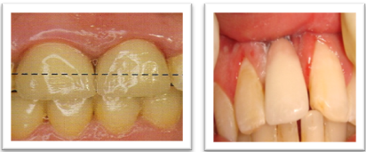
Prosthetic methods of restoring interpapillary esthetics are:
- Less invasive
- More convenient to the patient
- Do not regenerate soft tissue but help to mask the defects
- Can be used as second line treatment when other options are not possible, or have failed.
UNDER FURTHER RESEARCH
Regeneration of inter implant papillae following 1-3 years after surgery due to inflammatory enlargement.

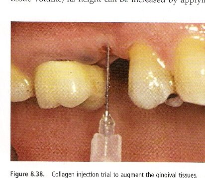
Collagen injection trial to augment gingival tissues showed statistically significant increase from baseline in papillary height
CONCLUSION
The prognosis of surgical attempts to restore interproximal papilla has not been very encouraging due to the delicate peri-implant soft tissues, compromised vascularity, and post operative remodeling. Thus at a stage, where the methods of regeneration of gingival fibres has not been perfected, it is considered wisest to preserve rather than attempt to reconstruct
As they say, Natures beauty can seldom be recreated in human hands, prevention is yet, better than cure!
Disclaimer from Guident
The views and opinions expressed in articles published in Guident are solely those of the authors and do not necessarily reflect the views of the editors, the editorial board or the publisher, Ivory India. Guident does not endorse any product, technique or treatment described. Clinical content is intended for qualified dental and medical professionals and is not a substitute for individual clinical judgement. Guident, its editors and the publisher accept no liability for any consequence arising from the use of information published in the journal.
How to cite this article
Dr. Manesh Lahori, Dr. Rahul Nagrath, Dr. Siddharth Sisodia. Papilla Preservation- a Journey to Pink Health. Guident. 2013 Feb;6(4). Available from: https://ivoryindia.in/articles/implantology/papilla-preservation-a-journey-to-pink-health.html