
Reconsconstruction and Rehabilitation of a Resected Mandible for Ameloblastoma with Oral Implants -a 20 Year Follow Up
Oral & Maxillofacial Prosthodontist, Prosthodontist, Surgical Prosthodontist & Chief Implantologist
Abstract
Ameloblastoma is a slow growing benign tumor of the jaw and patients usually present late after the tumor achieved considerable size to cause facial disfigurement. Diagnosis mainly from tissue biopsy and characteristic findings on plain X-rays does assist in differentiating between types of ameloblastoma. The management of this patient was done by marsupialization in 1990 and in 2003 she was followed up with dental implants; moreover, patient revisited after 20 years and remarkably showed no signs of recurrence.
Key words: Ameloblastoma, marsupialization, dental implants
INTRODUCTION
- Definition: A true neoplasm of enamel organ type tissue which does not undergo differentiation to the point of enamel formation3.
- As described by Robinson ‘usually unicentric, nonfunctional, intermittent in growth, anatomically benign and clinically persistent’3.
- Types: there are 3 forms that are common the peripheral, unicystic, and multicystic tumors. The histological variants (follicular, plexiform and acanthomatous) show no correlation with either the clinical appearance of the lesion or its behavior and variation may be seen between different sections of the same tumor. A unicystic variant of the ameloblastoma tends to occur during teenage years and has been reported to exhibit a less aggressive behavior4.
- Its peak incidence is in the 3rd to 4th decades of life and the male to female ratio is 1:1. It is often associated with an unerupted third molar3.
- It may present as a result of routine radiographic examination finding. Eighty percent of ameloblastomas occur in the mandible and majority is found in the angle of mandible and ramus region.
- The challenge in managing ameloblastoma is in achieving complete excision and reducing the risk of recurrence as maximally as possible.
- We present a case with a mandibular ameloblastoma, a review of its literature and follow up with dental implant treatment.
CASE REPORT
In 1990 a 13 year old Indian girl, came to the dental clinic with a swelling, discomfort over the right jaw along with disfigurement on the same side. It was first noticed a while ago but progressively increased to the size when it was first shown to the doctor. However, it was not large enough to cause malocclusion. An OPG and a lateral cephalogram were taken prior to surgery.
Physical examination: There was a medium firm swelling over the right mandibular region which was non tender extending from the left angle of mouth anteriorly to the anterior border of sternocliedomastoid muscle posteriorly and causing right facial asymmetry. The skin overlying the mass was normal in color. There was no enlarged lymph node over the neck region. There was no trismus and intraorally, she had good dental hygiene and all the present teeth were vital.
An OPG (fig.1) revealed a cystic lesion in the right mandibular region extending mesially up to right parasymphisis, laterally up to right border of the angle, inferiorly up to border of the ramus and superiorly up to the roots of the involved teeth, which are numbers #31 #30 #29 #28 #27.
Though an incisonal biopsy, which was done under local anesthesia, result did not show specific features of ameloblastoma, the clinical history and physical examination along with radiological findings were highly suggestive of ameloblastoma.
Histopathological result revealed sheets of odontogenic epithelium with basal layer of cuboidal cells and suprabasal stellate reticulum like cells exhibiting cystic degeneration (Fig. 3).
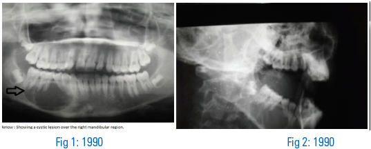

Figure 3-1990
Management: Considering the young age of the patient, the cyst being non aggressive and unicystic or unilocular, marsupialization was done instead of radical dissection of the cyst (Fig. 4).
Slight paraesthesia was reported immediately following surgery. Lower border was left intact and time was given for the healing of bone. Six months postoperatively, paraesthesia had subsided and normal function had returned.

Figure 4-2000
In 2003, patient revisited complaining of mal-alignment of teeth and inability to chew properly. Thus, fixed orthodontic appliance was placed for correction.

Fig 5: 2003
After achieving alignment, patient was consulted for implant surgery. However, she feared recurrence of paraesthesia in the lower border and hence, implant surgery was only placed in the premolar region. Along with the patient’s apprehension, there was an area of radiopactiy, in the lower right border, which denoted incompetent bone formation. The decision was made to only restore the premolar regions which lead to a regain of function for mastication and normal aesthetics.

Figure 6-2005

Discussion: Ameloblastoma in the mandible can progress to great size and cause facial asymmetry, displacement of teeth, loose teeth, malocclusion, and pathologic fractures. Tumor size may ranges from 1 to 16 cm at presentation which result from expansion of bone and invasion into soft tissue5.
Typically ameloblastoma present as painless slow growing mass. Becelli et al. (2002) studied 60 patients whom were confirmed with mandibular ameloblastoma and found out that about half of them showed typical symptoms such as swelling of the affected region (38.3%), paraesthesia of the innervated region of the mandibular nerve (13.3%) and alteration in dental occlusion in 10% of the cases1.Radiographically, ameloblastoma appear as radiolucent lesion that may have either a unilocular or multilocular appearance.
The primary concern for this patient was esthetics followed by replacement lost teeth and restore optimal function. With the goal in mind, marsupialization was done to treat the ameloblastoma.
Postoperatively, patient suffered slight paraesthesia on the right side and she was advised for rest. And because we could not locate the alveolar canal along with the patient’s fear of paraesthesia returning and radiopacity in the lower right border; the decision was made to not include implants in the molar region.
In consideration of the postoperative events, only two implants were placed in the premolar region in the lower right mandible (fig. 7). Becker and Wong reported an early functional loading case in the fully edentulous mandibular resection and reconstruction due to an ameloblastoma and inserted five implants two years after the removal of the tumor and they concluded that the implants are stable and the patient eating comfortably2.
As a conclusion, this monolocular lesion was managed by chemical cautery and marsupialization followed by osseointegration with implants which showed good primary bone healing and support. There were no signs of recurrence after a follow up appointment; even after long term follow up showed no signs. Marsupialization followed by the use of osseointegrated implants is a possible treatment plan in a monolocular ameloblastoma in the mandible.
Disclaimer from Guident
The views and opinions expressed in articles published in Guident are solely those of the authors and do not necessarily reflect the views of the editors, the editorial board or the publisher, Ivory India. Guident does not endorse any product, technique or treatment described. Clinical content is intended for qualified dental and medical professionals and is not a substitute for individual clinical judgement. Guident, its editors and the publisher accept no liability for any consequence arising from the use of information published in the journal.
How to cite this article
Dr. M.S. Gowd, Dr. T.Shankar, Dr.Vikas Gowd. Reconsconstruction and Rehabilitation of a Resected Mandible for Ameloblastoma with Oral Implants -a 20 Year Follow Up. Guident. 2013 Feb;6(4). Available from: https://ivoryindia.in/articles/implantology/reconsconstruction-and-rehabilitation-of-a-resected-mandible-for-ameloblastoma-with-oral-implants-a-20-year.html


