Surface Modification of Dental Implants
Department of Prosthodontics A.B. Shetty Memorial Institute of Dental Sciences, Nitte University, Deralakatte, Mangalore, India
ABSTRACT
Surface modifications of dental implants have been proved to improve the bone to implant contact and osseointegration. This article reviews themacrotopography, microtopography and nanotopography of dental implants established by various techniques and includes the advantages and disadvantages of each. This may enable the clinician and manufacturer to select an appropriate dental implant for a particular clinical situationas, ultimately a thorough knowledge of material science along with choice of a good clinical technique enables the clinician to deliver good treatment options.
KEYWORDS: Surface modification , Nanotopography , Microtopography , Dental implants
INTRODUCTION
The use of endosseous dental implants is a well- accepted treatment modality for oral and craniofacial reconstruction serving as transmucosal structures to support single tooth, fixed partial denture prosthesis, complete arch reconstructions and removable complete dentures or to reconstruct maxillofacial defects¹. Success of dental implants can be attributed to varioustheoriespropagated in the past century, of which P.I Branemark’sOsseointegration theory is the most important. In 1985, Branemark defined osseointegration at a light microscopic level as “ a direct structural and functional connection between ordered living bone and surface of a load carrying implant”². The main clinical advantage was the osseous interface formed and maintained at the titanium surface of load bearing dental implant in contrast to the fibrous encapsulation seen in osseopreservation mode of tisse integration which was a negative and undesired result.³Success of dental implants could be due to various factors i. e clinician related factors, implant factors , local or anatomic factors , biologic factors , systemic or functional factors4,5 . Implant surface character is an implant design factor affecting the rate and extent of osseointegration. Precisely, how much of the implant surface directly contacts bone , how rapidly osseointegration occurs and the mechanical nature of bone implant connection is influenced by the nature of implant surface itself6. Surface modifications of dental implants have been proposed to achieve better osseointegration and in turn higher implant success rates. Surface modification of dental implants may be broadly studied under 3 headings depending on the level at which surface topography has been altered viz. macrotopography, microtopography and nanotopography.
MACROTOPOGRAPHY
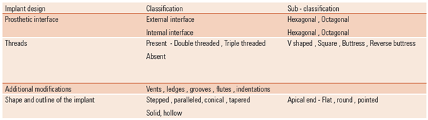
Macrotopography of an implant surface refers to the implant design; the 3 dimensional structure of the implant, with all the elements and characteristics that compose it. This could refer to the prosthetic interface, presence of threads and their shape, additional modifications, shape and outline of the implant1.
MICROTOPOGRAPHY
Ito et al had classified the various methods of obtaining microtopography of the surface into :7
Physicochemical method – It involves the alteration of surface energy , surface charge and surface composition . The method employed is glow discharge method. It increases the cell adhesion properties and is conducive to tissue integration. On the contralateral side , it does not help in adhering selective cells and tissues and it has not shown to increase bone implant strength.
Biochemical method – They are an alternative or adjunct to physicochemical and morphological methods. It endeavours to utilize current understanding of biology and biochemistry of cellular function and differentiation to develop favourable implant surfaces. It aims to immobilize proteins , enzymes or peptides on biomaterial for the purpose of inducing specific cells or tissue response. One approach describes controlling cell –biomaterial interaction utilizing cell adhesion molecules. ( eg : RGD – Arginine – Glycine – Aspartidin has been known as a mediator of attachment of cells to several plasma and extra cellular matrix protein including osteopontin ). The other approach utilizes biomolecules which demonstrate osteotropic effects (eg : interleukin , growth factor 1 and 2, plasma growth factor , bone matrix protein).
Morphological methods – These methods refer to an alteration of the surface morphology and roughness. The advantages are that it prevents epithelial down growth on dental implants, increases the surface area of implant adjacent to bone and thus increases the bone to implant contact, increases the bone present at implant interface, improved cell attachment to bone and increases the biochemical attachment of bone to implant. The roughness attained has been classified by Wennerberg and co workers as:
Minimally rough (0.5 -1 µ)
Intermedially rough (1-2 µ)
Rough (2-3 µ)
Implant surfaces that are subject only to decontamination after machining are known as turned surfaces. They are also called ‘machined ‘or ‘ smooth ‘ surfaces. The term smooth is not preferred anymore because microscopic observation reveal the presence of slight roughness due to grooves formed during turning. Turned surfaces exhibit roughness in the range of 0.9- 1.3 µ.8
Sandblasting the core of the implant biomaterial with gritting agents like alumina and titanium dioxide with particle size ranging from 25- 250 µ creates sandblasted surfaces of implants. The roughness attained depends on particle size , duration of blasting exposure , distance between particle source and implant surface and number of speed of rotation of implants. The particle residues on the implant surface, variability in texture and uncontrolled loss of implant biomaterial led to chemical etching of implants.
Metallic implants are immersed in baths of acidic solution like hydrochloric acid , hydrofluoric acid , nitric acid or a combination of acids for a time period of 12 hours followed by 6 hours intermittently until it completes a total exposure time of 72 hrs. Concentration of acidic solution , time of exposure of implant to the acidic medium and temperature of the acidic bath are factors that determine the amount of etching achieved. Using a combination of 2 acids produces a dual acid etched surface which has a finer texture that improves platelet adhesion and adsorption of extracellular genes.
Large grit sand blasting (250-500µ ) followed by etching with hydrochloric acid and sulfuric acid solutions produces SLA surfaces which are commercially very popular these days. It helps attain surface roughness by sandblasting and removal of the residues by using acid solution, which also refines the microtexture produced. It has higherosteoconductive property and a higher capacity to induce cell proliferation.
Another technique of modification aims at altering the oxide layer by applying voltage to the titanium implants placed in an electrolyte. These anodized surfaces have an added advantage of being free of deposited particle.
Plasma spraying molten metal on the titanium base results in an irregular surface which has the benefit of improving the primary stability, increasing the tensile strength of bone –implant interface, resisting shear forces and improving load transfer. Plasma sprayed surfaces are non uniform and detach from titanium after implant insertion. This has been further modified by placing the implants in a bath of sodium hydroxide after the plasma spraying where the oxide layer formed is 150 nm compared to the 20 nm of oxide layer formed in plasma spraying.
Coating implants with materials like hydroxyapatite and titanium increase the surface area of implants, increases surface roughness for initial stability and stronger bone implant contact. Due to the composition of hydroxyapatite, a direct bone to bone bond (Bonding osteogenisis) is seen. Gap healing is seen at a faster rate with hydroxyapatite which indicates faster healing bone to implant interface. There is less corrosion of metal seen and a stronger interface when compared to plasma sprayed titanium implants. Flaking and cracking of the coating is a primary drawback seen. It leads to increase in plaque retention, increase in bacterial accumulation and forms a nidus for infection. This introduces complications due to failing treatment and may lead to increased cost due to replacement of the implant. Hydroxyapatite coated implants are usually placed in Type IV bone due to lesser density.
Pulsed laser deposition is an advanced method in which a thin film is deposited on the metallic implant where a high power pulsed laser beam is focused inside a vacuum chamber to strike a target (metal to be deposited). This material is vapourized from the target and gets deposited on the substrate which is the metallic core of the implant. The procedure is done in ultra high vacuum, in the presence of a background gas such as oxygen or a Nd: YAG laser beam of 355 λ is used.
Sputtering is an additive method of deposition of particles onto the core of implant. Various methods of sputtering are diode sputtering, ion sputtering, radiofrequency sputtering, magnetron sputtering and reactive sputtering. It is much preferred due to the ease of the technique, the highly pure and uniform film which is deposited in high amounts. A heat sensitive substrate can also be modified by this method.
ADVANCES IN FIELD OF SURFACE MODIFICATIONS
As in every other field, various advances have been introduced in the field of surface modification of implants. Laser lok technology introduces implants with a 2 mm wide collar where the uppermost 0.5 mm is smooth below which 0.7 mm of the implant surface has grooves of 8 µ / 5µ. This helps in preventing epithelial downgrowth. The lowermost 0.8 mm of the collar has grooves of 12 µ / 10µ which helps in developing a strong bone to implant interface and retains crestal bone.
Titanium implants with zirconia collars ( termed hybrid implants) enhance the fibroblast and osteoblast adhesion and proliferation and demonstrate lesser level of crestal bone loss as compared to titanium collared implants.
Plasma nitriding is a technique by which Titanium implants are exposed to a gas atmosphere containing a mixture of 20% Nitrogen and 80% hydrogen at low pressure and ionized by a continuous current. This leads to deposition of nitride onto the metallic surface. It has the advantages of shorter treatment time , lower treatment temperature , increases the environmental cleanliness and is less expensive. It produces surfaces with hydrophilic characteristics and increased wettability and cell adhesion apart from modifying the surface topography and chemical characteristics.10
Flouride modified titanium implants are those which undergo additional cleaning procedure in hydrofluoric acid after the process of blasting. In calcified tissues, it leads to the formation of fluoridated hydroxyapatite and fluoroapatite which leads to increase in bone implant contact. Increase in the levels of alkaline phosphatase, increase in collagen in the matrix ,increased retention of the implant , greater shear strength are other factors which popularize these implants today.9
NANOTOPOGRAPHY 6
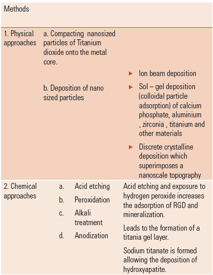
Attainment of surface roughness values of 1-100 nm led to the introduction of nanotopography of dental implants.
CONCLUSION
Patients come to us with a complex array of wants, needs and desires. The ability to provide a predictable life-restoring service, such as dental implant therapy is an enormous advancement for dentistry, an advancement that has really only been accepted in our generation. The work of health care pioneers such as Professors‟ Branemark, Steinmann and Albrektsson and others has led to the ability to offer a form of tooth replacement therapy that is minimally invasive and rather predictable. This only occurred through insightful assessment, demand and rigorous clinical assessment of outcomes before marketing to the dental community. This is an enormous legacy and responsibility for the current and future generation of clinicians and dental educators. As our implant therapy continues to evolve we must continue to demand that the innovations are grounded in science, rigorously tested and clinically evaluated before being marketed.
A thorough knowledge of the material aspect of dentistry is a must for every clinician to enable him / her to choose the right treatment modalities. As many studies have been conducted and variable results have been attained in some cases, these implants should be further tested and an implant with optimum surface modification is yet to be found. Surface modifications of implants is an important aspect of implantology along with the clinician‟s skill and understanding of implantology. At the end of the day, we owe it to our patients and the reputation of our profession to demand from manufacturer‟s reliability, stability and predictability in providing tooth replacement therapy for our patients.
REFERENCES
- NikitasSkyras, Iacopino ,Markes , Triplette, Woody. Implant materials, designs and surface topographies : their effect on osseointegration. A literature rivew. Int J Oral Maxillofacial Implants 2000 ; 15: 675-90.
- Branemark. P.I. Introduction to osseointegration .In :Branemark . P.I, Zarb G A, Alberktsson .T .tissue integrated prosthesis : Osseointegration in Clinical Dentistry. Chicago .Quintessance , 1985 :11-76.
- L. LeGuehennec ,Soueiden , Layrolle , Amouriq. Surface treatments of Titanium dental implants for rapid osseointegration. Dental Materials 2007 : 23 ; 844-854
- Adell R, Lekholm ,Rockler B, Branemark P.I . A 15 year study of osseointegrated dental implants in the treatment of the edentulous jaws. Int J Oral Surgery 1981 ; 10: 387-416
- Zarb G A , Schmitt A. The longitudinal clinical effectiveness of osseointegrated dental implants : The Toronto study Part III : Problems and complications encountered.
- Mendonca. G ,Mendonca Daniela B.S , Aragao. F, Cooper. C. Advancing dental implant surface technology – From micron to nanotopography . Biomaterials 29 (2008) 3822- 3835.
- A. Gupta , M. Dhanraj , G. Sivagami : Implant surface modification : review of literature . The Internet Journal of Dental Science. 2009 Volume 7 Number 1.
- Adriano Piattelli , Carl E Misch , Ana Emilia Farias Pontes, Giovanna Iezzi, Antonio Scarano , Marco Degidi. Carl Misch: Contemporary Implant Dentistry. 599-620.
- Ellingsen, Johansson, Ann Wennerberg , Anders Holmen. Improved Retention and Bone to Implant Contact with Fluoride – Modified Titanium Implants. Int J Oral Maxillofacial Implants 2004 ; 19: 659- 66.
- Pereira Da Silva, Campos Amico, NevesRodrigues ,Barboza, TesconiCroci. Osteoblastlike Cell Adhesion on Titanium Surfaces Modified by Plasma Nitriding. Int J Oral Maxillofac Implants 2011; 26: 237- 44.
Disclaimer from Guident
The views and opinions expressed in articles published in Guident are solely those of the authors and do not necessarily reflect the views of the editors, the editorial board or the publisher, Ivory India. Guident does not endorse any product, technique or treatment described. Clinical content is intended for qualified dental and medical professionals and is not a substitute for individual clinical judgement. Guident, its editors and the publisher accept no liability for any consequence arising from the use of information published in the journal.
How to cite this article
Dr. Manoj Shetty, Dr. Krishna Prasad, Dr. S. Rajalakshmi. Surface Modification of Dental Implants. Guident. 2013 Sep;6(11). Available from: https://ivoryindia.in/articles/implantology/surface-modification-of-dental-implants.html