Evaluation of Direct Sinus Lift and Simultaneous Implant Placement
Department of Maxillofacial & Oral Surgery Government Dental College and Hospital, Civil Hospital, Ahmedabad, India
ABSTRACT
Aim: To evaluate the efficacy of the lateral approach for sinus membrane elevation and floor augmentation in increasing the residual ridge height in the partially edentulous posterior maxilla and simultaneous implant placement.
Method: Study comprised of 10 patients in whom residual alveolar bone height was in range of 4.8 mm to 7 mm (avg. 5.63 mm). Lateral approach for sinus membrane elevation and grafting with simultaneous implant placement was done. Implants were loaded at 6 months postoperatively. Follow up was done to assess for the amount of bone height gain, implant stability, prosthesis loosening or fracture and any signs of maxillary sinusitis.
Results: At 6 months after sinus floor augmentation increase in residual bone height was in the range of 3.7 to 6.4 mm (avg. 4.93 mm). There was small perforation in sinus membrane in three patients (30%). Two patients (20%) had postoperative pain due to local inflammation which was managed conservatively. At 6 months follow up none of patients showed loosening of implant. Radiographic evaluation at 6 months follow up post loading of implants showed no evidence of peri implant radiolucency and good clinical stability in all patients except one who had prosthesis loosening at 4th month due to abutment loosening which required refitting of abutment.
Conlusion: From our study it can be concluded that Sinus floor augmentation by lateral approach along with simultaneous placement of an implant improves both the residual alveolar ridge dimension and the osseointegration of implants and is viable option in restoring partially edentulous dorsal maxilla. However a larger sample size and a longer follow up are essential to reinforce the above conclusion.
Keywords: sinus lift, bone graft, augmentation, sinusitis.
INTRODUCTION
Loss of tooth not only causes disharmony in the dynamic occlusal equilibrium but is also intriguing and psychologically disturbing on part of the patient, as it compromises both esthetic and masticatory component of the occlusion1.
When missing teeth are to be replaced treatment options includes a removable dental prosthesis, a conventional fixed dental prosthesis and an implant supported prosthesis. Some of these methods are, however associated with drawbacks such as loss of the tooth substance and a potential loss of vitality especially in young individuals. In some patients, where there is distally missing teeth, there is severe problem of retention and stability in removable partial denture. In addition, the prognosis for fixed bridge can be complicated by carious lesion progression in abutment, periodontal disease, technical failures such as loss of abutment and fracture of bridge components or abutment teeth.
Considering the above factors Implant has emerged as an excellent option whenever possible. However in partially edentulous patients especially in the posterior maxilla, implant placement is most challenging and frequently complicated by unfavourable post extraction bone patterns, pneumatisation of the maxillary sinus, poor quality of the remaining alveolar bone and higher occlusal forces2-4; making it insufficient for holding the implant; there by arises the need to increase the vertical dimension of dorsal maxilla by surgical technique.
Subantral augmentation method with simultaneous implant placement was first presented by Tatum in 1976 at Alabamas implant conference and published by Boyne and James in 19805,6. It intended to increase the vertical bone dimension in the posterior maxilla where access to the maxillary sinus is obtained by drilling a bony window in the lateral sinus wall using a small round bur, while ensuring that the sinus membrane remains intact.
AIMS & OBJECTIVES
A study of 10 patients was conducted in the Department of Oral and Maxillofacial Surgery, Government Dental College and Hospital, Ahmedabad to evaluate both, clinically and radiographically, the efficacy of the lateral sinus lift alongwith simultaneous implant placement for the rehabilitation of the partially edentulous posterior maxilla deficient in residual alveolar bone height with following aims and objectives:
1. To evaluate the efficacy of the lateral approach for sinus membrane elevation and floor augmentation in increasing the residual ridge height in the partially edentulous posterior maxilla.
2. To evaluate the efficacy of the lateral approach for the sinus floor augmentation to enable simultaneous implant placement and evaluation of the gain in the bone height.
MATERIALS & METHOD
Sample of the study:
The study consisted of sample of 10 patients who were selected randomly irrespective of the sex, creed and socioeconomic status, with a residual alveolar bone height between 4-8 mm at the edentulous posterior maxillary region.
Following inclusion criteria for patients were taken into consideration:
- Patients having partially edentulous posterior maxilla, between the ages of 20-60 years.
- A delay of at least 6 months between tooth extraction and an implant placement.
- Good systemic health.
- Absence of maxillary sinusitis.
Following exclusion criteria were taken into consideration:
- Uncontrolled diabetes, hematologic disorders, pregnancy, prior radiotherapy of the surgical site, chemotherapy.
- Patients with habits of smoking and tobacco chewing.
- Inadequate mouth opening, which cannot allow placement of instruments necessary for implant insertion.
- Active or recurrent maxillary sinusitis or any sinus related pathology.
Presurgical dimensions of the residual alveolar ridge were determined using digital radiographs CT Scans (Dentascans). (Fig. 1,2)

Preoperative prophylactic antibiotics which included Tab. Amoxycillin 500mg three times a day, were started one day before surgery. Antihistaminics in the form of tablet was started twice a day, a day before surgery, nasal decongestant drops (Otrivine nasal drops) was started as 2 drops twice a day, a day before surgery.
Lignocaine with adrenaline was injected to block the Posterior superior alveolar nerve, Infraorbital nerve and Greater palatine nerve as per the requirement. An incision was made 2-3 mm on the palatal side of the crest of the ridge with a releasing incision at least 15 mm mesial to the antral opening that was judged on the basis of X-ray. Full thickness mucoperiosteal flap was reflected exposing the anterior wall of maxilla. With the help of 3mm diameter round bur and surgical handpiece at 40000 rpm speed, elliptical bony window was marked with the bur at 45 degree angulation to the bone surface under copious irrigation of the saline. With delicate paintbrush strokes, the bony surface was scored, taking extreme caution not to touch the underlying schneiderian membrane. Window was deepened till the bluish hue of the sinus membrane appeared (Fig. 3). As the plate was weakened a surgical curette was engaged under the inferior margin between the bone and schneiderian membrane. With a sliding motion, the window of bone was pushed in and at the same time the underlying membrane was peeled from inside wall of the sinus. Then the osteotomized segment was stabilized with the membrane elevators to prevent perforation of the sinus membrane during drilling for implant insertion. 1:20 reduction handpiece was used at the low speed (800-1200 rpm) high torque (35Ncm) along with copious irrigation of normal saline to prevent thermal injury to the bone. Having prepared the osteotomy site the implants were placed maintaining the elevated membrane in

Fig. 3: Preparation of bony window and lifting of sinus membrane
its position (Fig. 4). Alloplastic bone graft (Bioactive glass, Novabone putty) was placed in the lateral window and the implant surface was covered with the same (Fig. 5). The lateral window was then covered with the collagen membrane (PerioCol – GTR) to avoid the fibrous adhesion between the inner surface of the flap and bone graft. The mucoperiosteal flap was repositioned and sutured with the help of 3-0 Vicryl material. There was small perforation during

sinus membrane elevation in 3 (30%) patients. This was managed by placement of collagen membrane at the perforation site followed by insertion of implants and bone grafting followed by placement of collagen membrane in usual manner.

Fig. 6: Post operative CT scan at 6 months showing implants in position and gain in bone height around the implants (approx. 10.3mm)

Fig. 7: Prosthesis in position
6 months following the sinuslift and implant placement(Fig.6), the implants were uncovered and cover screw was removed and a healing cap was placed. 1 week later the abutment was screwed and prosthesis given. Followup was done at 1, 3 and 6 months.
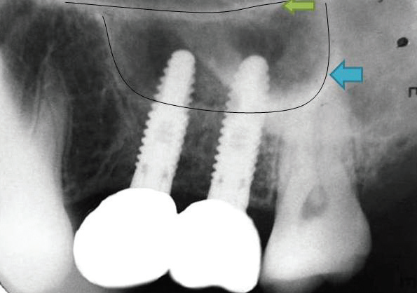
Fig. 8: Radiograph with final prosthesis in position

Cropped OPG image
RESULTS
Study comprised of 10 patients of age range 21 to 60 years, 6(60%) of which were females and 4(40%) were males. The cause of tooth loss in all patients was caries.
The residual alveolar bone height was in the range of 4.8 mm to 7 mm, on an average it was 5.63 mm. The alveolar ridge height after sinus lift was in the range of 8.8mm to 12mm; on an average 10.56 mm at the period of 6 months after sinus floor augmentation.
There was significant increase in residual bone height following sinus floor augmentation, in the range of 3.7 to 6.4 mm (on an average 4.93 mm) over the period of 6 months. Hence, 87.56% bone gain was noted on an average after surgery.
Table I


Implants of diameter 3.75mm and length 11.5mm were placed in seven sites while in three patients implant of diameter 3.75mm and length 10mm was placed.
Postoperatively patients were evaluated for complications like local inflammation, pain, soft tissue dehiscence and maxillary sinusitis. Most patients had an uneventful postoperative healing. There was small perforation in sinus membrane in three patients (30%) and two patients (20%) had some postoperative pain due to local inflammation. The same patients had features of maxillary sinusitis as well, which were however managed with nasal decongestant drops, oral antihistaminincs and benzoine steam inhalation.
Table II:

Radiographic evaluation at 6 months followup showed no evidence of peri implant radiolucency in any of the patients. No patient had any signs of implant loosening. One patient showed loosening of the prosthesis due to loosening of the abutment at 4th month. This was managed by removal of the prosthesis followed by retightening of the abutment and recementation of prosthesis.

DISCUSSION
Tooth replacement with dental implants is a standard and well established treatment modality. In a large majority of cases, threaded implants are used, and have shown outstanding long-term results when placed with adequate bone support7,8.
The discrepancy between the need and availability of bone is highest in posterior maxilla where the alveolar bone is reduced by the alveolar resorption following tooth extraction and due to pneumatization of the maxillary sinus. In order to overcome this discrepancy supplementation of the bone with an augmentation procedure known as the “sinus lift” has been a very effective treatment. There are 2 basic variations of the sinus floor augmentation technique i.e. Tatum’s Lateral approach (Direct technique) and Summer’s Crestal approach (Indirect technique). If the residual alveolar bone height is less than 6mm in the posterior maxilla, then the Crestal approach (Indirect technique) may not provide adequate stability to the implant placed simultaneously, so it is preferred when the residual alveolar bone height is more than 6mm9.
Implants are placed either simultaneously with the graft (1-stage lateral antrostomy) or after a delayed period of up to 12 months to allow for graft maturation (2-stage lateral antrostomy)10. The initial bone thickness at the alveolar ridge seems to be a reliable indicator in deciding between these 2 methods. If the bone thickness is 4 mm or less, initial implant stability would be jeopardized. Therefore, a 2-stage lateral antrostomy should be carried out. The reverse holds true for a 1-stage procedure10.
To overcome the limitations of other methods used for restoring the partially edentulous posterior maxilla, the present study was conducted to determine and evaluate the efficacy of lateral approach (Direct technique) for an augmentation of the antral floor with simultaneous placement of an implant to restore the dorsal maxilla; and also to evaluate the implants, both clinically and radiographically following the antral floor augmentation.
In this study, an alloplastic bone graft material, a Bioactive glass (Novabone putty containing Calciumphosphosilicate (CPS-55%), a smaller CPS particulate, Polyethylene glycol (PEG) and Glycerin as a binder) was used for the sinus floor augmentation. Bioactive glass has several advantages like its cohesive & graft retentive properties, easy manipulation during surgery, no risk of immunogenic response and infection transmission and very low chances of graft infection because antibiotics readily penetrates into it due to its hydrophilic nature. The only disadvantage of it is its higher cost values11,12.
At 1st, 2nd and 3rd follow up at the period of 1 month, 3 months and 6 months respectively; significant raise in the bone level above the initial sinus floor was noted in the OPGs. The residual alveolar bone height was 4.8 to 7mm (Avg. 5.63 mm) pre operatively and after sinus floor augmentation, the residual alveolar bone height was in the range of 8.8 to12mm (Avg.10.56 mm) (TABLE – I). The final bone gain was very significant in the range of 3.7-6.4 mm (Avg. 4.93 mm) after sinus floor augmentation at the period of 6 months following surgery. Hence, 87.56% bone gain was noted on an average after sinus floor augmentation with alloplastic bone graft. These findings were compatible with Ziv Mazor, Michael Peleg and Martin Gross13 who performed sinus floor augmentation for a single tooth replacement in the posterior maxilla which can provide the necessary bone support for holding the implant.
Perforation of the Schneiderian membrane has been reported as a complication occurring 10% to 60% of the time during sinus augmentation14. In three out of ten patients (30%), schneiderian membrane perforation occurred during surgery which was of approximately 0.5 cm in size and was repaired by sealing the perforation with resorbable collagen membrane. Two patients (20%) out of ten had experienced pain and showed signs of mild maxillary sinusitis initially. This was treated conservatively with the help of antibiotics, analgesics and decongestants. The next follow up visits were asymptomatic. It had not adversely affected the outcome during the follow up and the postoperative healing was uneventful. In rest of the eight patients (80%), there was absence of local inflammation/infection, pain and soft tissue dehiscence after surgery. This data were compatible with Ziv Mazor et al13, Leon Ardekian et al15, Pikos MA16, Ardekian L et al17 and Karabuda C et al18.
Sinus floor augmentation by lateral approach (Direct technique) alongwith simultaneous implant placement can be an excellent method for restoring the partial edentulism, if performed by the experienced hands. Complications occurring during treatment require an understanding of the biomechanical principles involved in surgical management of schneiderian membrane during sinus lift, perfect attention to the minor details involved in a diagnosis and treatment planning and encouraging the patient towards maintaining strict oral hygiene to increase the longevity of the implant.
BIBLIOGRAPHY
- J Fiske, D M Davis, C Frances & S Gelbier. The emotional effects of tooth loss in edentulous people. Br Dent J 1998; 184: 90-93
- Jem T., Pettersson P. A 3-year follow up study on single implant treatment. J. Dent 1993; 21: 203-208
- Jaffin RA, Berman CL. The excessive loss of Branemark fixtures in type IV bone: A 5- year analysis. J. Periodontol 1991; 62: 203-208
- Bergendal T., Engquist B. Implant supported overdentures: A longitudinal prospective study. Int J Oral Maxillofac Implants 1998; 13: 253-262
- Tatum OH: Maxillary and sinus implant reconstruction. Dent Clin North Am. 1986; 30:207–229
- Boyne P, James RA. Grafting of the maxillary sinus floor with autogenous marrow and bone. J Oral Maxillofac Surg. 1980; 17:113–116
- Karoussis IK, Brägger U, Salvi GE, Bürgin W, Lang NP. Effect of implant design on survival and success rates of titanium oral implants: a 10-year prospective cohort study of the ITI Dental Implant System. Clin Oral Implants Res. 2004; 15: 8-17
- Oliva J, Oliva X, Oliva JD. Five-year success rate of 831 consecutively placed Zirconia dental implants in humans: a comparison of three different rough surfaces. Int J Oral Maxillofac Implants 2010; 25: 336-44
- Zitzmann NU, Scharer P. Sinus elevation procedures in the resorbed posterior maxilla: Comparison of the crestal and lateral approaches. Oral Surg Oral Med Oral Pathol Oral Radiol Endod. 1998; 85:8–17
- I Woo, B.T.Le: Maxillary sinus floor elevation: Review of anatomy and two technique. Implant Dent 2004; 13:28–32
- Imran Farooq, Zonera Imran, Umer Farooq, Ali Leghari, Humera Ali. Bioactive glass: A material for the future. World J Dent 2012; 3: 199-201
- Salonen JI, Arjasmaa M, Tuominen U, Behbehani MJ , Zaatar EI. Bioactive Glass in Dentistry. J Minimum Intervention in Dentistry 2009; 2: 208-218
- Ziv Mazor, Michael Peleg and Martin Gross. Sinus Augmentation for Single-Tooth Replacement in the Posterior Maxilla: A 3-Year Follow-up Clinical Report: The International Journal of Oral & Maxillofacial Implants 1999; 14: 55-60
- Eric OH, Richard AK. Effect of Sinus Membrane Perforation on Dental Implant Integration: A Retrospective Study on 128 Patients. J Implant Dent 2011; 20: 13-16
- Leon Ardekian, Efrat Oved-Peleg, Eli E. Mactei and Micha Peled. The Clinical Significance of Sinus Membrane Perforation During Augmentation of the Maxillary Sinus. J Oral Maxillofac Surg 2006 64:277-282
- Pikos MA: Maxillary sinus membrane repair: Report of a technique for large perforations. Implant Dent 8:29, 1999
- Ardekian L, Oved-Peleg E, Mactei E,et al. The clinical significance of sinus membrane perforation during augmentation of the maxillary sinus. J Oral Maxillofac Surg. 2006;64:277-282
- Karabuda C, Arisan V, O¨ zyuvaci H. Effects of sinus membrane perforations on the success of dental implants placed in the augmented sinus. J Periodontol. 2006; 77:1991-1997
Disclaimer from Guident
The views and opinions expressed in articles published in Guident are solely those of the authors and do not necessarily reflect the views of the editors, the editorial board or the publisher, Ivory India. Guident does not endorse any product, technique or treatment described. Clinical content is intended for qualified dental and medical professionals and is not a substitute for individual clinical judgement. Guident, its editors and the publisher accept no liability for any consequence arising from the use of information published in the journal.
How to cite this article
Dr. Parmar Babu S., Dr. Bhatt Utsav U., Dr. Jodia Kunal M.. Evaluation of Direct Sinus Lift and Simultaneous Implant Placement. Guident. 2013 Dec;7(1). Available from: https://ivoryindia.in/articles/implantology/evaluation-of-direct-sinus-lift-and-simultaneous-implant-placement.html