Imaging Modalities in Implantology
Department of Prosthodontics A.B. Shetty Memorial Institute of Dental Sciences, Nitte University, Deralakatte, Mangalore, India
ABSTRACT
Diagnostic imaging plays an important role in evaluating the dental implant patient. Many types of radiographic imaging are recommended for treatment planning for implants. These include panoramic, periapical and occlusal radiographs, conventional and computed tomography (CT) and MRI. The clinician needs to identify the best method for each clinical situation. This article gives an overview of the various available imaging modalities at our disposal with the advantages and limitations of each.
Keywords: Dental implants, Implant imaging, Dental radiography
INTRODUCTION
The placement of dental implants requires meticulous planning and careful surgical Procedures. Excellent theoretical knowledge and clinical skills provide a firm foundation for success in implant dentistry. Pre-operative assessment of the proposed surgical sites as well as the condition of adjacent teeth and osseous structures is essential to ensure that their status is consistent with the proposed treatment plan. Hence, a radiographic prescription is often needed to provide a more complete visualization of the current clinical situation and to guide in further clinical steps. Radiographs can be utilized at various steps during implant treatment planning, the treatment itself, and the follow-up. The objectives of radiographic analysis and the types of radiographic imaging modalities vary at each phase of therapy.
There are many imaging modalities that have been employed for implant imaging. These modalities can be described as either analog or digital and two dimensional or three dimensional.1
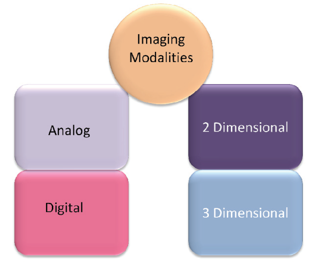
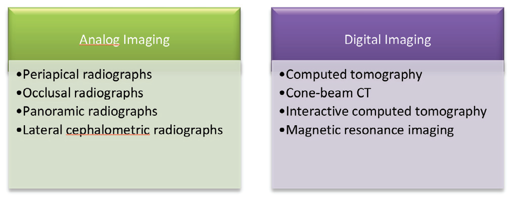
Analog imaging modalities are two dimensional systems that employ X-ray film and/or intensifying screens as the image receptors. Digital imaging include the computed tomography, tuned aperture computed tomography, cone-beam CT, magnetic resonance imaging. These create a three dimensional image which is described not only by its width, height and pixels, but additionally by its depth and thickness.1
ANALOG IMAGING
-
Periapical Radiographs :
The intraoral radiograph, when correlated with the case history and clinical examination, is one of the most important diagnostic aids available to the dental practitioner. Intraoral periapical radiography is one of the most commonly employed radiographic modality during the initial phases of patient evaluation to assist in treatment planning. When the technique is correctly implemented, images with greatest details may be expected. Periapical radiographs commonly are used to evaluate the status of adjacent teeth and remaining alveolar bone in the mesiodistal dimension. In addition they are also commonly used for determining vertical height, architecture and bone quality , bone density, amount of cortical bone and amount of trabecular bone.2 Other advantages include its ease of availability, low radiation dose and cost effectiveness.
Indications:
- Evaluation of small edentulous spaces – single-tooth implants in regions with abundant bone width
- Alignment and orientation during surgery
- Recall/maintenance evaluation
However, periapical radiography has geometric and anatomic limitations. The most accurate intraoral radiographic technique used for implant planning is the paralleling technique. If the paralleling technique is not used, periapical radiographs create an image with foreshortening and elongation. When the x-ray beam is perpendicular to the film, but the object is not parallel to the film, foreshortening is seen. If the x-ray beam is oriented perpendicular to the object but not the film, elongation will occur. Because the film plane can rarely be placed parallel particularly in edentulous areas, the target film distance is difficult to standardize. Hence, periapical radiographs do not provide an accurate assessment of vertical bone dimension or precise position of critical anatomic structures.3 When compared to the 94% of all measurements made from CT images, only 53% of measurements from alveolar crest to superior wall of mandibular canal in periapical radiographs were accurate within 1 mm.3
An alternative to film radiography is the Direct Digital Intraoral Imaging which makes use of a sensor to collect the data. Its advantages include1:
- Significantly less radiation
- Superior resolution
- Rapid acquisition of intraoral images
- Easy storage, retrieval and transmission to remote sites.
- Easy enhancement – Helps measuring bone density
- Color enhancements : human eye distinguishes color better than shades of grey
- Inversion : switching between negative and positive images
2. Occlusal Radiographs:
Occlusal radiography is also used during the initial stages as an aid in treatment planning. An occlusal radiograph is a radiograph designed to be placed between the occlusal surfaces of the teeth with the central beam directed at 90o or at 50 -60o to the plane of the film depending on what is required to be viewed. One of the limitations of periapical radiography is that cross-sectional information cannot be obtained. Hence, occlusal radiographs are sometimes used to determine the faciolingual dimensions of the mandibular alveolar ridge. However, the greatest drawback of occlusal radiography is that it records only the widest portion of the mandible which is located inferior to the alveolar ridge. This often provides a wrong impression to the clinician that greater amount bone is available in the cross-sectional dimension than that actually exists. Furthermore, its application in the maxillary arch is limited due to anatomic constraints.4 It is the projection of choice when several implants in the anterior part of the jaws are to be viewed.
3. Panoramic Radiographs:
Panoramic radiography is a curved plane tomographic radiographic technique used to depict the body of the mandible, maxilla and the lower one half of the maxillary sinuses in a single image. Panoramic radiographs have been used frequently as a radiographic method for pre-implant evaluation and the preparation of treatment protocols. Although the resolution and sharpness of panoramic radiographs are less than those of intraoral radiographs, panoramic radiographs is an excellent tool in providing a two-dimensional overview of both jaws, teeth, foreign bodies, cysts, tumours and other conditions of the jaws in a single film including many of the vital structures, such as maxillary sinus, inferior alveolar nerve and nasal fossa.5
However, information acquired from panoramic radiographs must be applied judiciously because this technique has significant limitations as a definitive preoperative planning tool. Panoramic radiographs are subjected to nonuniform distortion which often leads to inaccurate interpretation and measurements. The magnification of panoramic radiographs can be >30%,
especially when patients are not in the optimal position. Angular measurements on panoramic radiographs tend to be accurate, but linear measurements are not. Vertical measurements are unreliable because of foreshortening and elongation of the anatomic structures because the x-ray beam is not perpendicular to the long axis of the anatomic structures or to plane of the image receptor. The negative vertical angulations of the X-ray beam also may cause lingually positioned objects such as, mandibular tori to be projected superiorly on the film which may result in an overestimation of vertical bone height.2 Besides, a panoramic image cannot provide clinicians with information about the buccolingual cross-sectional dimension or the inclination of the alveolar ridge.
However, the magnification factor can be calculated at the given site by dividing the actual diameter of the object by the diameter measured on the radiographs. Diagnostic templates that have ball bearings or wires incorporated around the curvature of the dental arch and worn by the patient during the panoramic examination enable the dental specialist to determine the amounts of magnification in the radiograph.6
- Opposing landmarks are easily identified.
- The vertical height of bone initially can be assessed
- The procedure is performed with convenience, ease, and speed in most dental setups.
- Gross anatomy of the jaws and any related pathology findings can be evaluated.
- Does not demonstrate bone quality/ mineralization
- Distortions are inherent
- Errors in patient positioning
- No third dimension
- No spatial relationship between structures

Although panoramic radiographs may provide a useful overview and may be used in conjunction with ridge mapping or other diagnostic tools, they are unlikely to meet the strict criteria set for primary imaging tests for implant planning.8
4. Cephalometric Radiographs:
Lateral Cephalometric radiographs are produced with the patient’s midsagittal plane oriented parallel to the image receptor by using a cephalometer. The cephalometer physically fixes the position of the skull with projections into the external auditory canals. A cross-sectional image of the alveolus of both the mandible and the maxilla in the midsagittal plane is demonstrated by this radiograph. These projections provide image of known magnification (7-12%).4 The geometry of cephalometric imaging devices results in a 10% magnification of the image with a 60 inch focal-object and 6 inch object to film distance.
Lateral cephalometric radiographs have been recommended for evaluating the anterior maxilla and mandible for dental implant placement.9 They can accurately measure the height and width of the residual bone at the anterior midline of both the maxilla and mandible. Lateral cephalometric radiography also allows analysis of the quality of the bony host site (ratio of compact to cancellous bone), particularly that in the anterior region of the mandible. Position and relationship of mental foramen, nasal fossa and maxillary sinus to the adjacent structures can be assessed. The soft tissue profile is also apparent on this film and can be used to evaluate profile alterations after prosthodontic rehabilitation.
Limitations:4
- A 7-12% image magnification -undesirable - leads to measurement errors of implant site.
- Reduced resolution and sharpness
- Not useful for demonstrating bone quality
- Image information limited to midline
- Superimposition on the contralateral side - complicating the evaluation of other implant sites.
- Technique sensitive
Indications:
- Used in combination with other radiographic techniques for anterior implants.
- Symphysis bone graft evaluation
DIGITAL IMAGING
1. Computed Tomography:
It is a three-dimensional scanning which allows the clinicians to visualize the bony architecture, nerves, joints, sinuses and other structures much more completely than traditional flat radiographs. Computed Tomography is an advanced imaging technique in which images are acquired digitally and subsequently reformatted into any plane, such as axial, sagittal or coronal.10
A narrow X-ray beam is used to scan a patient at right angles to the long axis of the body.11,12 The axial slices should be oriented parallel to the inferior border of the mandible for mandibular imaging and parallel to the hard palate for maxillary imaging.These axial images are 1-2 mm thin and overlapping, resulting in approximately 30 axial images per jaw. The image information of these sequential axial images can be manipulated into various planes using computer based process called multiplanar reformatting (mpr).
Computed tomographic scanning for dental implant surgery planning has been well described. It allows exact preoperative analysis of the available bone volume and helps to determine the appropriate position, angulation, number, and length of the planned implants.13 This modality also gives a high-density resolution, and the soft tissues can also be visualized to some degree. The reformatted CT images provide axial, panoramic, and cross-sectional images that are all cross-referenced to one another, allowing rapid correlation of the different views.14

2. Cone Beam Computed Tomography (CBCT):
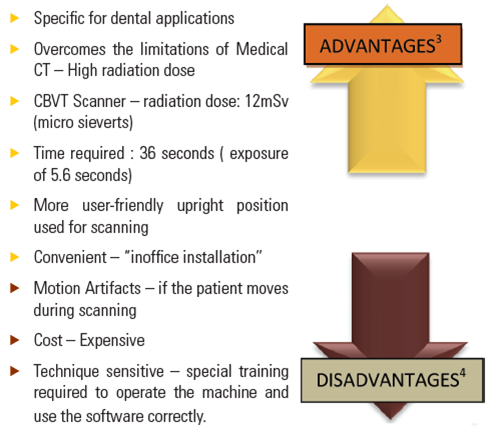
Because of higher radiation exposure, higher cost, huge footprint, and difficulty in accessibility associated with CT, CBCT was developed. As the name implies, CBCT employs a cone-shaped X-ray beam rather than the flat fan shaped beam used in conventional CT. During a single rotation of the source and receptor, the receptor captures the entire volume of anatomy within the field of view by an image intensifier of flat panel detector.15 As the source and receptor rotate once around the patient, many exposures are made, ranging in duration between 8.9 and 40 seconds. These images are in the Digital Imaging and Communications in Medicine (DICOM) data format.
DICOM is a standard for handling, storing, printing and transmitting information in medical imaging. Advantage of using a DICOM data format is that the dental specialist can make precise measurements in any plane within the viewing software. The software incorporates computer-aided design/computer-aided manufacturing replicas of dental implants for the clinician to place into the region of interest. The clinician sends the file to manufacturing facility, which creates a surgical guide through a process termed “stereolithography.” The guide includes metal cylinders that direct osteotomy drills into precise locations in the maxilla and/or mandible, as planned by the software.15
The overall effective radiation dosage in CBCT is 0.035 to 0.10 mSv, which is equivalent to between two and eight panoramic radiographs. Individual voxels are much smaller than conventional CT voxels, resulting in greater resolution. Examples of such machines include NewTom DVT 9000 (Quantitative Radiology, Verona, Italy), i-CAT (Imaging Sciences International, Hatfield, USA), and 3D Accuitomo (J. Morita, Kyoto, Japan.7
3. Interactive Computed Tomography (ICT):
Interactive Computed Tomography was developed to bridge the gap in information transfer between the radiologist and the clinician. This technique enables the radiologist to transfer the imaging study to the clinician as a computer file and enable the clinician to view and interact with the imaging study on their own computer. The clinician’s computer becomes a diagnostic radiologic workstation with tools to measure the length and the width of the alveolus, measure bone quality and level of the gray scale of the study to enhance the perception of critical structures. It helps to determine the number and size of implants accurately along with the density of bone at the proposed implant sites.6
An important feature of ICT is that the clinician and radiologist can perform “electronic surgery” (ES) by selecting and placing arbitrary size cylinders that stimulate root form implants in the images. With an appropriately designed diagnostic template, ES can be performed to electronically develop the patient’s treatment plan in three dimensions.6
At this time ICT is the most accurate imaging technique for implant imaging and surgery but suffers some limitations. ES enables placement of electronic implants in the imaging study but the refinement and exact relative orientation of the implant positions is difficult and cumbersome.16
4. Magnetic Resonance Imaging (MRI):
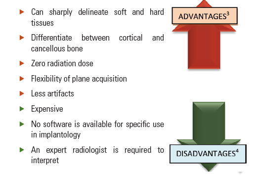
Magnetic resonance imaging was first introduced by Lauterbur. Magnetic fields and radiofrequencies, which are used by electromagnetic detectors to provide electronic images of body protons, are processed by a computer and digital images are generated. There is no radiation exposure and soft tissues can be visualized when MRI is used. It can be used as a secondary imaging technique when primary imaging techniques such as complex tomography and CT scanning are not satisfactory.17 However, MRI does not yield cortical bone images, so estimation of bone quality may be problematic.
Devge et al.18 found that MRI images are not affected by implants of the Branemark system. On the other hand, MRI is contraindicated for patients with ferromagnetic metallic implants in their bodies because of the potential risks associated with movement or dislodgment of such objects. Non-precious ferromagnetic alloys (cobalt-chromium) produce large image deformations, whereas precious alloys (Au, Ag, Ti, amalgam), which are mainly non-ferromagnetic, have no effect.19
DISCUSSION
The objectives of diagnostic imaging depend on a number of factors, including the amount and type of information required and the time period of the treatment rendered. The decision to image and determine which imaging modality to use depends on the phase of the treatment.
Peripaical radiographs (PA), panoramic radiographs (Pan), lateral cephalographs (Ceph), conventional/computed tomography (Tomo/CT) are the recommended imaging modalities for treatment planning. During the surgical placement of implants, PA, Ceph or CT imaging may be used in order to analyse the placement of the implants and carry out any corrections in the angulation or depth of the implant. PA, Pan, tomo, CT or ceph are the recommended treatment modalites to analyse how well the healing and osseointegration of the implant has taken place following the first 3 months of implant placement. If no complications are encountered during the first 3 months, Periapical radiograph or Panaromic radiograph may suffice during the remodelling phase of 4-12 months and the maintenance phase i.e follow up approximately every 3 years post implant placement. However, incase of any complications, CT imaging may be indicated.20
Although, there are many options of the imaging modalities available, as mentioned before, each of them have some or the other limitations. For example, inconsistent image magnification is a drawback of panoramic radiography. This Magnification can be calculated by the use of templates with incorporated metal spheres of known diameter in situ when the radiograph is taken. The distance between the alveolar crest and the mandibular canal is measured on the film. The actual distance between the canal and the crest is then calculated using the following formula :

A limitation of lateral cephalographs is that superimposition of the image on the contralateral side takes place, obstructing the view of the implants in the posterior region. Hence, they are the recommended modality when placing an implant in the anterior maxilla or mandible, but not a modality of choice for the posterior region. Periapical radiographs, panoramic radiographs or CT may provide with a much clear and precise view.
The major limitation of computed tomography remains the high radiation exposure which alarms a judicious use of the modality, particularly in preganant ladies. Elective dental implant surgery should be preferably avoided during pregnancy. However, if necessary efforts to minimize the radiation dose must be made. Conventional radiography is preferred over computed tomography as it is the least radiating modality. Thyroid collars, lead aprons and leaded glasses should be used wherever possible. High speed films and digital imaging is preferred over analogue imaging. CBCT with smallest field of view that will cover only the region of interest may be used.
CONCLUSION
Diagnostic imaging is an integral part of dental implant therapy for preoperative planning, intraoperative and postoperative assessment by use of variety of techniques. In general, good starting point would be to proceed with panoramic radiograph and possibly intraoral radiographs if greater image detail is required. If images are required of all of the maxilla and mandible to evaluate possible implant sites, cross-sectional images assists to clinician.
Two-dimensional diagnostic imaging has served dentistry well and will continue to do so for the foreseeable future. However, the advent of CBCT allows complete visualization of the oral and maxillofacial complex. Today, CBCT is the best modality for the ease of acquisition and relatively low radiation risk even for single implants.
REFERENCES
- Lingeshwar D, Dhanasekar B, Aparna IN.Diagnostic Imaging in Implant Dentistry. Int J Oral Impl Clin Res. 2010;1(3):147-153.
- Benson BW , Shetty V . Dental implants . In: White SC , Pharoah MJ editor. Oral radiology: principles and interpretation . St. Louis: Mosby Elsevier; 2009;597–612.
- Donald A. Tyndall, Sharon L Brooks, Chapel Hill NC, Ann Arbor, Mich. Selection criteria for dental implant site imaging: A position paper of the American Academy of Oral and Maxillofacial Radiology. Oral Surg Oral Med Oral Pathol Oral Radiol Endod 2000;89:630-37.
- Shetty V, Benson BW. Oraofacial implants. In White SC, Pharoah MJ (Eds): Oral Radiology: Principles and Interpretation.4th ed. Saint Louis: Mosby, 2000;623-35.
- Chan H-L., Misch K., Wang H-L.Dental Imaging in Implant Treatment Planning Implant Dent.2010;19:288-298.
- Resnik, R.R., Kircos, L. and Misch C.,E.Diagnostic Imaging and Techniques, In:Contemporary Implant Dentistry, C.E. Misch. Mosby, Elsevier.2008; 38-67.
- Siu, ASC; Chu, FCS; Li, TKL; Chow, TW; Deng, F. Imaging modalities for preoperative assessment in dental implant therapy: an overview. Hong Kong Dental Journal, 2010;7(1): 23-30.
- Reiskin AB. Implant imaging. Status, controversies, and new developments. Dent Clin North Am 1998;42:47-56.
- Strid KG. Radiographic results. In: Branemark PI, Zarb G, Albrektsson T, editors. Tissue-integrated prostheses: osseointegration in dentistry. Chicago, Quintessence; 1985: 187-98.
- Todd AD, Gher ME, Quintero G, et al. Interpretation of linear and computer tomograms in the assessment of implant recipient sites. J Periodontol 1993;64:1243-49.
- Allan B Reiskin. Implant imaging status, controversies and new developments. Dental Clinics of North America 1998;42(1): 47-56.
- Frederiksen NL, Benson BW, Sokolowski T. Effective dose and risk assessment from computed tomography of the maxillofacial complex. Dentomaxillofacial Radiology 1995;24:55.
- Schwarz MS, Rothman SL, Chafetz N, Stauts B. Preoperative diagnostic radiology for the tissue-integrated prosthesis. In: Laney WR, Tolman DE, editors. Tissue integration in oral, orthopedic, and maxillofacial reconstruction. Chicago, Quintessence; 1990: 68-79.
- Schwarz MS, Rothman SL, Rhodes ML, Chafetz N. Computed tomography: part I. Preoperative assessment of the mandible for endosseous implant surgery. Int J Oral Maxillofac Implants 1987;2:137-41.
- Arai, Y., Tammisalo, E., Iwai, K. Development of a compact computed tomographic apparatus for dental use. Dentomaxillofac Radiol.1999; 28: 245-8.
- Petrikowski CG et al. Presurgical radiographic assessment for implants. J Prosthet Dent 1989;61(1):59-64.
- Zabalegui J, Gil JA, Zabalegui B. Magnetic resonance imaging as an adjunctive diagnostic aid in patient selection for endosseous implants: preliminary study. Int J Oral Maxillofac Implants1990;5:283-7.
- Devge C, Tjellström A, Nellström H. Magnetic resonance imaging in patients with dental implants: a clinical report. Int J Oral Maxillofac Implants 1997;12:354-9.
- Lissac M, Coudert JL, Briguet A, Amiel M. Disturbances caused by dental materials in magnetic resonance imaging. Int Dent J 1992;42:229-33.
- Jayadevappa BS, Kodhandarama GS, Santosh SV, Rashid WT. Imaging of dental implants.Journal of Oral Health Research.2010;1(2):50-61.
Disclaimer from Guident
The views and opinions expressed in articles published in Guident are solely those of the authors and do not necessarily reflect the views of the editors, the editorial board or the publisher, Ivory India. Guident does not endorse any product, technique or treatment described. Clinical content is intended for qualified dental and medical professionals and is not a substitute for individual clinical judgement. Guident, its editors and the publisher accept no liability for any consequence arising from the use of information published in the journal.
How to cite this article
Dr. Krishna Prasad, Dr. Divya Mehra, Dr. Anupama Prasad. Imaging Modalities in Implantology. Guident. 2013 Nov;6(13). Available from: https://ivoryindia.in/articles/implantology/imaging-modalities-in-implantology.html