Coronoid Hyperplasia : A Rare Condi Tion and an Unusual Cause of Restricted Mouth Opening
Department of Orthodontics and Dentofacial Orthopedics, Department of Orthodontics and Dentofacial Orthopedics, Manipal College of Dental Sciences, Manipal University, Manipal, Karnataka, India
Abstract
Several disorders affecting the masticatory system can cause limited mouth opening, of which, coronoid hyperplasia is a rare yet plausible cause. It may be congenital or developmental in origin, and leads to excessive growth of the coronoid process which impinges against the posterior portion of the zygomatic bone, causing restricted mouth opening. The current paper discusses a case of coronoid hyperplasia in a young male, the etiology of which was diagnosed to be hyperactivity of the temporalis muscle. Although this is an uncommon finding, yet, through proper clinical examination and radiographic imaging, this condition may be diagnosed accurately.
Keywords:Coronoid Hyperplasia, Temporo mandibular disorder
INTRODUCTION
Coronoid hyperplasia is defined as “an abnormal bony elongation of histologically normal bone in which there is progressive difficulty in mouth opening” .1 The coronoid processes may enlarge to such an extent that they impinge upon the zygomatic bones, thereby causing limited mouth opening. It usually commences in the second decade of life, however, due to its slow rate of onset and lack of pain, the patient may not present with symptoms until years later.1,2
Coronoid process hyperplasia usually develops progressively, and the clinical symptoms are often similar to those of temporo-mandibular joint disorders (TMD). Even the American Academy of Orofacial Pain classifies coronoid process hyperplasia as a congenital or developmental temporomandibular joint disorder.3 The decrease in mouth opening is gradual, and many a times it has been overlooked while determining the patient’s diagnosis.1,4 The most frequent diagnoses considered for mouth opening restrictions include masticatory muscle contraction disorders, TMJ internal derangements or ankylosis. Therefore, when examining such patients, the practioners must systematically review all of the possible causes as well as utilize the appropriate imaging methods for the definitive diagnosis. The etiology is yet to be confirmed, however, several hypotheses have been proposed, such as trauma, endocrine disorders, hyperplasia of muscles, genetic components and cartilage growth center persistence.5
This paper presents a case of coronoid hyperplasia secondary to temporalis muscle hyperactivity in a young male. The diagnosis with regards to the hyperplasia and the etiology is described.
CASE REPORT
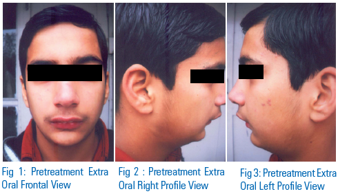
A 22-year-old male patient presented with complaint of forwardly placed upper anteriors with clicking and pain in the temporomandibular joint region during function since past four years. Patient reported good general health with no previous history of TMJ disease, otitis media, or facial trauma. Clinical examination revealed limited condylar process movement with no lateral deviation. Bilateral clicking was present and was found to be more severe on the right side. The temporalis muscle on the right side was also tense on palpation. The increased overjet of the incisor teeth was apparent. The maximal inter incisal distance while mouth opening was found to be 28mm. An orthopantogram examination revealed bilateral coronoid processes hyperplasia which was more prominent on right side. The coronoid process head on right side
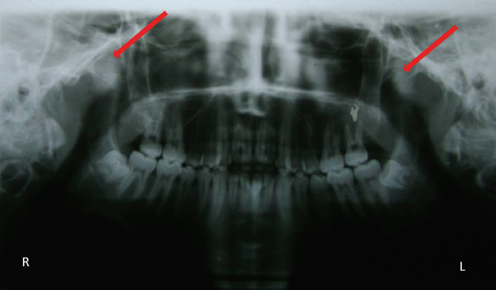
Fig 1: Panoramic Radiograph showing bilateral coronoid hyperplasia, more significant on the right side.
was as high as the condylar head (Figure 1). Further, the ratio of the length of the coronoid process to the condylar process was calculated bilaterally using the Levandoski panographic analysis and found to be more than 1.1 indicating coronoid hyperplasia.6
In order to diagnose the etiology related to temporalis muscle hypertrophy, we conducted a bilateral electromyographic evaluation of the temporalis muscle. The needle electrode of 1.5mm diameter and Neuropack (NIHON KHODEN) electromyogram was used in the procedure (Figure 2). This particular needle electrode was used due to better quality of the electromyogram obtained with it. Also, there were lesser technical artifacts while using needle electrode, because distance between the muscle and the electrode remains more constant.7

Fig 2: The figure showing the NEUROPACK(NIHON KOHDEN) electromyogram used to record the temporalis muscle activity
The temporalis muscle was carefully located & divided into anterior, middle and posterior fibres and the 3 regions were marked (Figure 3). The patient was then made to lie in a supine position. The needle electrode was carefully inserted into the anterior, middle and posterior fibres on left side, followed by the right side – both at rest and then at maximum intercuspation position (Figure 4). The results showed increased activity of temporalis muscles bilaterally which was more intense on the right side compare to the left, with maximum activity seen in the posterior fibres. Presence of progressive limitation of the mouth opening, excessive enlargement of coronoid process and increased activity of the temporalis muscle led to the diagnosis of coronoid hyperplasia. Further, a comprehensive treatment plan including fixed orthodontic treatment and surgical correction involving coronoidectomy was planned, but could not be completed as the patient did not consent for the same.


Fig 4: The figure shows the needle electrode insertion into the right posterior temporalis muscle fibres.
DISCUSSION
Symptomatic enlargement of the coronoid process is a rare condition. Although there is not enough epidemiological data for this condition, it can be assumed that asymptomatic cases are more frequent than symptomatic.8 This was first described by Von Langenbeck in 1853.9 It may be unilateral or bilateral. In the existing literature, mainly two different descriptions of the elongated coronoid process have been described – one being ‘Jacob disease’ and second being an elongation of the coronoid process based on normal or hyperplastic bony histology without cartilage components.9
A number of hypotheses have been proposed for the enlargement of coronoid process which includes hyperactivity of the temporalis muscle - causing reactive elongation of the coronoid process, trauma, endocrine stimuli, genetic and family factors and dysfunction of the temporomandibular joint caused by chronic disc displacement.10-12 The literature establishes no relationship between a specific cephalometric pattern and coronoid hyperplasia.
As mentioned earlier, the clinical signs and symptoms of coronoid hyperplasia are often overlapped with those of temporomandibular disorders which leads to clinicians often overlooking this rare entity However, few studies have also reported a relationship between TMD and coronoid process hyperplasia. Farrar and McCarty through an experimental study found that coronoid process hyperplasia was one of the consequences of disk displacement.13 Isberg and Eliasson have reported eight cases of TMD with coronoid process hyperplasia.14
Early diagnosis of this rare condition is important in order to avoid future complications such as secondary ankylosis. In addition to progressive limitation of the mouth opening, possibly evolving over years, the disorder can be diagnosed by means of radiology: an initial orthopantomography and, if the suspicion persists, 3D computed tomography(CT), which will give an exact indication of the length of the coronoid process and its situation in relation to the zygomatic bone and/or arch .15
In the case discussed here, the patient presented with progressive limitation of the mouth opening with tenderness of the temporalis muscle especially on the right side. Further, coronoid process enlargement was well evident in panoramic radiograph. Tavassol et al;, reported that a coronoid/condyle ratio of <1.0 could indicate normal anatomic dimensions, and that under normal anatomic conditions the length of the coronoid is about 20% shorter than the condyle in both adults and adolescents.15 However in the present case, Levandoski panoramic analysis showed increased coronoid-condylar ratio (>1.1), thereby suggesting coronoid hyperplasia. Further, by systematically eliminating all the other etiological factors, temporalis muscle hypertrophy was determined to be the underlying cause.
It has been reported that 3D CT is useful in the diagnosis of this disorder, but it is important to find a simple diagnostic method for patients with limited mouth-opening because it might be difficult to use CT as a routine examination for all patients.16
The use of panoramic radiographs has been recommended in the diagnosis of facial asymmetries. Because the locking of the coronoid process occurs solely as a result of hyperplastic enlargement of the process without any contributing aberrant dimensions of the upper facial skeleton, panoramic radiographs can be used to study the enlarged coronoid processes on both sides.14 In the current case, the patient was unable to afford a CT scan, hence, final diagnosis was made on the basis of the panoramic radiograph.
The elongated coronoid process, although a rare cause of restricted mouth opening, can significantly affect a patient’s quality of life. Therefore, clinicians should include this entity into their regular diagnostic procedures while examining a patient with limited mouth opening. Once diagnosed, surgical treatment using various intraoral and extraoral techniques, is the only definitive treatment modality. This case report will enhance our knowledge about the rare nature of coronoid hyperplasia, secondary to temporalis muscle hyperactivity.
REFERENCES
- McLoughlin PM, Hooper C, Bowley NB. Hyperplasia of the mandibular coronoid process: an analysis of 31 cases and a review of the literature. J Oral Maxillofac Surg 1995; 53: 250 -55.
- Shultz RE, Thiesen FC. Bilateral Coronoid Hyperplasia. Oral Surg Oral Med Oral Pathol 1989; 68: 23-26
- De Leeuw R. Orofacial pain: guidelines for classification, assessment, and management. 4th ed. Chicago: Quintessence Publ. Co.; 2008
- Loh HS, Ling SY, Lian CB, Shanmuhasuntharam P. Bilateral coronoid hyperplasia – a report with a view on its management. J Oral Rehabil 1997, 24: 782-787
- Zhong SC, Xu ZJ. Zhang ZG, Zheng YH, Li TX, Su K. Bilateral coronoid hyperplasia (Jacob disease on right and elongation on left): report of a case and literature review. Oral Surg Oral Med Oral Pathol Oral Radiol Endod 2009; 107:64-67.
- Kubota Y, Takenoshita Y, Takamori K, Kanamoto M, Shirasuna K. Levandoski panographic analysis in the diagnosis of hyperplasia of the coronoid process. Brit J Oral Maxillofac Surg 1999;37:409-11.
- Iyer M, Valiathan A. Electromyography and its application in orthodontics. Current science 2001; 80: 503-08.
- Hernandez-Alfaro F, Escuder O, Marco V. Joint formation between an osteochondroma of the coronoid process and the zygomatic arch (Jacob disease): report of case and review of literature. J Oral Maxillofac Surg 2008: 58: 227-232.
- Lagenbeck B. Angeborene Kleinert der Unterkiefer. Langenbeck’s Arch 1861;1:451
- Lyon LZ, Sarnat BG. Limited opening of the mouth caused by enlarged coronoid processes. J Am Dent Assoc 1963; 67:644–650.
- Rowe NL. Bilateral developmental hyperplasia of the mandibular coronoid process. Br J Oral Surg 1963; 1: 90–104.
- Tucker MR, Guildford WB, Howard CW. Coronoid process hyperplasia causing restricted mouth opening and facial asymmetry. Oral Surg Oral Med Oral Pathol 1984; 58:130–133.
- Farrar WB, McCarty WL Jr. Inferior joint space arthrography and characteristics of condylar paths in internal derangements of the TMJ. J Prosthet Dent 1979;41:548-55
- Isberg A, Eliasson S. A cephalometric analysis of patients with coronoid process enlargement and locking. Am J Orthod Dentofacial Orthop 1990;97:35-40.
- Travassol F, Spalthoff H, Essig M, Bredt N.C., Gellrich H, Kokemuller. Elongated coronoid process: CT-based quantitative analysis of the coronoid process and review of literature. Int J Oral Maxillofac. Surg. 2012; 41: 331-338
- Costa YM, Porporatti AL, Stuginski-Barbosa J, Cassano DS, Bonjardim LR, Conti PC. Coronoid process hyperplasia: an unusual cause of mandibular hypomobility. Braz Dent J. 2012;23(3):252-5.
Disclaimer from Guident
The views and opinions expressed in articles published in Guident are solely those of the authors and do not necessarily reflect the views of the editors, the editorial board or the publisher, Ivory India. Guident does not endorse any product, technique or treatment described. Clinical content is intended for qualified dental and medical professionals and is not a substitute for individual clinical judgement. Guident, its editors and the publisher accept no liability for any consequence arising from the use of information published in the journal.
How to cite this article
Dr. Chand Sawhney Ex., Dr. Santosh Kumar, Dr. Kanika Sabhlok. Coronoid Hyperplasia : A Rare Condi Tion and an Unusual Cause of Restricted Mouth Opening. Guident. 2013 Nov;6(13). Available from: https://ivoryindia.in/articles/orthodontics/coronoid-hyperplasia-a-rare-condi-tion-and-an-unusual-cause-of-restricted-mouth-opening.html