Total Transformation: Exploring Full Mouth Rehabilitation Using Implant Supported Prosthesis Using Malos Bridge- a Case Series
Department of Prosthodontics and Crown & Bridge, Sardar Patel Post Graduate Institute of Dental and Medical Sciences, Lucknow, Uttar Pradesh, India
ABSTRACT
Implant-supported prosthetic solutions have revolutionized dental rehabilitation, offering patients enhanced functionality and aesthetics. Malo's prosthesis, a fixed, full-arch implant-supported restoration, represents a significant advancement in this field.
This abstract explores the principles, advantages, and outcomes associated with Malo's prosthesis, emphasizing its biomechanical stability, patient satisfaction, and long-term reliability. This case highlights the efficacy of Malo's prosthesis in restoring oral function and improving quality of life for patients with complete edentulism.
Furthermore, it discusses the importance of interdisciplinary collaboration between prosthodontists, oral surgeons, and dental technicians in achieving optimal results with Malo's prosthesis. This article hopes to serves as a valuable resource for dental professionals seeking to enhance their understanding and utilization of implant-supported Malo's prostheses in clinical practice.
INTRODUCTION
Patients who are completely edentulous generally arrive at the clinic complaining of a variety of issues, including poor phonetics, compromised masticatory systems, and aesthetic issues. The most common type of treatment is making a complete denture, but the success of a full arch rehabilitation completely depends on the patient's ability to follow the dentist's instructions over the next three months as adaptive changes occur during this phase and to follow up on a regular basis. However, if the patient is financially stable, has retention issues in addition to having full palate coverage from the denture base, and has good ridge quality and quantity, we can plan an implant-supported denture prosthesis.1
Due to its ability to meet all of the requirements for retention, stability, support, comfort, shape, and aesthetics, dental implants are the most appealing and cutting-edge treatment option available in dentistry today. Patients' and physicians' expectations for aesthetics have escalated, united with the higher success rate of implant-supported prostheses. Malo Implant Bridge is an alternative to screw-retained prosthesis.1
The Malo’s bridge has 2 components. 1st component is the metal framework which is seated over the implants and 2nd components are the crowns, which can be individual or splinted and these are cemented over the framework. The primary factor that determines this restoration type is the amount of inter-arch space:
- Along with clinical parameters, esthetic parameters such as lip support, high maxillary lip line during smiling, a low mandibular lip line during a speech, and the patient with greater esthetic demands should be accurately evaluated. The prostheses come with increased advantages which include being less expensive to fabricate, less invasive, and highly esthetic restorations.
- Malo concept may be successfully used by a combination of tilted and axially placed implants in complete edentulous in the anterior and posterior part of resorbed maxilla.
- Problems like food impaction, speech problems, or difficulties in dealing with hygiene were reported by numerous authors. The purpose of this article is to present the clinical experience and positive outcomes of treating completely edentulous patients with the Malo implant bridge prosthesis.
CASE REPORT
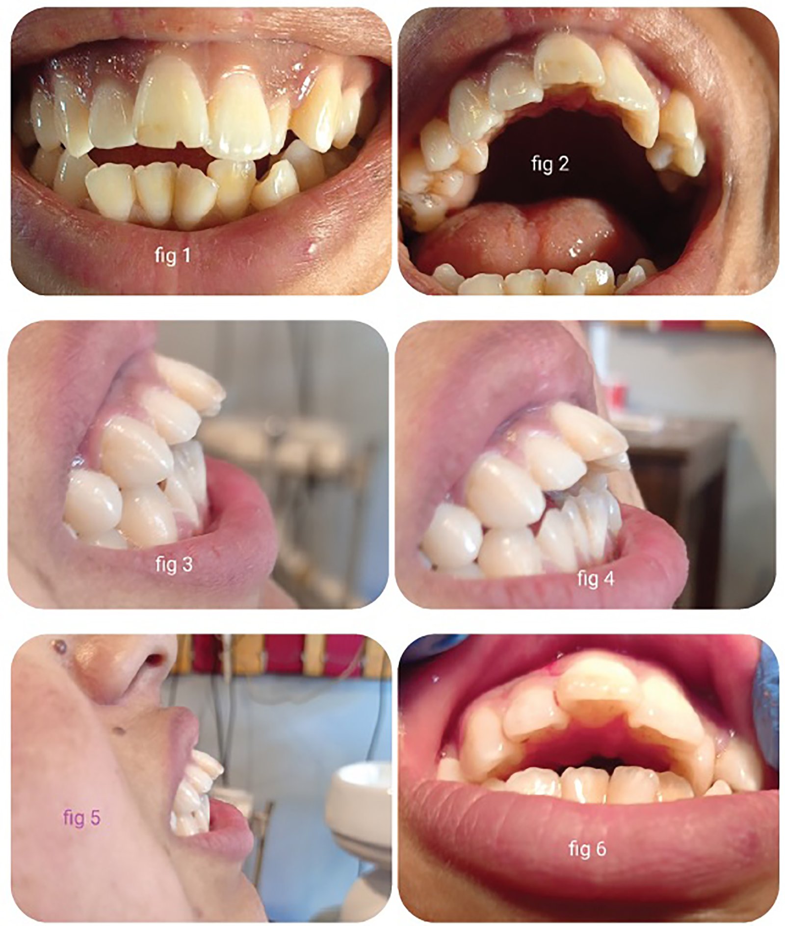
42 years old male patient reported with a chief complaint of mobile and missing teeth in both the arches. Patient also gave a history of Type II Diabetes Mellitus and was under medication for the same.
Clinical Findings
On intraoral examination, the patient was partially edentulous w.r.t. 17, 25 and 37. The remaining natural teeth were periodontally not sound.
Past dental history revealed the extraction of multiple teeth due to periodontal disease.

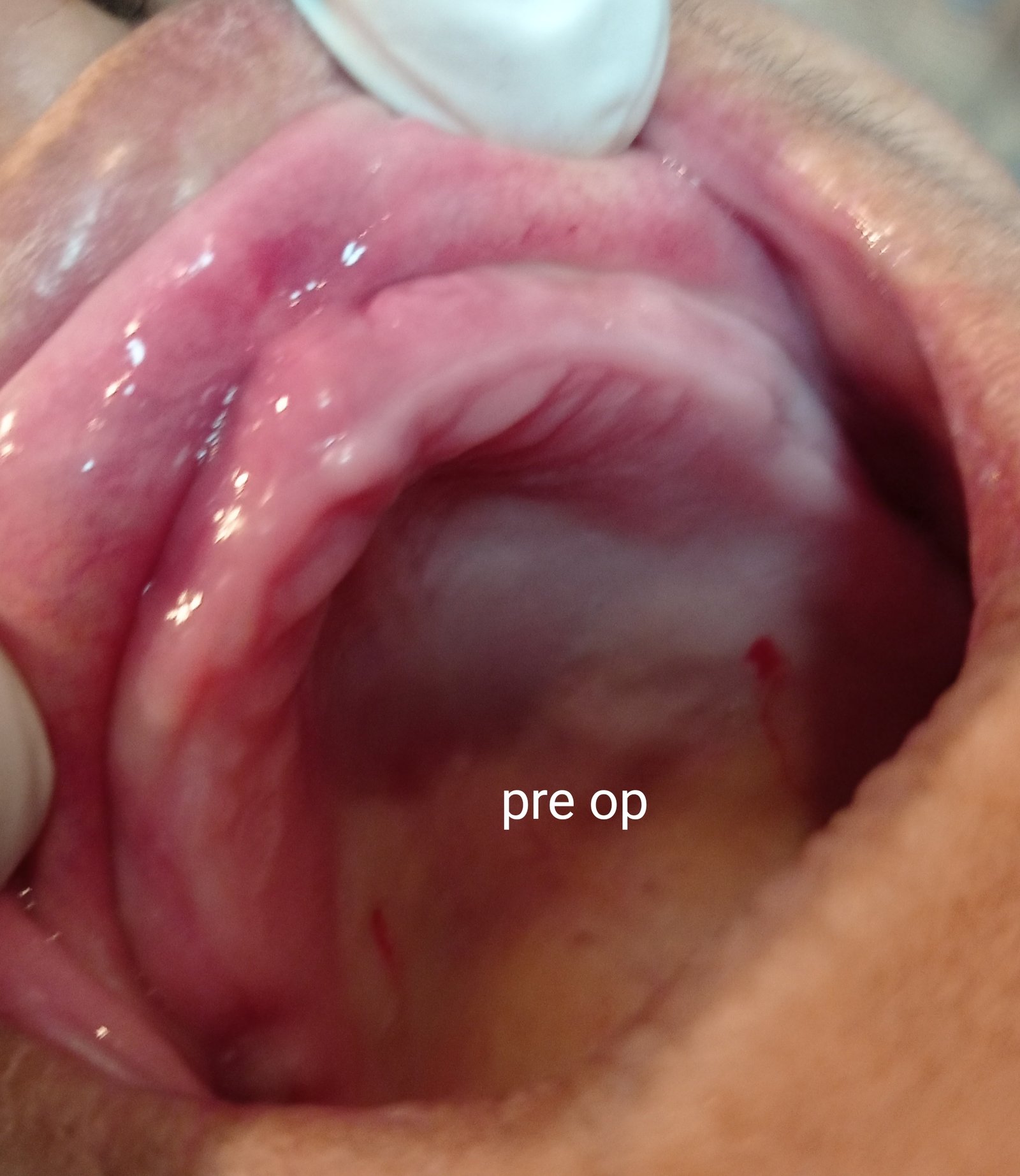
Fig 1: Pre-op photograph of the patient (The loss of tonicity of the facial muscles can be seen)
Pre-operative analysis and treatment planning
The patient was advised for extraction as the remaining periodontally weak teeth. After complete healing of the extracted socket, a conventional complete denture was fabricated and the patient was recalled after 6 months for the definitive treatment. Then the patient was advised for Orthopantomography (OPG) and Cone beam computer tomography (CBCT) of the maxillary and mandibular arch.
After evaluating the radiographs, it was concluded that there was a good amount of bone height, bone width, bone density, and adequate inter-arch space. So, the treatment plan was decided as an implant-supported Malo implant prosthesis.
Surgical phase
After radiographic analysis of CBCT and OPG we decided to place six endosseous root form implant (MIS-7 implant) in the maxillary arch with 11, 13, 16, 21, 23 and 26 positions and six implants in the mandibular arch with 33, 35, 36, 43, 45 and 46 positions respectively. The surgical procedure was planned in two phases.
- In the first phase of treatment, the surgical placement of six maxillary implants followed by placement of the cover screws and two-stage Branemark protocol was followed.
- After the one-week patient was recalled and the surgical placement of six mandibular implants was done.
OPG was advised post-surgically. After completion of the healing period patient was kept under regular follow up for one month.
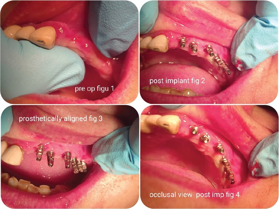
After 6 months the patient was recalled and second stage surgery was performed and suitable healing abutment were placed.
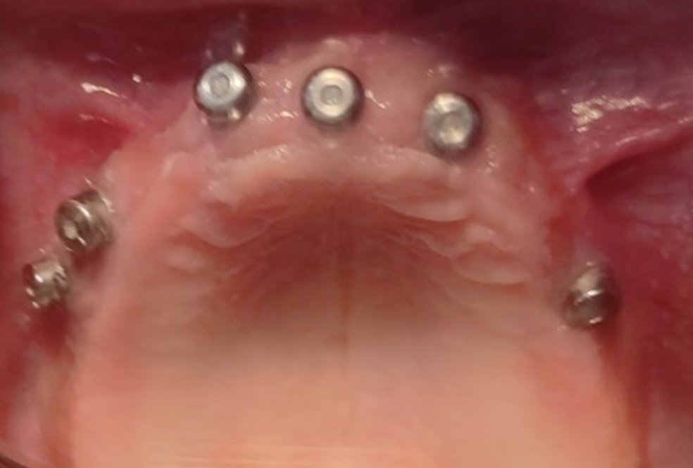
Fig 2: Post Stage 2 surgery with healing caps (maxilla)

Fig 3: Post Stage 2 surgery with healing caps (mandible)
Prosthetic phase
After the three weeks of second-stage surgery, the steps for definite prosthesis was commenced, healing abutments were removed. The closed tray impression was made.
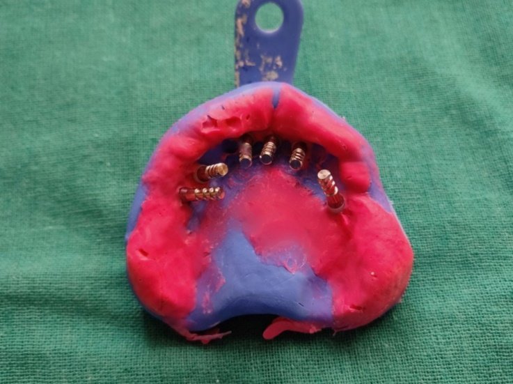
Fig 4: Closed tray impression (maxilla)
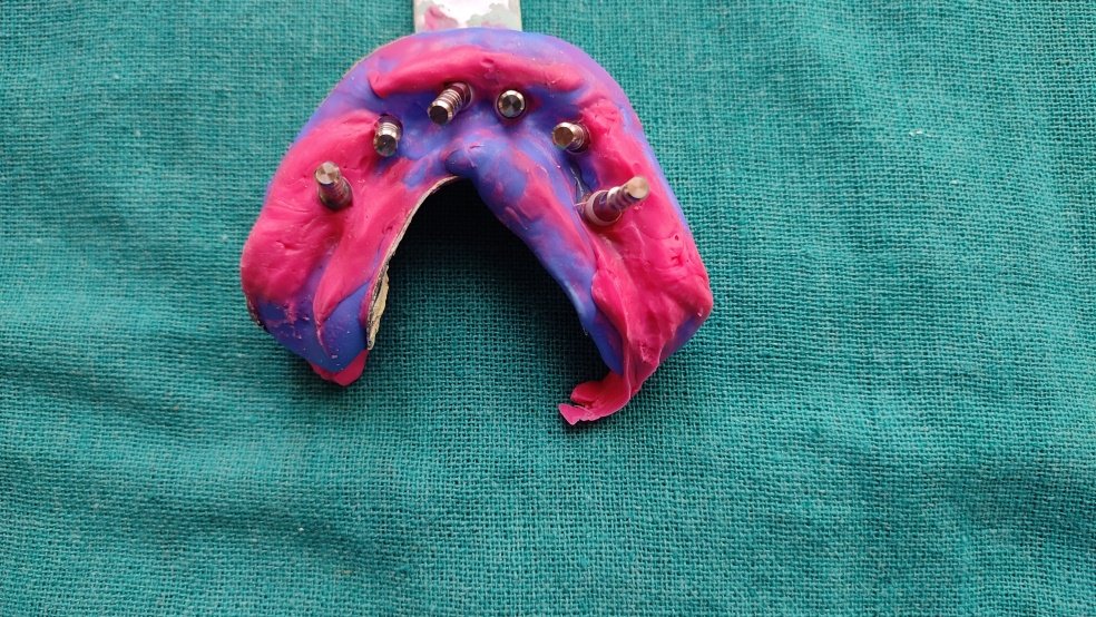
Fig 5: Closed tray impression (mandible)
Attachment of laboratory analogs, pouring off the gingival mask (Gingifast Elastic- Zhermack) was done around the neck of the impression of implants and die-stone (Ultra rock Kalabhai, Mumbai, India) was poured for fabricating the master cast. Record bases and occlusal rims were fabricated and the facebow transfer was done to the Hanau wide viu articulator. Then the maxillomandibular relationship was recorded.
Open-tray impression
The prepared acrylic custom tray was marked according to the impression coping and for the proper placement with impression copings the tray was adjusted intra-orally.
The prepared jig trial was done to evaluate the parallelism intra-orally.
Tray adhesives were applied and addition silicon (Photosil India) was used in medium body. Once the material set impressions copings were incorporated into impressions and screw was unscrewed and impressions were carefully removed in the single stroke to prevent the distortion.
Records were sent to the laboratory for fabrication of metal framework which was later evaluated intra-orally.
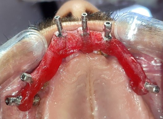
Fig 6(a): Fabrication of intra oral Jig
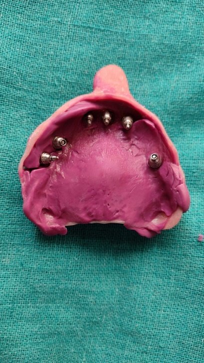
Fig 7(a): Open Tray impressions

Fig 6(b): Fabrication of intra oral Jig
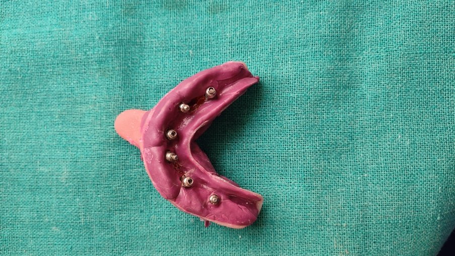
Fig 7(b): Open Tray impressions
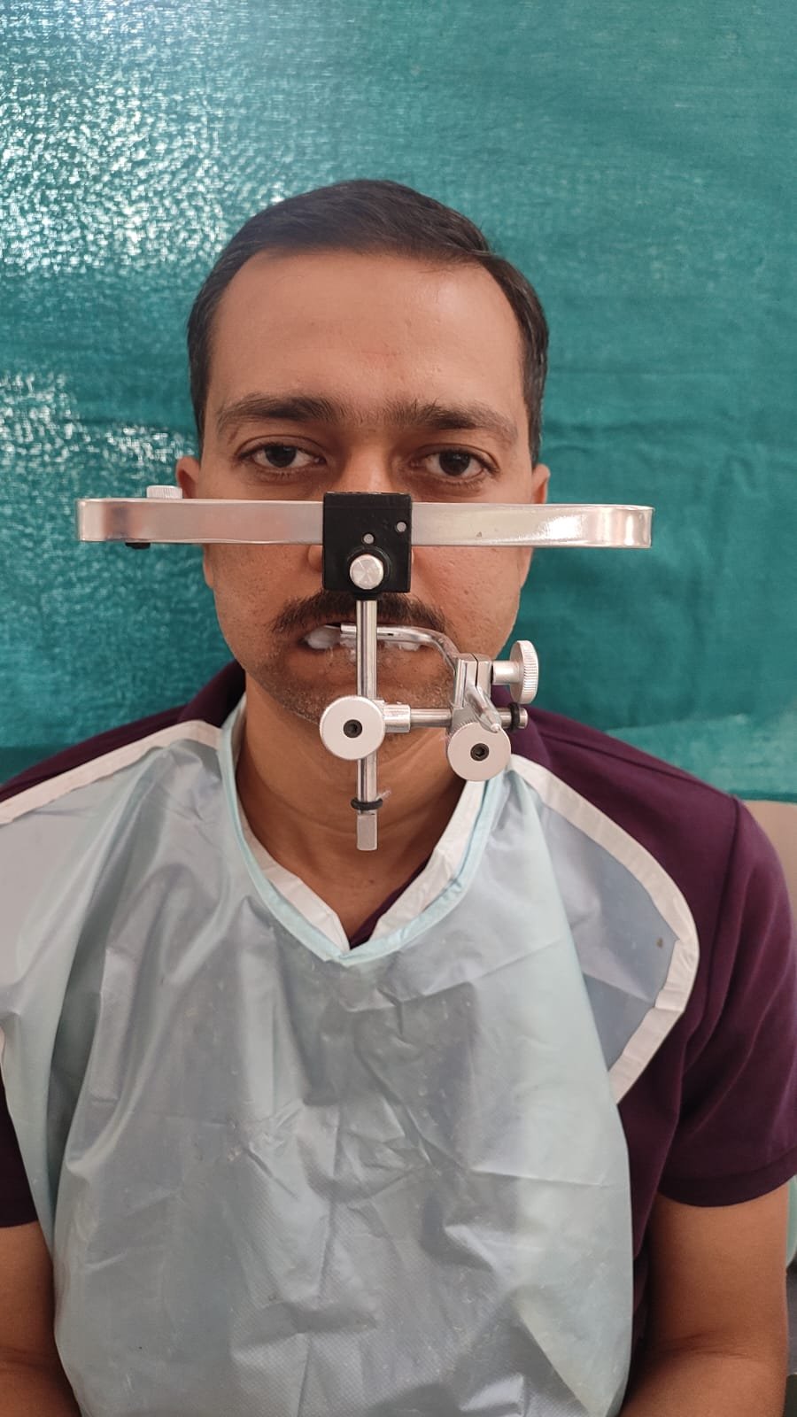
Fig 8: Facebow record (Front view)
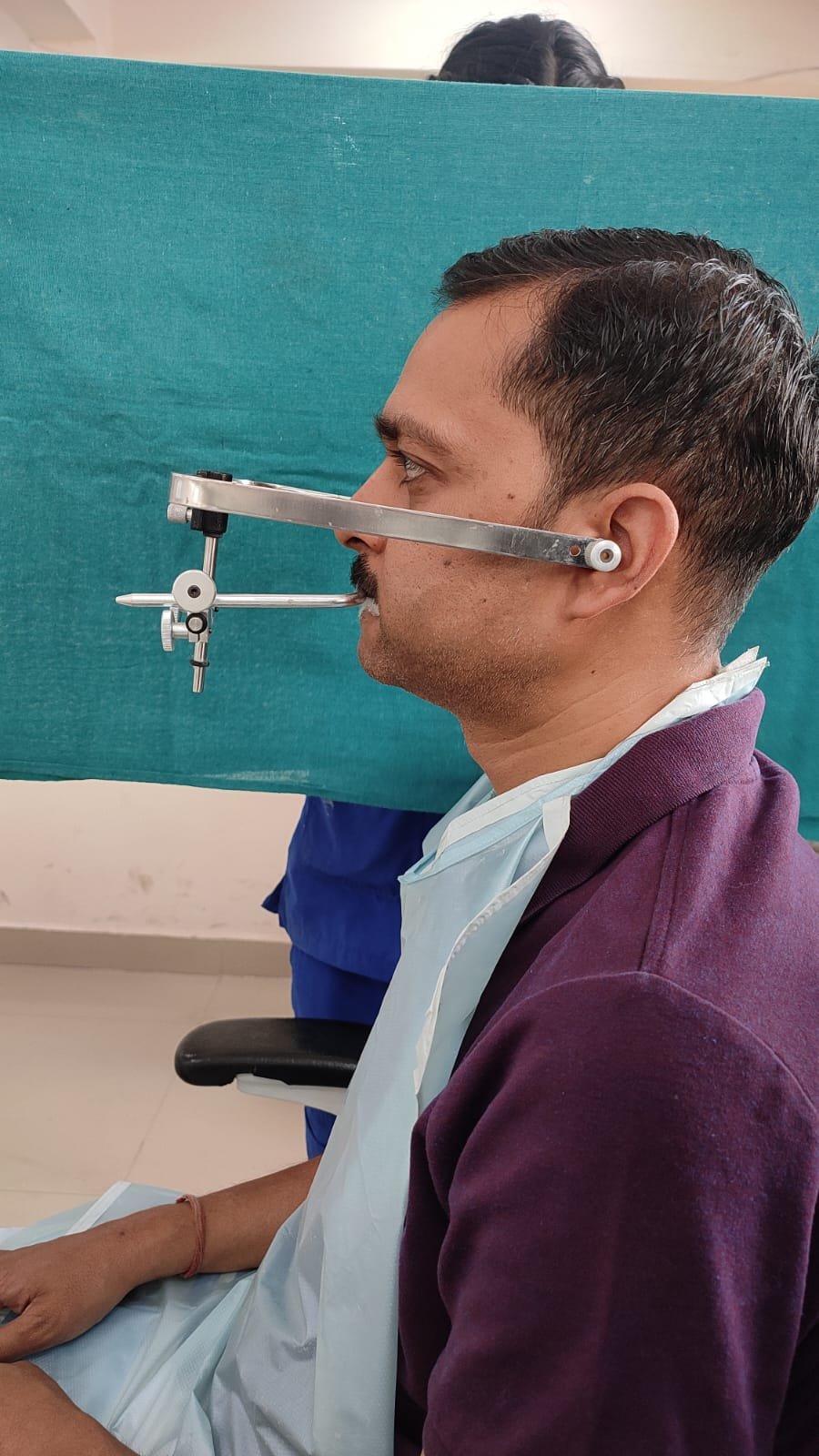
Fig 9: Facebow record (Side view)
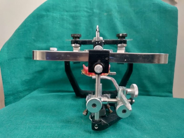
Fig 10: Facebow record (On articulator with rims)
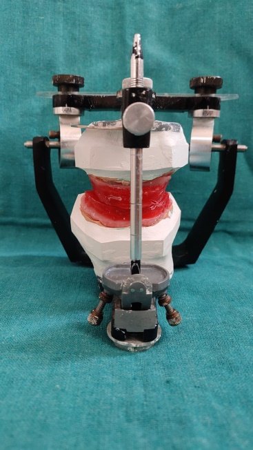
Fig 11: Jaw relation (Hanau wide viu articulator)
Then maxilla-mandibular relationship was evaluated with fabrication of occlusal rims.
The Jaw relation was then mounted of semi-adjustable articulator and the records were sent to the laboratory.
The designing was done on 3-Shape by Trios and DMLS printing was done followed by the fabrication of the framework.

Fig 12

Fig 13
The framework was again sent to the laboratory for porcelain build up over metal framework after adjustments (Fig 12-13). Bisque trial was done and was evaluated. Minor adjustments were done and final glazing was done.

Fig 14a

Fig 14b
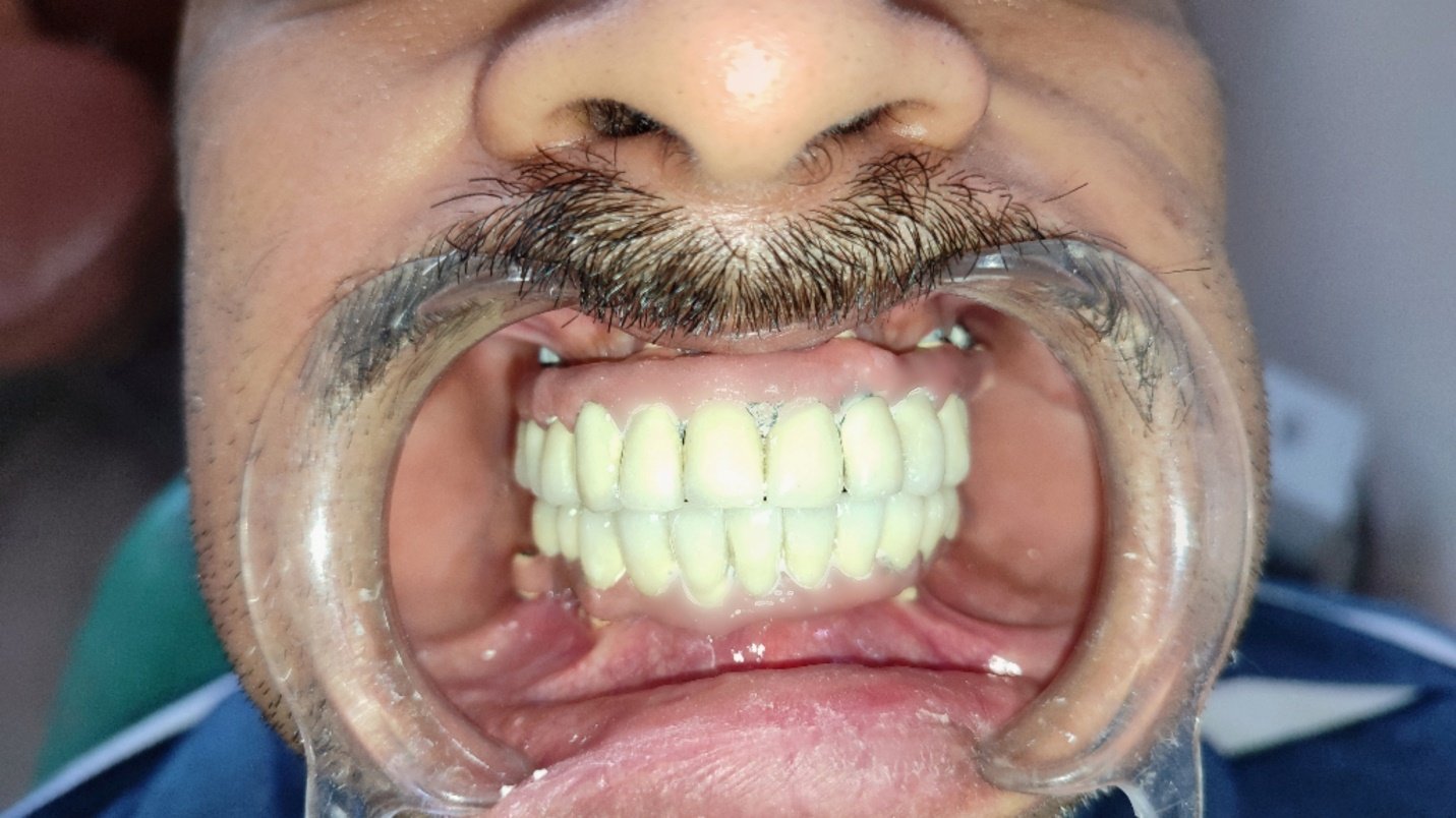
Fig 14 (a-c): Intra oral Images of the final prosthesis
The prosthesis was inserted with minor adjustments with the passive fit, after the occlusal evaluation was done post-operative instructions were given to the patient. A soft splint of 1 mm was giver post 24 hrs of insertion.
The patient was kept under regular follow up for 2 weeks.
DISCUSSION
With all modifications in the techniques, the primary need for the prosthesis is to produce a passive fit for the fixed screw-retained prosthesis is arguably one of the most technically important phases in implant dentistry.
With the number of modifications for preventing distortion that occurs during various laboratory procedures followed in the fabrication of the framework. To overcome the errors the disclosing media used to adjust the internal aspect of the casting can result in the non-bonding and passive fully seated prosthesis.2
The rehabilitation of edentulous patients with a Malo implant bridge has been observed to achieve greater masticatory function and psychological satisfactions then with conventional overdentures. Also, the occlusal force has been noticed to increase after the placement of the implant-supported prosthesis.
CONCLUSION
Every patient is different and has the unique treatment needs; proper diagnosis and treatment plan are important but cannot be all-inclusive. Careful integration and sequencing of the different areas of treatment needed to enhance the final result.
The procedure in this clinical procedure is for the rehabilitation of edentulous patients results in accurately fitted, esthetic, and functionally efficient prosthesis. Patients were previously restored with the conventional complete denture and reveal his dissatisfaction with his removable prosthesis.3
On the other hand, this type of prostheses eliminates the screw access openings in the occlusal surface of the crowns and also, makes it possible to remove and repair the fractured porcelain of the individual crown without removing the whole structure.
In the cases where there is increased inter-arch space, concern for esthetics, young age of the patient and reduced bone volume one can consider a Malo’s bridge.
Better esthetics as there is no visibilty of access holes intra-orally also; customization of the patients gingival color can be done and replicated to the metal framework.
Easy and convenient removal of the prosthesis when required. Easy repair of the chipped margins of the crowns as there is no requirement of removing the whole framework only the concerned crown can be retrieved and repaired easily which is not possible in a conventional screw or cement retained prosthesis.
Therefore, for this patient use of full arch implant-supported fixed Malo implant bridge prosthesis provided the best option for a prosthetic solution. Occlusion and articulation were found to be good along with very good retention and stability was found during follow up visits. It was concluded that the implant-supported Malo bridge enhances the psychological support and more functionally efficient as compared to conventional complete dentures.
REFERENCES
- Sha, Raj & Bhayana, Rahul & Kumar, Sonu & Shill, Manela & Sharma, Akash. (2020). Full Mouth Reconstruction- Malo Implant Bridge: A Case Report. International Journal of Scientific Research. 9. 1-2. 10.36106/ijsr/0618149.
- Jain AR, Nallaswamy D, Ariga P, Philip JM. Full mouth rehabilitation of a patient with mandibular implant screw retained Fp-3 prosthesis opposing maxillary acrylic removable over-denture. Contemp Clin Dent. 2013 Apr;4(2):231-5.
- Prithviraj, D. R.; Gupta, Ankit. Full-mouth rehabilitation of completely edentulous patient using implant-supported fixed prosthesis. The Journal of Indian Prosthodontic Society 8(1):p 44-47, January–March 2008.
- Ponnanna AA, Maiti S, Rai N, Jessy P. Three-dimensional-Printed Malo Bridge: Digital Fixed Prosthesis for the Partially Edentulous Maxilla. Contemp Clin Dent. 2021 Oct-Dec;12(4):451-453.
- Gulnar, A & Altintas, S & YILMAZ, Onur & Ate?, Gül. (2020). Rehabilitation of the completely edentulous young patient with the "Malo Bridge": A case report. Nigerian journal of clinical practice. 23. 1328-1331. 10.4103/njcp.njcp_170_20.
Disclaimer from Guident
The views and opinions expressed in articles published in Guident are solely those of the authors and do not necessarily reflect the views of the editors, the editorial board or the publisher, Ivory India. Guident does not endorse any product, technique or treatment described. Clinical content is intended for qualified dental and medical professionals and is not a substitute for individual clinical judgement. Guident, its editors and the publisher accept no liability for any consequence arising from the use of information published in the journal.
How to cite this article
Dr. Akanksha Dobhal, Dr. Ayush Saluja, Dr. Shitij Srivastava. Total Transformation: Exploring Full Mouth Rehabilitation Using Implant Supported Prosthesis Using Malos Bridge- a Case Series. Guident. 2024 Jun;17(7). Available from: https://ivoryindia.in/articles/implantology/total-transformation-exploring-full-mouth-rehabilitation-using-implant-supported-prosthesis-using-malos.html