Congenital Hypothyroidism
Department of Oral Medicine And Radiology K.D Dental College and Hospital, Mathura, India
ABSTRACT
Thyroid disorders have a widespread effect on the ossification of cartilage, growth of teeth, facial contour, and overall body proportions. Deregulations in thyroid homeostasis can result in various orofacial and skeletal abnormalities. This report describes a case of congenital hypothyroidism in a 16 year old female patient, presenting with short stature, multiple retained deciduous teeth, delayed fusion of intracranial sutures and epiphysis and diaphysis with absence of secondary ossification center. Based on various biochemical and radiographic investigations, diagnosis of congenital hypothyroidism was established.
KEY WORDS: congenital hypothyroidism, multiple retained deciduous teeth, oral manifestations, radiography.
INTRODUCTION
Congenital hypothyroidism is one of the most common preventable causes of mental retardation. In most cases, the disorder is permanent and results from an abnormality in thyroid gland development(dysgenesis or agenesis) or a defect in thyroid hormogenesis. Less commonly, the altered neonatal thyroid function is transient, attributable to the transplacental passage of maternal blocking antibodies, or iodine deficiency or excess. In rare cases,
hypothyroidism may result from a pituitary or hypothalamic abnormality(secondary/tertiary hypothyroidism).5
Fetal thyroid development can be divided into three distinct phases: embryogenesis, hypothalamic maturation and finally development of the thyroid-pituitary feedback inhibitor system. It is during intrauterine life that the lack of adequate TH production determines more damaging consequences, since these hormones have a fundamental role in normal fetal brain development. For an adequate TH production, it is important that the hypothalamic-pituitary-thyroid axis be maintained whole so as to ensure the sequence of activities of the hypothalamic releasing hormone (thyrotropin releasing hormone-TRH) over the pituitary gland, producing thyroid stimulating hormone (TSH), which in turn acts on the thyroid, producing TH.[4]
Thyroid hormone plays an important role in growth and development. Before a baby is born and upto 2 to 3 years of life, thyroid hormone is very important for brain development. After this, thyroid hormone is important for growth as well as enabling the body to use energy and stay warm(metabolism) and to help the brain,heart,muscles, and other organs work as they should.[1]
When thyroid hormone deficiency is detected at birth it is called “congenital hypothyroidism”.
Congenital hypothyroidism is characterized by dwarfism, overweight, a broad , flat nose,wide set eyes, thick lips, a large protruding tongue;poor muscle tone;pale skin;stubby hands;retarded bone age;delayed eruption of teeth;malocclusions;a hoarse cry;an umbilical hernia;and mental retardation.[2]
CASE REPORT
A 16-year-old female patient reported to the Outpatient Department (OPD) with a chief complaint of pain in the lower left mandibular posterior tooth region since one month. The patient’s medical history revealed long-standing tiredness and lethargy, along with increasingly severe symptoms of somnolence, cold intolerance, constipation, and muscle stiffness with pain. She presented with poor fine-motor skills,hearing impairment,delayed cognizance. The developmental milestones were delayed. She was born at full term, in a normal delivery and was diagnosed with congenital hypothyroidism at the age of four years and since then was taking levothyroxine 10μg.
The general examination revealed that the patient had a short stature (114 cm) and subnormal weight for her age with a body mass index (BMI) of 28.6 [Figure 1]. Other features included, hoarseness of
voice, proportional skeletal immaturity, absence of secondary sexual characters, and a coarse, dry skin.
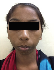
Extraoral examination revealed that patient was bracheocephalic with skeletal immaturity, puffiness of the face and neck, periorbital swelling, prominent supraorbital ridge with a widened nasal bridge,frontal bossing hypertelorism, flat foot.[Figure 2].
Intraoral examination showed mixed dentition was present, with multiple retained deciduous teeth irt 12,53,54,55,16,61,21,22,63,24,65,26,31,32,73,74,35,36,41,42,83,84,85,46.
On the basis of the above clinical findings a provisional diagnosis of congenital hypothyroidism was made.
Dwarfism, juvenile hypo thyroidism, achon droplasia, Secondary hypo thyroidism, hypo pituitarism, vitamin D deficiency and hypo phosphatemia were considered for differential diagnosis.
Since it was already an established case of congenital hypothyroidism a series of investigations were done to further strengthen the diagnosis.
Laboratory investigations were performed. The hemogram revealed a low hemoglobin level of 9.9 gm%. [figure 4].
Renal function profile was normal suggestive of no renal disorder[figure 5].
Liver function profile revealed an increase in the levels of SGOT,SGPT,ALP levels suggestive of liver dysfunction[figure 6].
The levels of S.T4 was < 6 mmol/l which was significantly decreased and the level of S.TSH was >60 which was significantly increased indicative of hypoplasia of the thyroid gland or presence of rudimentary thyroid gland[figure 7].
The radiographic survey included a panoramic view, anterioposterior (AP) skull view, lateral skull view, and a hand wrist radiograph, chest radiograph, anterioposterior view of the legs, and anterioposterior view of the pelvis.
The panoramic radiograph demonstrated multiple retained deciduous teeth. Unerupted permanent teeth .Reduced height of alveolar bone. Mandibular condylar growth was less..Antegonial notch was obscured.Angle of mandible not well developed. Teeth visible were were 11, 12, 53, 54, 55, 16, 61, 21, 22, 63, 24, 65, 26, 31, 32, 73, 74, 35, 36, 41, 42, 83, 84, 85, 46.
Unerupted teeth present were: 13, 15, 17, 23, 25, 27, 33, 34, 37, 43, 44, 45, 47 [figure 8].
The anterioposterior skull view showed delayed closure of intracranial sutures [Figure 9].
The lateral cephalogram showed elongated skull. Relative Frontal bossing.Pseudo prognathism of the mandible. Skeletal and dental class iii malocclusion.Increased intervertebral disc space[figure 10] .
The chest radiograph revealed failure of fusion of clavicle to that of the scapula[figure 11].
The hand wrist radiograph revealed epiphysis was of equal width to that of diaphysis in the middle phalynx of the third finger(acc.to fishman’s skeletal maturity index).Capping of epiphysis to that of diaphysis was absent.Absence of sesamoid bone.No fusion of epipyhsis to diaphysis [figure 12].
The anterioposterior view of the pelvis revealed that ossification centers were defective and they appeared in an irregular and mottled pattern with multiple foci that join together to give a porous or fragmented appearnace known as “stippled epiphyseal dysgenesis. Unfused femoral head epiphysis[figure 14].
The patient was referred to an endocrinologist, who prescribed increased dose of levothyroxine 75 μgm once daily, and a comprehensive, multidisciplinary treatment was planned followed by root canal treatment irt 36.
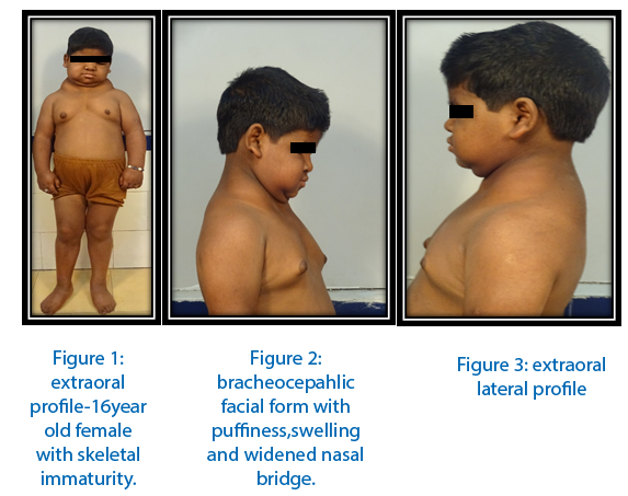


Figure 7: thyroid function test

Figure 8:Multiple retained deciduous teeth. Unerupted permanent teeth .Reduced height of alveolar bone. Mandibular condylar growth was less. Antegonial notch was obscured. Angle of mandible not well developed.
Teeth visible were11,12,53,54,55,16,61,21,22,63,24,65,26,31,32,73,74,35,3 6,41,42,83,84,85,46.
Unerupted teeth present were: 13,15,17,23,25,27,33,34,37, 43,44,45,47.

Figure 9:AP skull VIEW. DELAYED FUSION OF INTRACRANIAL SUTURES

Figure 10:LATERAL CEPHALOGRAM. Skull appears elongated. Relative Frontal bossing.Pseudoprognathism of the mandible.Skeletal and dental class iii malocclusion.Increased intervertebral disc space.

Figure 11: Chest radiograph:Failure of fusion of clavicle to scapula

Figure 12:HAND WRIST RADIOGRAPH:Epiphysis is of equal width to that of diaphysis in the middle phalynx of the third finger(acc.to fishman’s skeletal maturity index).Capping of epiphysis to that of diaphysis is absent.Absence of sesamoid bone.No fusion of epipyhsis to diaphysis.

Figure 13:anterioposterior view of the legs.

Figure 14: anterioposterior view of the pelvis revealed that ossification centers were defective and they appeared in an irregular and mottled pattern with multiple foci that join together to give a porous or fragmented appearnace known as “stippled epiphyseal dysgenesis. Unfused femoral head epiphysis
DISCUSSION
The ‘mouth is a mirror of systemic diseases’, as many metabolic and genetic disorders can be established by a dental professional, based on their oral manifestations.[8] Thyroid disorders are the most common among all endocrine diseases.
Primary hypothyroidism is the most common, with dysfunction occurring at the level of the thyroid gland. In secondary or tertiary hypothyroidism (central hypothyroidism), the defect is at the pituitary, hypothalamus or hypothalamic portal circulation level, respectively. If left untreated congenital hypothyroidism can catapult into severe mental and growth retardation.[9]
Skeletal growth is mainly influenced by growth hormones. Thyroid hormones have a widespread effect on ossification of the cartilage, growth of teeth, facial contour, and overall body proportions.[10] In the present case, the presence of short stature (dwarfism) prompted us to initiate growth hormonal assays and routine radiographic investigations, comprising of hand-wrist, and skull radiography. Low T3 and T4 levels with raised thyroid stimulating hormone (TSH) levels and a normal growth hormone assay supported the diagnosis of hypothyroidism secondary to a pituitary cause. Both growth hormones and thyroid hormones show permissive action, that is, both cannot produce normal growth individually, but when they present together, they stimulate growth.
The manifestations of hypothyroidism result from a reduction in metabolic activity and deposition of glycosaminoglycans.[13] Facial puffi ness with periorbital edema, proportional skeletal immaturity, cold extremities, coarse and dry skin, with sparse hair, voice hoarseness, and muscular hypotonia are the findings commonly observed in hypothyroidism. The presence of anemia, high serum cholesterol, and reduced BMR contribute toward the diagnosis of hypothyroidism.
Anemia in patients of hypothyroidism may represent iron deficiency due to menorrhagia.
The oral manifestations include characteristic macroglossia, dysgeusia, delayed eruption, poor periodontal health, and altered tooth morphology,
making it susceptible to dental caries.[14]
A radiographic survey revealed an absence of sesamoid, hook of hamate ossification along with delay in fusion of epiphysis and diaphysis of phalanges, metacarpals, radius, and ulna in the hand-wrist radiograph.[17] Findings of open sutural markings in skull radiography support the findings of a delayed growth pattern in the present case.[18]
Congenital hypothyroidism is associated with an increase in the risk of associated malformations with high proportions of renal and heart abnormalities, including septal defects, impaired diastolic function, low exercise capacity.
After confirmation of diagnosis, immediate treatment with levothyroxine 10-15microgram/kg normalizes t4 and TSH within 7-30 days.
Thus ,congenital hypothyroidism if not detected and not promptly treated, then it leads to an irreversible mental retardation.
It is a disease that is expressed in multiple forms concerning physical ,mental and social level and therefore should be treated early on as soon as the child is born
CONCLUSION
Many inherited disorders manifest in the form of alterations in the anatomy and morphology of the orofacial complex, which can be first detected on a dental examination. Knowledge of such disorders leads to an evolved screening process and treatment of such patients. This case report emphasizes on the fact that congenital hypothyroidism should be considered in patients with severe dental anomalies and growth retardation.
REFERENCES
- Jain V, Agarwal R, Deorari AK, Paul VK. Congenital hypothyroidism. Indian J Pediatr 2008;75:363-7.
- LaFranchi S. Congenital hypothyroidism: Etiologies, diagnosis, and management. Thyroid 1999;9:735-40.
- Rastogi MV, LaFranchi SH. Congenital hypothyroidism. Orphanet J Rare Dis 2010;5:17.
- Abduljabbar MA, Afi fi AM. Congenital hypothyroidism. J Pediatr Endocrinol Metab 2012;25:13-29.
- Buket A, Demet ST, Sema C, Behiye B. Dental treatment way of congenital hypothyroidism: Case report. Int Dent Med Disorders 2008;1:34-6.
- Li le JW. Thyroid disorders. Part II: Hypothyroidism and thyroiditis. Oral Surg Oral Med Oral Pathol Oral Radiol Endod 2006;102:148-53.
- Jameson JL, Weetman AP. Disorders of the thyroid gland. In: Kasper DL, Braunwald E, Fauci AS, Hauser SL, Longo DL, Jameson JL, et al., editors. Harrison’s Principles of Internal Medicine 16th ed. New York: Mc Graw-Hill; 2005. p. 2104-7.
- Pinto A, Glick M. Management of patients with thyroid disease: Oral health considerations. J Am Dent Assoc 2002;133:849-58.
- Edwards CR, Baird JD, Frier BM, Shepherd J, Toft AD. Endocrine and metabolic diseases including diabetes mellitus. In: Edwards CR, Bouchier IA, Hasleô€„´ C, Chilvers ER, editors. Davidson’s Principles and Practice of Medicine. 17th ed. New York: Churchill Livingstone; 1995. p. 685-97.
- Jain AK. Endocrine system. In: Jain AK, editor. Human Physiology for BDS. 2nd ed. New Delhi: Avichal Publishing; 2004. p. 340-1.
- Connelly KJ, Boston BA, Pearce EN, Sesser D, Snyder D, Braverman LE, et al. Congenital hypothyroidism caused by excess prenatal maternal iodine ingestion. J Pediatr 2012;161:760-2.
- Schuld A. Is dental fluorosis caused by thyroid hormone disturbances? Fluoride 2005;38:91-4.
- Guha B, Krishnaswamy G, Peiris A. The diagnosis and management of hypothyroidism. South Med J 2002;95:475-80.
- Young ER. The thyroid gland and the dental practitioner. J Can Dent Assoc 1989;55:903-7.
- Ruchała M, Szczepanek E, Sowiński J. Diagnostic value of radionuclide scanning and ultrasonography in thyroid developmental anomaly imaging. Nucl Med Rev Cent East Eur 2011;14:21-8.
- Kempers MJ, van Trotsenburg AS, van Rijn RR, Smets AM, Smit BJ, de Vijlder JJ, et al. Loss of integrity of thyroidmorphology and function in children born to mothers with inadequately treated Graves’ disease. J Clin Endocrinol Metab 2007;92;2984-91.
- Bhalajhi SI. Skeletal maturity indicators. In: Bhalajhi SI, editor. Orthodontics: The Art and Science. 3rd ed. New Delhi: Arya (MEDI) Publishing House; 2006. p. 161-74.
- White CS, Pharoah JM. Systemic diseases manifested in jaws. In: White CS, Pharoah JM, editors. Oral Radiology Principles and Interpretation. 5thed. New Delhi: Elsevier; 2006. p. 522-3.
Disclaimer from Guident
The views and opinions expressed in articles published in Guident are solely those of the authors and do not necessarily reflect the views of the editors, the editorial board or the publisher, Ivory India. Guident does not endorse any product, technique or treatment described. Clinical content is intended for qualified dental and medical professionals and is not a substitute for individual clinical judgement. Guident, its editors and the publisher accept no liability for any consequence arising from the use of information published in the journal.
How to cite this article
Dr. Rimmi Narula, Dr. Gopa Kumar R. Nair. Congenital Hypothyroidism. Guident. 2018 Dec;12(1). Available from: https://ivoryindia.in/articles/oral-medicine/congenital-hypothyroidism.html