Treacher Collins Syndrome
Department of Pedodontics and Preventive Dentistry K. D. Dental College, Mathura, U.P. India
INTRODUCTION
Treacher Collins syndrome (TCS, OMIM number 154500) is a rare condition of craniofacial morphogenesis affecting only 1 in 50,000 live births. Being genetic in origin, it is inherited in an autosomal dominant fashion affecting structures originating from first and second pharyngeal arches which may affect the size and shape of the ears, eyelids, cheek bones, and jaws. Patients have normal intelligence but often have to deal with social challenges due to their physical appearance
TCS was describe for the first time by Thompson in 1846. George Andreas Berry described TCS as a congenital neonatal deformity with coloboma of lower eyelid in 1889. Later in 1900, an ophthalmologist Edward Treacher Collins explained the condition with its two key clinical features. But it was in 1940 when two colleagues Adolphe Franceschetti and David Klein explained the condition in detail under the heading Mandibulofacial Dysostosis. As of now, this condition is known by several eponyms—Treacher Collin syndrome is famous in United States and United Kingdom; Franceschetti-Klein syndrome in Europe, and mandibulofacial dysostosis otherwise.
GENETICS
Treacher Collins syndrome is an autosomal dominant disorder which shows no predilection toward any sex. The gene responsible for TCS has been named as TCOF1which exhibit linkage to the human chromosome 5q32 locus. TCOF1 is found to encode a low complexity, serine/alanine-rich, nucleolar phosphoprotein-termed Treacle. It helps in neuroepithelial survival and neural crest cell proliferation that is central to normal craniofacial development. Individuals who have the TCOF1 mutation have a 50% chance of passing it on to their children. TCS shows high penetrance and extreme variation in expressivity of phenotype. Multiple exons have been found within the gene and so far 120 mutations have been detected. Mutations in TCOF1gene leads to high degree of neuroepithelial apoptosis and consequent loss of neural crest cells. General cranioskeletal hypoplasia occurs due to generation of insufficient neural crest cells. Every mutation leads to a premature termination codon, and mutation can be spliced or deletion type.
It is hypothesized that loss of function mutation of TCOF1 leads to haploinsufficiency. Haploinsufficiency of TCOF1/Treacle results in deficient ribosome biogenesis, which is incapable of meeting the proliferative needs of the neuroepithelium. Deficient ribosome biogenesis leads to nucleolar stress activation and stabilization of p53, which causes the high degree of neuroepithelial apoptosis and consequent loss of neural crest cells
ETIOLOGY
Craniofacial morphogenesis starts early during embryonic life. It is a complex process and most critical step takes place between 3rd and 8th week of pregnancy. The process starts with three layers: ectoderm, mesoderm and endoderm, which is followed by formation of neural crest cells. These cells arise from neural ectoderm and travel to distant location to form various tissues like cartilage, connective tissue, bones, etc.
Neural crest cells are responsible for the scaffold on which construction of head and face takes place deciding about its shape and variations. Mesoderm is responsible for the facial muscles and the lining of the oral cavity of the mouth is derived from endoderm.
As craniofacial birth defects are typically attributed to abnormality in the underlying structures which provide the shape to face and head. Therefore, neural crest cell abnormally is the most promising cause of the condition. Treacher Collins syndrome is a genetic condition caused by mutations of certain genes.
TCS is caused by mutations in one of three genes: TCOF1, POLR1C, POLR1D. A mutation in the TCOF1 gene causes the syndrome in 90–95 percent of people with TCS. A mutation in the POLR1C or POLR1D gene accounts for around 8 percent of people with TCS. All these genes play vital roles in the early development of facial bones and tissues. When they are mutated, certain cells involved in the development of bones and tissues of the face self-destruct
TYPES
TCS can be classified into four forms - complete form showing all known features; incomplete form with less severe features; abortive form where only hypoplastic zygoma and pseudocoloboma of the lower eyelid are seen; unilateral form with features seen only on one side of the face and atypical form where other abnormalities that are not usually a part of the syndrome are also present
CLINICAL MANIFESTATIONS
The clinical presentation of TCS is well distinctive. Most of the characteristics are due to first and second pharyngeal arch defect. Despite being a bilateral anomaly, skeletal and soft tissue structures are rarely symmetrical.
A fully expressed case of Treacher Collin’s syndrome will have typical anti mongoloid features of start palpebral fissures micropthalmia, hypoplastic facial bones convex facial profile prominent nasal dosium with retrongnathia (Figs1&2).
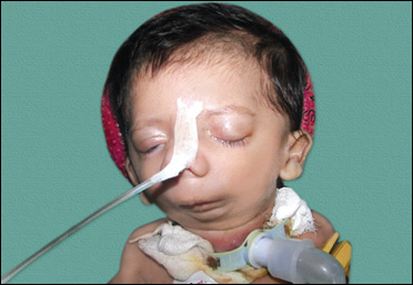

Figs1&2: The classical presentation of TCS in a small child exhibiting micrognathia, antimongoloid slanting eyes, hypoplasia of the facial bones and low-positioned auricles where the tracheostomy is performed on the newborn as the child has breathing difficulty
Ocular
Ocular and periorbital findings are most prominent features of TCS. Patient presents with skeletal dimorphism of orbits, malar hypoplasia and ellipsoid shape. Nasal and maxillary prominences are relatively normal and give exophthalmic appearance. Downstanting “antimicrobial” palpebral tissue is key presentation. Surrounding soft tissues are hypoplastic along with thinning of lower lid skin. Meibomian and orbicularis oris muscles are either absent or attenuated. Lacrimal duct atresia, lid notching can also be present. Lack or absence of eyelashes from medial third of lower eyelid is the characteristic features. Amblyopia from corneal screening, strabismus and refractory error can be present. Some patients may show congenital cataract but most of the patients retain at least one eye with normal vision.
Auricular
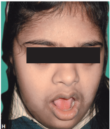
Treacher Collins syndrome patients show notably deformed external ear. Auricular remnants are usually malpositioned at a lower location (Fig 3). Stenosis or atresia of external auditory meatus, which is accompanied by misshapen tympanic membrane. Middle ear is mostly absent or deformed. Inner ear is the only exception with normal morphology; however, pathological ossicular chain leads to hearing loss. Degree of hearing loss varies from patient to patient and may go up to maximum conductive hearing loss of 60 dB.

Fig 3: Gross facial asymmetry, maxillary prognathism and micrognathia along with low set ear lobes
Zygoma and Malar Region
Malar hypoplasia is the characteristic feature of TCS. Zygomatic hypoplasia was demonstrated anthropometrically by Posnick et al. with the help of axial CT scan. Varied zygomatic hypoplasia in different patients leads to a classification based on morphology and volume of zygoma. Type-I denoting minor, Type-II denoting moderate and Type-III denoting severe.
Maxilla and Mandible
Maxilla and mandible are also involved, characteristically pronounced retrognathic leads to convex facial profile. Mandibular angle is either absent or significantly underdeveloped. Patients also show decreased height of lower third of face along with high antegonial notching. In severe cases, ramus/condyle unit may be lacking altogether. Studies have documented that proximal mandibular hypoplasia is most common, i.e. condyle followed by ramus and body. Condyle involvement affects normal functioning of temporomandibular joint (TMJ) and zygomatic relation is also affected, which further leads to ankylosis of TMJ in TCS patients.
Other Important Features
Absent parotid gland, Beaked nose, Cervical spine malformation, Choanal atresia , Cleft palate, Congenital heart disease , Cryptorchidism, Renal anomalies.
ORAL MANIFESTATIONS
Oral manifestations are common because of involvement of maxilla and mandible. Hypoplasia of the facial bones, particularly the mandible (78% of cases) and zygomatic complex (81%), is an extremely common feature of TCS.
Hypoplasia of the facial bones may result in dental malocclusion such as Class II and III malocclusion, retrognathia, microstomia, abnormal positioning and anterior open bite a common finding. Hypoplastic dental anomalies are commonly seen followed by heterotopic and hyperplastic dental alterations, enamel opacity, ectopic eruption, supernumerary teeth. The teeth may be widely spaced, malpositioned or reduced in number. In a large proportion of cases, the palate is high, arched and occasionally cleft (28%) and in severe cases, the zygomatic arches may be completely absent (Poswillo, 1975).
DIAGNOSIS
Ultrasonography: 3D sonography and Doppler sonography are useful to reveal microcephaly, micrognathia, cleft lip palate and skeletal defects. As it is very difficult to see facial features with traditional 2D sonography until 30 weeks of gestation.
Amniocentesis and chorionic villous sampling: Amniocentesis at 16–19 weeks or chronic villous sampling at 10–11 weeks can be performed to identify mutations and differential diagnosis.
Once born, diagnosis is straight forward due to characteristic orofacial features.
MANAGEMENT
Management of TCS is widely divided into two phases:
1. Neonatal management
2. Surgical management.
Neonatal Management
Neonatal management is crucial and airway management, feeding and growth remain at priority. Oxygen saturation monitoring may be required in same patients. While assessing, determining the cause of apnea is very important as it may be caused by multiple factors, e.g. choanal atresia, micrognathia, glossoptosis, laryngomalacia, etc. Mild cases are easily managed through conservative treatment using suctioning and nasopharyngeal trumpet along with positioning. However, sever cases with anatomical anomaly early tongue lip adhesion or mandibular advancement (Figs 26.7A and B) is required. Further severe cases may need intubation or tracheostomy.
Surgical Management
Surgical management is planned by a multidisciplinary craniofacial team. A multidisciplinary approach is important to cover all aspects of deformities, i.e. ocular, oral, dental, pediatric and craniofacial. Preoperative planning should start as soon as possible and preoperative assessment should be done with the help of advanced diagnostic imaging modalities. Surgical treatment starts early in life and goes on still adolescence (Fig. 26.8).
Oral Surgery
Nearly all patients of TCS show malocclusion. In severe cases, there is a sleep occlusal plane, aberrant dentition and an anterior open bite. Normal anterior maxillary, height, shortened mandible, prominent nose and retruded lower third of face give rise to a bird like appearance.
Lowering of mandibular plane is argued by few surgeons to allow descent and growth of posterior maxilla; however, this treatment is not good enough to deal with residual posterior open bite. After attaining the skeletal maturity, Le Fort I with bilateral sagittal split osteotomy is performed to level the occlusal plane in late adolescence.
Dentition is abnormal and maintenance of oral hygiene is problematic. Teeth may be absent, malrotated or malpositioned. High plaque builds up further compromise the oral hygiene. Orthodontist can help the patient in attaining improved bite and intercuspation. Repeated surgical procedures with poor bone stock makes extraction and implants necessary due to potential devascularization and injury to roots.
CONCLUSION
In most TCS patients, a spectrum of affected features is observed; in fact, rarely if any single abnormality alone sufficient to lead a diagnosis of TCS. More usually, the entire facial appearance is considered when trying to arrive at a diagnosis, particularly, in mildly affected patients.
Many clinicians and scientists have the subjective impression that patients with TCS have a tendency toward short stature, at least early in life, and generally have average or above-average intelligence.
Disclaimer from Guident
The views and opinions expressed in articles published in Guident are solely those of the authors and do not necessarily reflect the views of the editors, the editorial board or the publisher, Ivory India. Guident does not endorse any product, technique or treatment described. Clinical content is intended for qualified dental and medical professionals and is not a substitute for individual clinical judgement. Guident, its editors and the publisher accept no liability for any consequence arising from the use of information published in the journal.
How to cite this article
Dr. Priya Verma. Treacher Collins Syndrome. Guident. 2018 Dec;12(1). Available from: https://ivoryindia.in/articles/pedodontics/treacher-collins-syndrome.html