Mucormycosis in COVID-19 Patient: A Case Report with Review
Department of Oral Medicine and Radiology, K.D. Dental College and Hospital, Mathura India
ABSTRACT
After candidiasis and aspergillosis, mucormycosis is an opportunistic deep fungal infection that comes in third place. It is brought on by a Mucorales-order angioinvasive fungus. The hallmark symptoms of this illness, which causes rapid progression, are tissue necrosis brought on by blood vessel invasion and subsequent thrombosis. Rhinocerebral mucormycosis is the most prevalent type of this condition in the maxillofacial region, affecting the oral cavity, maxilla, palate, nose, paranasal sinuses, orbits, and central nervous system extensively. The treatment of mucormycosis is mainly based on antifungal and surgical debridement. The increase of ketoacidosis in diabetics, transfusion of hematopoietic growth factors in long-term neutropenia and hyperbaric oxygen therapy may be useful. The morbidity and mortality of mucormycosis are high due to the invasive nature of the frequent underlying malignancy, hence the importance of early and appropriate management. Impairment of senses, such as taste, smell, and hearing, along with functional disabilities and compromised esthetics, promotes a negative impact on the patient after surgical debridement and maxillectomy. It adversely affects not only function and aesthetics but also results in emotional disturbances.
Keywords: Rhinocerebral mucormycosis, mucormycosis, surgical debridement, antifungal drugs, maxillectomy
INTRODUCTION
Mucormycosis is an opportunistic fungal infection that comes in third in all fungal infections. It is brought on by a Mucorales-order angioinvasive fungus. Mucormycosis can be classified into at least six clinical groups based on the clinical presentation and the involvement of a specific anatomic site: (i) rhinocerebral, (ii) pulmonary, (iii) cutaneous, (iv) gastrointestinal, and (v) disseminated. The hallmark symptoms of this illness, which causes rapid progression, are tissue necrosis brought on by blood vessel invasion and subsequent thrombosis. Rhinocerebral mucormycosis is the most prevalent type of this condition in the maxillofacial region, affecting the oral cavity, palate, paranasal sinuses, orbits, and central nervous system extensively.1,2
Paltauf first defined mucormycosis as an infection caused by nonseptate, wide, branching hyphae typical of molds in 1885. A case of pulmonary mucormycosis reported by Sluyter in 1847 was the first known human infection with mucorales. Gregory originally identified the symptoms of acute orbital mucormycosis in 1943, including uncontrolled hyperglycemia, unilateral internal and external ophthalmoplegia, proptosis, meningoencephalitis, and sudden death.3,4
Early signs include nasal inflammation, cellulitis, and periorbital edema. Later signs include extensive tissue necrosis. Post-COVID infections, such as mucormycosis, are more likely to affect patients taking corticosteroids or those with coexisting conditions like diabetes mellitus. They have a high mortality rate, so early identification, antifungal therapy, complete surgical excision, and sufficient prosthodontic rehabilitation are essential for successful treatment.
Mucormycosis infection in the compromised host occurs from impaired immunity, which leads to the fast proliferation and invasion of fungal organisms in deeper tissues. Uncontrolled diabetes (especially in patients with ketoacidosis), cancers like lymphomas and leukaemias, renal failure, organ transplant, long-term corticosteroid and immunosuppressive therapy, cirrhosis, burns, protein-energy malnutrition, and acquired immune deficiency syndrome are some of the predisposing factors for mucormycosis.5-8 Early disease detection, vigorous, and rapid medical and surgical therapies are necessary for the successful management of this lethal infection in order to reduce the significant morbidity and mortality linked to this disease process.12-15 We describe a case of report of mucormycosis in COVID-19 patient and the radiographical importance to diagnose the case of mucormycosis.
CASE REPORT
A 60-year-old male patient reported to out patient department with the chief complaint of missing tooth in his left side of mouth and wants replacement of the same. The patient gave a history of mucormycosis in left side of palate for which he underwent treatment in the past. He also gave a history of development of post surgical defect in his left side palate, which led to difficulties in speech, eating, and drinking.
Patient had undergone treatment of mucormycosis affecting left side of maxilla in which there was surgical excision done along with maxillectomy and extraction of involved tooth. patient was diabetic since 20 years with a positive history of covid-19 .
On intra oral examination, intraoral communication was present on left side of palate which was erythematous with nasal discharge present. White yellowish lesion present on the left side of excised maxilla.
On the basis of history and examination provisional diagnosis was given as surgical defect on left side of palate. OPG reveals radiolucency seen on left side of maxilla involving maxillary sinus. horizontal bone loss seen in maxilla and mandible. infected root stump i.r.t 18.(figure 4) Preoperative facial CT showing facial swelling on the left side of face and obliteration of the left maxillary sinus.(figure 3).
On the basis of history, examination and radiological assessment a final diagnosis was given as post surgical morbidity caused due to surgical treatment of mucormycosis associated with COVID -19.

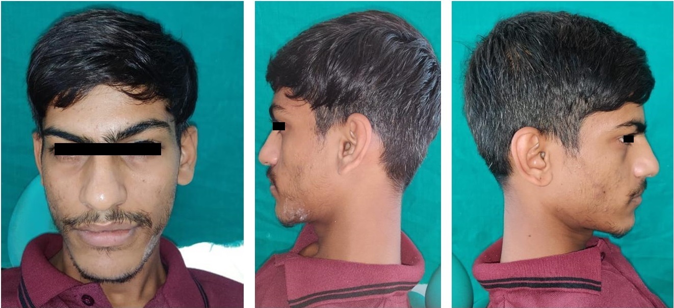
Fig 1: Nodular swelling below left eyelid with facial asymmetry.

Fig 2: Oro-antral communication present on left side of palate

Fig 3: Pre operative CT radiograph on left side shows hyperdensity.

Fig 4: Post operative OPG of patient shows hemi maxillectomy done on left side.
DISCUSSION
Based on the morphology of the mycelium and the manner of spore generation, the kingdom of fungi is separated into numerous classes, including Phycomycetes, Oomycetes, Zygomycetes, Deuteromycetes, Ascomycetes, and Basidiomycetes. Zygomycetes, a group of fungi that develop branching, ribbon-like hyphae and reproduce sexually by producing zygospores, are responsible for a variety of illnesses that are referred to as mucormycosis. In addition to being widely distributed in fruits, soil, and human waste, pathogens can also be cultivated in the throat, nasal passages, and oral cavity of healthy, disease-free people. Zygomycetes' subtype, Mucorales, causes a specific pattern of clinical infection. The fungi are typically non-pathogenic and only become so when the host resistance is extremely low. In the maxillofacial region, mucormycosis can enter through an extraction wound in the mouth or an ulcer in the mucosa, especially if the patient is immunocompromised.4-7
The treatment of mucormycosis is mainly based on antifungal drugs and surgical debridement. Morbidity and mortality of mucormycosis are high; hence the importance of early and appropriate management is required for the treatment of impairments of the senses, such as taste, smell, and hearing, along with functional disabilities and compromised esthetics. Maxillectomy adversely affects not only function and aesthetics but also results in emotional disturbances.5-8
The case presented with a common history of diabetes and COVID-19 infection that warranted extensive surgical resection of the maxilla, alveolar process, teeth, soft tissue along with complications like microstomia. We have presented a case of post surgical morbidity caused due to surgical treatment of mucormycosis associated with COVID -19 which according to authors have not been discussed earlier
Mucormycosis, which is brought on by fungus of the Zygomycetes class, is the third most serious invasive mycosis after candidiasis and aspergillosis.4 Rhizopus arrhizus is the most significant species in order of frequency (oryzae). Mucormycosis can be classified into at least six clinical groups based on the clinical presentation and the involvement of a specific anatomic site: I rhinocerebral, (ii) pulmonary, (iii) cutaneous, (iv) gastrointestinal, (v) disseminated, and (iv) miscellaneous. In their retrospective investigation over ten years in India, Chakrabarti et al. found that the rhino-orbito-cerebral type (44.2%) was the most frequent presentation, followed by cutaneous (15.5%) and renal (14.0%) involvement.5 A susceptible person inhales the fungus, which triggers the onset of the rhinomaxillary form of the disease, a subdivision of the rhinocerebral type.6 The initial line of defence, mononuclear and polynuclear phagocytes, are able to neutralise the spores once they have entered the lungs or subcutaneous tissues.4 By producing oxidative metabolites and defensins (cationic peptides), the phagocytes of the healthy host can kill Mucorales spores.4
Ketoacidosis brought on by uncontrolled diabetes mellitus can change a patient's immune system's natural response to infections. These individuals exhibit altered polymorphonuclear leukocyte response and reduced granulocyte phagocytic capacity.7 Rhizopus arrhizus can use the ketone bodies produced by diabetes people because they produce the enzyme ketoreductase.8 The release of iron bound to proteins may also contribute to the increased risk of mucormycosis in patients with ketoacidosis.
Consistent symptoms, sinusitis or periorbital cellulitis, and facial numbness are brought on by fungal invasion of the oronasal cavity or paranasal sinuses of a susceptible host. These symptoms are then followed by the development of conjunctival suffusion, blurred vision, and soft tissue swelling, which leads to the necrosis of the nasofacial region. A rapidly progressing infection typically moves from the ethmoid sinus to the orbit, causing proptosis with obvious chemosis, loss of extraocular muscle function, cavernous sinus thrombosis, carotid artery thrombosis, or jugular vein thrombosis (Lemierre syndrome), and eventually death. A radiographic examination, especially a CT scan of the maxilla and orbit, is required to confirm a clinical suspicion of mucormycosis if bone disruption and membrane or periosteal thickening are present.9,10
Imaging findings could be general and could show unilateral or bilateral para nasal sinus inflammatory alterations, like thickening of the polypoid mucosa. In CT scans, foci of hyperdensity in the afflicted sinus strongly imply a fungus-related condition. Sinonasal mycosis can be diagnosed by CT with 100% sensitivity and 78% specificity. The most prevalent opportunistic infections in immunocompromised patients are phycomycetes and Aspergillus. Hence, we must distinguish between aspergillosis and mucormycosis. Aspergillosis exhibits radiological concretions on radiographs, whereas mucormycosis exhibits opacification as in the present case. An infected tissue sample is used to make the diagnosis. The distinctive wide, ribbon-like, aseptate hyphal components with right-angled branching should be visible in the biopsy. Many pieces of massive necrotic material frequently surround the organisms.11,12
Rapid identification, reversal of the underlying predisposing conditions (if possible), adequate surgical debridement of infected tissue, and suitable antifungal medication are essential for eliminating mucormycosis. Early diagnosis allows for the combination of surgical removal of the diseased region and systemic Amphotericin B treatment for up to three months to cure mucormycosis.6 The proper management of the underlying disease is a critical factor impacting the treatment's eventual result. Treatment for mucormycosis has also included hyperbaric oxygen therapy. High morbidity and death are the prognosis; however, it may improve with quick diagnosis, early treatment, and reversible underlying risk factors. If the infection extends to the brain, case fatality ratios surpass 80%. Survival rates among groups of patients with invasive sinus illness without cerebral involvement may be as high as 50-80%. The authors suggest that in order to diagnose and treat patients early, oral physicians should be informed of innovative viewpoints on the disease.12-16
CONCLUSION
Mucormycosis is an opportunistic fulminant fungal infection, which has the ability to cause significant morbidity and mortality in the patients. The authors propose that oral health care workers should be aware of the novel perspectives of the disease for early diagnosis and management.
REFERENCES
- Viterbo S, Fasolis M, Garzino-Demo P, Griffa A, Boffano P, Iaquinta C, et al. Management and outcomes of three cases of rhinocerebral mucormycosis. Oral Surg Oral Med Oral Pathol Oral Radiol Endod. 2011;112:e69–74. [PubMed] [Google Scholar]
- Torres-Narbona M, Guinea J, Muñoz P, Bouza E. Zygomycetes and zygomycosis in the new era of antifungal therapies. Rev Esp Quimioter. 2007;20:375–86. [PubMed] [Google Scholar]
- Goel S, Palaskar S, Shetty VP, Bhushan A. Rhinomaxillary mucormycosis with cerebral extension. J Oral Maxillofac Pathol. 2009;13:14–7. [PMC free article] [PubMed] [Google Scholar]
- Salisbury PL, 3rd, Caloss R, Jr, Cruz JM, Powell BL, Cole R, Kohut RI, et al. Mucormycosis of the mandible after dental extractions in a patient with acute myelogenous leukemia. Oral Surg Oral Med Oral Pathol Oral Radiol Endod. 1997;83:340–4. [PubMed] [Google Scholar]
- Neville WB, Damm D, Allen CM, Bouquot JE. 2nd ed. Philadelphia: W.B.: Saunders; 2001. Text Book of Oral & Maxillofacial Pathology; p. 16. [Google Scholar]
- Mohindra S, Mohindra S, Gupta R, Bakshi J, Gupta SK. Rhinocerebral mucormycosis: The disease spectrum in 27 patients. Mycoses. 2007;50:290–6. [PubMed] [Google Scholar]
- Bakathir AA. Mucormycosis of the jaw after dental extractions: Two case reports. Sultan Qaboos Univ Med J. 2006;6:77–82. [PMC free article] [PubMed] [Google Scholar]
- Marx RE, Stern D, editors. Carol Stream III, USA: Quintessence Publishing; 2003. Inflammatory, Reactive and Infectious Diseases in Oral and Maxillofacial Pathology; pp. 104–6. [Google Scholar]
- Kajs-Wyllie M. Hyperbaric oxygen therapy for rhinocerebral fungal infection. J Neurosci Nurs. 1995;27:174–81. [PubMed] [Google Scholar]
- Lador N, Polacheck I, Gural A, Sanatski E, Garfunkel A. A trifungal infection of the mandible: Case report and literature review. Oral Surg Oral Med Oral Pathol Oral Radiol Endod. 2006;101:451–6. [PubMed] [Google Scholar]
- Artis WM, Fountain JA, Delcher HK, Jones HE. A mechanism of susceptibility to mucormycosis in diabetic ketoacidosis: Transferrin and iron availability. Diabetes. 1982;31:1109–14. [PubMed] [Google Scholar]
- Castrejon-Perez A, Welsh EC, Miranda I, Ocampo-Candiani J, Welsh O. Cutaneous Mucormycosis. Bras Dermatol. 2017;92:304–11. [PMC free article] [PubMed] [Google Scholar]
- Spellberg B, Edwards J, Jr, Ibrahim A. Novel perspectives on mucormycosis: Pathophysiology, presentation, and management. Clin Microbiol Rev. 2005;18:556–69. [PMC free article] [PubMed] [Google Scholar]
- Kim J, Fortson JK, Cook HE. A fatal outcome from rhinocerebral mucormycosis after dental extractions: A case report. J Oral Maxillofac Surg. 2001;59:693–7. [PubMed] [Google Scholar]
- Doni BR, Peerapur BV, Thotappa LH, Hippargi SB. Sequence of oral manifestations in rhino-maxillary mucormycosis. Indian J Dent Res. 2011;22:331–5. [PubMed] [Google Scholar]
- Burkets Textbook of Oral Medicine Diagnosis and Treatment. 10th edition. Canada: Elsevier; 2003. p. 79. [Google Scholar]
Disclaimer from Guident
The views and opinions expressed in articles published in Guident are solely those of the authors and do not necessarily reflect the views of the editors, the editorial board or the publisher, Ivory India. Guident does not endorse any product, technique or treatment described. Clinical content is intended for qualified dental and medical professionals and is not a substitute for individual clinical judgement. Guident, its editors and the publisher accept no liability for any consequence arising from the use of information published in the journal.
How to cite this article
Dr. Swati Dixit, Dr. Vinay Mohan, Dr. Anuj Gaur. Mucormycosis in COVID-19 Patient: A Case Report with Review. Guident. 2023 Oct;16(11). Available from: https://ivoryindia.in/articles/oral-medicine/mucormycosis-in-covid-19-patient-a-case-report-with-review.html