Giant Cell Tumor of Mandible: A Case Report
Wisdom Dental Clinics ®, Jaipur, Department of Periodontics, Institute of Dental Sciences, Bareilly, Uttar Pradesh, India
ABSTRACT
Giant cell tumor (GCT) of bone is a distinctive neoplasm characterized by abundance of multinucleated giant cells scattered throughout the stroma of mononuclear cells. Its importance lies in recognizing and differentiating the characteristic histology, which at times may mimic several other bone tumors and endocrine disorders ranging from locally aggressive giant cell granulomas to hyperparathyroidism to malignant tumors. The jaw bones account for less than 1% of this lesion. We present a rare case of GCT of the mandible which occurred in a 19 year old male.
Key words: Giant cell tumor, mandible, mononuclear cells
INTRODUCTION
Jaffe and Lichetenstein first described the lesion in the early 1940 in studies of unicameral bone cysts. They are considered as pseudocyst because of lack of epithelial lining.1 The GCT’s had been described under a variety of names viz: haemorrhagic osteomyelitis, ossifying haematoma, osteitis fibrosa cystica, atypical subperiosteal giant cell tumor, aneurysmal giant cell tumor, hemangiomatous bone cyst, subperiosteal bone aneurysm, expansile haemangioma and pulsating giant cell tumor.2
The incidence of these tumours in facial bone is infrequent, with a 2–12% of all giant cell tumours of the body.3 In case of craniofacial location, the mandible is more frequently affected than the maxilla with a proportion from 2:1 to 11:9.4,5 The body and the mandibular ramus are the main location with rare case reports in the coronoid process and the mandibular condyle.5
The age of presentation is the second or third decades of life.6 The median age of diagnosis is 13 years. 80% of the patients are under the age of 20, with greater sex predilection in female (62%).7 Although GCT is a benign lesion, it can behave locally in an aggressive manner because of its rapid growth and osteolytic capacity.
The best and the most accepted theory of relationship between GCT, Central giant cell granuloma (CGCG), and traumatic bone cyst (TBC) is presented by Hillerup and Hjorrting-Hansen,8 who proposed that these lesions are different manifestations of the same general process, the cause of which is a “vascular mishap” resulting from trauma, primary bone disease or malformation. The rapid growth may result in the erosion of the cortical plates of an asymptomatic slow growth lesion that then becomes symptomatic.1,3 The multilocularity with soap bubble or honey comb appearance radiographically, should be differentiated with ameloblastoma, giant cell granuloma, ossifying fibroma, sarcomas. The histologic features consist of a fibrous connective tissue stroma containing many cavernous or sinusoidal blood filled spaces. Surgical curettage and excision have been the treatment of choice. Various modalities have been used in the treatment of GCT including enucleation, curettage, cryotherapy, radiation, resection, amputation.9
CASE REPORT
A 19-year-old healthy male patient reported with chief complaint of swelling in front region of lower jaw, which has come to patient’s notice around 2 months back. (Figure 1) There was no past history of trauma, pain and mobility.

Figure 1 - Pre- operative image of patient
On oral examination, there was hard bony swelling i.r.t, lower anteriors (31,32,41,42) extending both buccally and lingually below tongue. Also, the roots of anterior teeth which are associated with the lesion, have migrated apart.
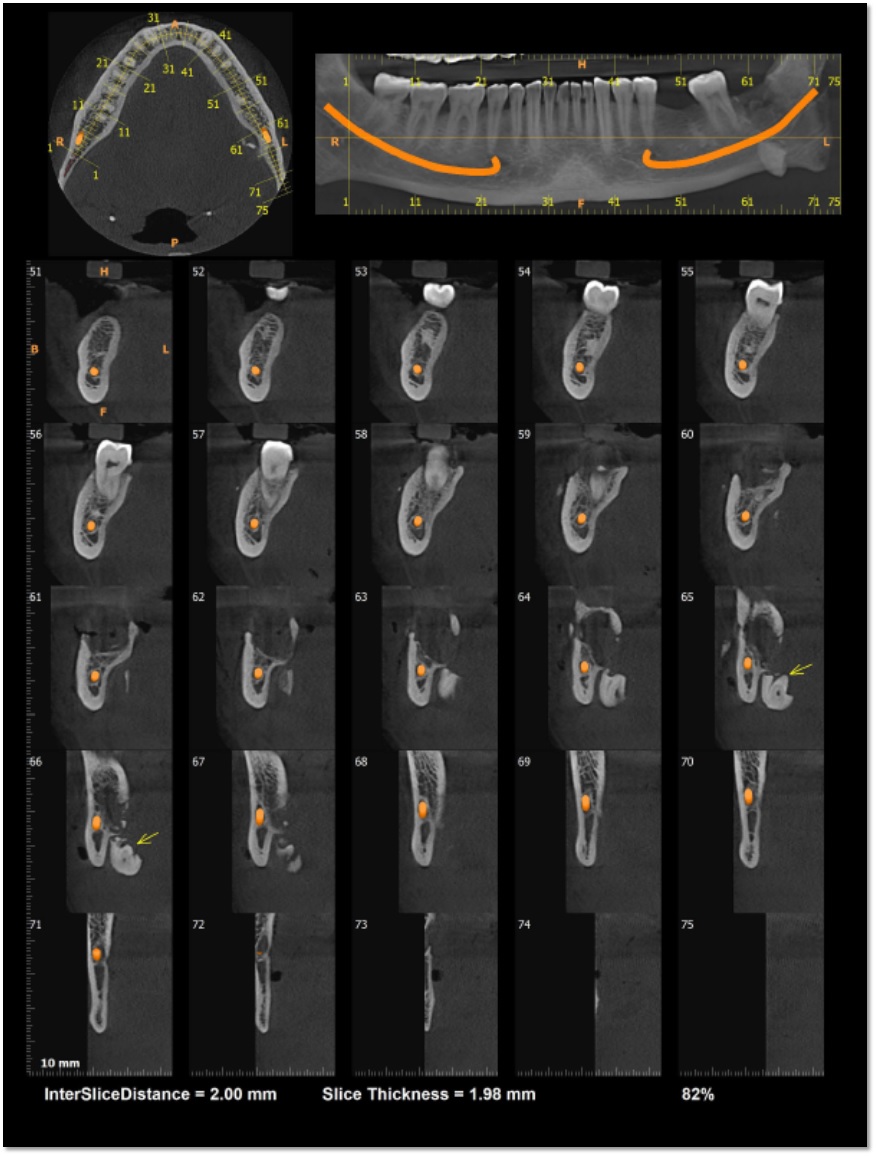
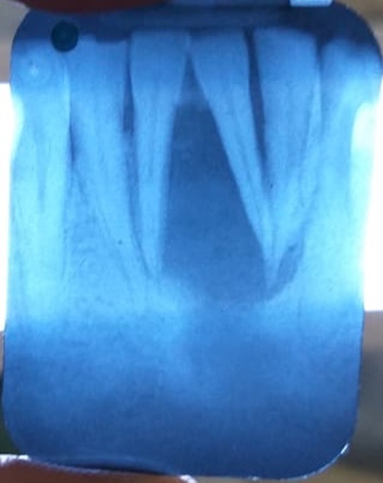
Patient was advised for OPG. (Figure.2) OPG revealed radiolucent lesion irt 31,32, 41, 42 which was inconclusive, so advised for 3d – CT scan. CT scan revealed marked localized expansile growth of the lesion both bucally and lingually, indicating its aggressive nature.

Figure 2 :OPG
Hence patient was advised for treatment which include enucleation of the lesion with all the four anterior teeth. Healing will be by secondary intention with periodic dressing by iodoform gauze.
The procedure is performed under local anesthesia. A crevicular incision was given from lower left canine to right canine (33,32,31,41,42,43) and a vestibular incision is given in buccal and lingual canine region. Subperiosteal flap was raised exposing the pathology. (Figure 3a, b)

Figure 3a,3b: Crevicular and vestibular incision given)
The lesion was separated from surrounding bone and soft tissue with the help periosteal elevators and curettes completely and then sent for biopsy.(Figure.4a,b,c,d)

Figure 4a,4b,4c,4d: Lesion separated from surrounding bone and sent for biopsy
Iodoform dressing was placed for healing of the wound with some sutures so to retain the dressing at that region. (Figure 5a,5b)

Figure 5: Iodoform dressing and sutures)
Dressing was changed every 10th day and after 3 months the wound healed uneventfully. (Figure 6a,6b)

Figure 6a,6b : Post operative images
DISCUSSION
GCT makes up 4.9–5% of all osseous neoplasms, 21.87% of the benign tumors of bone and head and neck region accounts for 2% of all GCTs. Most of the lesions occur in the ethmoid, sphenoid and temporal bones; the occurrence in the mandible is less than 2%. The case being discussed involves the mandible.
The age distribution of GCT is around 20–40 years. The sex distribution is 1.3:1 female:male. The case being discussed occurred in a 19-year-old male. This is in concurrence with the cases reported to have occurred in jaw bones in the literature (based on PubMed search engine). The symptoms were pain, swelling of the affected region, weakness and limitation of the jaw movement associated with elicitation of moderate tenderness and eggshell crackling.10 It may cause pathologic fracture or atrophy of the muscles. The case was painless with swelling of the affected region without limitation of the jaw movements. Eggshell crackling was elicited, but the case did not exhibit tenderness, pathologic fracture or atrophy of muscles.
The typical radiologic features are an expanding zone of radiolucency either eccentric or central with either cortical thinning or involvement of the soft tissue without a sclerotic margin. It may be either well or poorly marginated.11 Similar radiologic features are observed in our case which had a multilocular radiolucency with poor margination evincing a stretching of the lower border of the mandible.12
The gross appearance exhibits chocolate brown or greyish white foci with yellowish discoloration interspersed with hemorrhagic or cystic areas with a soft and friable consistency.
The differential diagnoses include central giant cell granuloma (CGCG), aneurysmal bone cyst, hyperparathyroidism and other giant cell lesions.
Though many options have been performed for the similar case but concerning the treatment, the gold standard is still the surgical excision and curettage of the cavity.13 There have been various treatment modalities used in the treatment of GCT, including enucleation, curettage, cryotherapy, radiation, resection.9 The gold standard of surgical enucleation and curettage was used in our case. Curettage used in combination with cryotherapy may reduce the rate of recurrence in nonfacial bones, which has been reported to be greater than 50% when curettage alone is used.9 Radiation has been used as a therapeutic modality, but the subsequent development of sarcoma is possible and has been reported. Segmental resection must be done only in case of multiple recurrences or extension to overlying tissues.1,3,4 The effort should be expended preoperatively to ensure that the lesion treated is GCT. Bleeding is expected and may be severe, therefore wide exposure for access to afferent vessels, rapid curettage of the lesion, and the liberal use of gauze packing and hemostatics should be employed. Recurrence rates range from 20 to 30% according to different sizes and seems to occur most frequently within the first year of surgery.3,4
CONCLUSION
Numerous bone tumors have multinucleated giant cells that must be distinguished from conventional GCT. These range from benign lesions such as ossifying fibroma to locally aggressive lesions like CGCG, aneurysmal bone cyst, high-grade sarcomas and also metabolic disorders such as hyperparathyroidism which is disguised by masses of reactive osteoclast-like giant cells.
Careful histopathological examination is emphasized on with exclusion of other possible lesions to arrive at the appropriate diagnosis. The case being discussed was in a 19-year-old male, the age being concurrent with cases occurring in the jaw and in accordance with the female predilection usually observed. Eggshell crackling was elicited, but there was no associated tenderness. Radiologic features were also concurrent with a multilocular radiolucency with poor margination. Histopathologically, the even distribution of giant cells throughout the cellular stroma composed of hyperchromatic and pleomorphic cells, the giant cells with central aggregation of nuclei close to 25–50 all led to the diagnosis being made in favor of GCT of the mandible.
REFERENCES
- Kiateeavorncharoen S, Joos U, Brinkschmidt C, Werkmeister R. Aneurysmal bone cyst of the mandible a case report of the mandible. Int PediatrOtorhinollaryngol. 2004;68:1339–1344. doi: 10.1016/j.ijporl.2004.05.001. [CrossRef]
- Becker R(1973) Zystenim Kiefer und Gesichtbereich. Paxis der Zahnheilkunde and 2. Urban und S chwarzenberg, Munchen, Berlin, Vein
- Rapidis AD, Vallianatou D, Apostolidis C, Lagogiannis G. Large lytic lesions of the ascending ramus, the condyle and the infratemporal region. J Oral Maxillofac Surg. 2004;62:996–1001. doi: 10.1016/j.joms.2004.04.009. [PubMed] [CrossRef]
- Bataineh AB. Aneurysmal bone cyst of the maxilla. A clinicopathologic review. J Oral Maxillofac Surg. 1997;55:1212–1216. doi: 10.1016/S0278-2391(97)90169-5. [PubMed] [CrossRef]
- Motamedi MH. Destructive aneurysmal bone cyst of the mandibular condyle:report of a case and review of literature. J Oral Maxillofac Surg. 2002;60:1357–1361. doi: 10.1053/joms.2002.35744. [PubMed] [CrossRef]
- Auclair LP, Arendt DM, Hellstein JW. Giant cell lesions of the jaws. Surgical pathology: Fibroosseous diseases. Oral Maxillofac Surg Clin North Am. 1997;9:655–678.
- Lopez-Arcas JM, Cebrian L, Gonzalez J, Burgueno M. Aneurysmal bone cyst of the mandible: case presentation and review of literature. Med Oral Patol Oral Cir Bucal. 2007;12:401–403. [PubMed]
- Hillerup S, Hjorting-Hansen E. Aneurysmal bone cyst-simple bone cyst, two aspects of the same pathological entity? Int J Oral Surg. 1978;7:16. doi: 10.1016/S0300-9785(78)80005-2. [PubMed] [CrossRef]
- Biesecker JL, Marcove RC, Huvos AG, et al. Aneurysmal bone cysts. Cancer. 1970;26:615–625. doi: 10.1002/1097-0142(197009)26:3<615::AID-CNCR2820260319>3.0.CO;2-I. [PubMed] [CrossRef]
- Pradhan E, Shrestha JK, Karmacharya PC. An unusual presentation of giant cell tumour (osteoclastoma).Kathmandu Univ Med J (KUMJ) 2003;1:190-2.
- Lanza A, Laino L, Rossiello L, Perillo L, Ermo AD, Cirillo N. Clinical practice: Giant cell tumour of the jaw mimicking bone malignancy on three-dimensional computed tomography (3D CT) reconstruction. Open Dent J 2008;2:73-7.
- Park SR, Chung SM, Lim JY, Choi EC. Giant cell tumor of the mandible. Clin Exp Otorhinolaryngol2012;5:49-52.
- Hernandez GA, Castro A, Castro G et al (1993) Aneurysmal bone cyst versus hemangioma of the mandible: Report of long term follow up of a self limiting case. Oral Surg Oral Med Oral Pathol 76:790.
Disclaimer from Guident
The views and opinions expressed in articles published in Guident are solely those of the authors and do not necessarily reflect the views of the editors, the editorial board or the publisher, Ivory India. Guident does not endorse any product, technique or treatment described. Clinical content is intended for qualified dental and medical professionals and is not a substitute for individual clinical judgement. Guident, its editors and the publisher accept no liability for any consequence arising from the use of information published in the journal.
How to cite this article
Dr. Gaurav Gupta, Dr. D.K. Gupta, Dr. Neeraj Chandra. Giant Cell Tumor of Mandible: A Case Report. Guident. 2023 May;16(6). Available from: https://ivoryindia.in/articles/oral-surgery/giant-cell-tumor-of-mandible-a-case-report.html