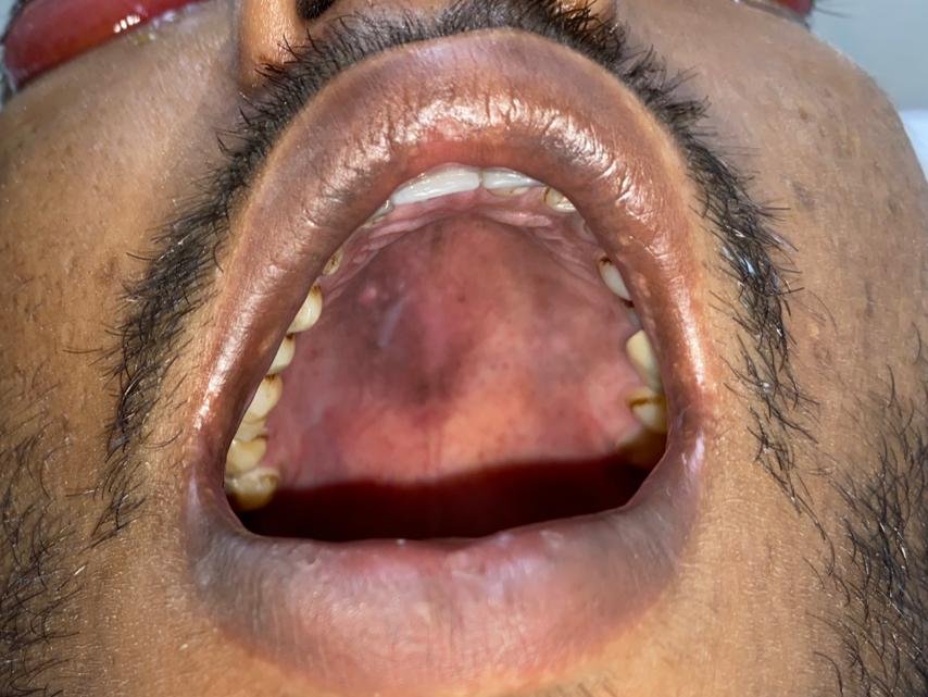
Lateral Periodontal Cyst Treated with Enucleation and Guided Bone Regeneration-a Case Report
Department of Oral and Maxillofacial Surgery, Department of Oral Medicine & Radiology, Faculty of Dental Sciences PDM University, Bahadurgarh
Odontogenic cysts are classified by the World Health Organization as inflammatory and developmental according to their epithelial lining. Lateral periodontal cysts (LPC) have been regarded as an independent condition.1
LPC is classified as an odontogenic cyst of developmental origin.2 Lateral periodontal cyst (LPC) is a noninflammatory cyst on the lateral surface of the root of a vital tooth.3 The etiology of LPC is unclear and the cell rests of Malassez, reduced enamel epithelium or remnants of dental lamina were suggested to cause formation and development of LPC.4,5 LPC was reported not to have a predilection for any race.6 LPC occurs more often in the mandible.7
The patients with LPC are generally asymptomatic and LPC is often identified during routine radiographical examination. It appears radiographically as Unilocular well defined a round or oval or tear drop, uniform radiolucency with well?defined borders <1 cm diameter.8
Histologically, the lateral periodontal cyst is a distinct type of developmental cyst characterized by a thin, nonkeratinized epithelium usually 1 to 5 cell layers thick, which resembles the reduced enamel epithelium. The epithelial lining exhibits focal thickenings or plaques, in which clear glycogen-containing epithelial cells have often been found. The connective tissue subjacent to the epithelium exhibits a zone of hyalinization.9
Untreated odontogenic cysts may cause root resorption, tooth displacement, expansion, and pain. Treatment of LPC, which provided that the lesion is unilocular on radiological examination, is by conservative surgical enucleation and following up the patient over a period of six months to one year, in order to monitor for recurrence. Nevertheless, simple surgical enucleation is associated with a residual bone defect which could potentially affect the periodontal health of the associated teeth.10 With the advent of tissue regeneration techniques in dentistry, several techniques including guided tissue regeneration (GTR) and guide bone regeneration (GBR) have been reported for the treatment of bone defects, following enucleation of LPC.11
The aim of this paper is to present a case of LPC treated with surgical enucleation and primary bone defect repair using GTR with Nova bone putty graft and collagen A-PRF, along with a recurrence-free follow-up period of one year.
CASE REPORT
A 27-year-old male patient reported at vivek oro dental care Bhiwani (Haryana) with a complaint of painless swelling in the mandibular Front region. Although the history revealed a presence of the swelling for more than two months without any associated symptoms, the patient preferred to obtain clinical consultation as he was concerned about the swelling. The patient had no previous history of dental treatment except for periodic oral prophylaxis.
Clinical and Radiographic Examination
Upon clinical examination, a well-circumscribed and fluctuant swelling, measuring about 9-12mm in diameter, was observed at the junction of the Labial attached and free gingival margins in between the mandibular incisors (31,41,32 ) (Figure 1). The swelling was nontender, and there was no dental focus of infection or inflammation in the associated teeth. Similarly, no abnormalities were detected clinically in the lingual aspect of the mandibular anterior teeth. Both teeth 31 and 41 were vital upon testing with a electric pulp testing. Extra oral examination revealed no clinically discernible asymmetry, swelling, or lymphadenopathy. Radiographic examination (IOPA) of the left mandibular body region revealed a well circumscribed radiolucency, with a sclerotic border in between the roots of teeth 31 and 41 as shown in FIGURE 1.
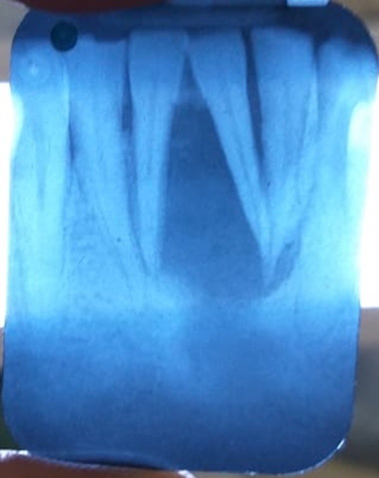
Figure -1 Pre-operative radiograph
Surgical Procedure and Follow-Up
Based on a clinical and radiographic diagnosis of a lateral periodontal cyst, the patient was consented for endodontic treatment, apicocetomy (Figure 2), enucleation and curettage of the cystic lesion and primary reconstruction of the defect site using guided bone regeneration. Following informed consent, under local anesthesia, the lesion was surgically approached through a gingival crevicular incision extending from tooth 33 to tooth 43(Figure-3). After mucoperiosteal flap elevation, an anterior vertical releasing incision was provided for optimum visualization was done superficial to the cystic wall in order to separate the mucoperiosteal flap without compromising its integrity.

Figure 2- Post Picture of endodontic treatment

Figure 3 – Flap Elevation
Following enucleation and curettage, the cystic cavity was thoroughly irrigated with sterile normal saline. Clinical examination revealed a bony wall with intact mucoperiosteum on the lingual aspect. The residual defect left behind by the cystic lesion was then filled with a Nova bone graft putty covered by A-PRF.(Figure-4) The mucoperiosteal flap was reapproximated and closed with Vicryl 4-0 interrupted. Postoperative radiographs revealed complete filling of the bone defect with graft material, and the patient was discharged with specific postoperative instructions. And antibiotic and analgesic medication. The immediate postoperative period was uneventful, and the surgical wound healed remarkably well. Follow-up examinations at six months and one year after cyst enucleation and guided bone regeneration reveal edno clinical or radiographic evidence of recurrence of the lesion. Moreover, teeth 33 and 34 were still vital during follow-up.

Figure 4- Placement of bone graft and A-PRF membrane
Post operative instructions
Patient was asked to maintain good oral hygiene, especially in the furcation area, because sites with minimal or no gingival inflammation have consistently better regenerative responses than sites with poor plaque control.12 Prescription For analgesic (Ibuprofen,600 mg every 6-8 hours for pain) and Amoxycillin (Thrice daily for 5 days) were given.
Follow up done at 7th day and suture was also removed at first week post operatively. At 1 month recall visit, healing was uneventful with the absence of mobility. Recall after 8 months indicated the absence of the periodontal ligament widening and bone formation at an extraction site. Healing of treated tooth was uneventful at recall visits. post-operative picture is shown below.

Figure 5- Follow up after 8 Months
CONCLUSION
Although LPC is an uncommon developmental odontogenic cyst, it should be considered a differential diagnosis in case of cystic lesions lateral to the surface of a tooth and without any associated inflammatory processes or symptoms. Unicystic LPC clinically and radiographically diagnosed based on the previously mentioned characteristic features can be managed with conservative surgical enucleation and GBR. The lateral periodontal cyst can be considered in the differential diagnosis when a radioloucent lesion appears adjacent to the roots of vital teeth. A histological study is essential in order to confirm the diagnosis. The treatment of choice is surgical removal and subsequent histological evaluation to confirm the diagnosis. Relapses are infrequent.
However, further long-term clinical studies in larger sample sizes should be mandated to as certain the efficacy of this treatment modality. Nevertheless, the role of HPE after surgical enucleation should not be discounted and histopathological con?rmation of diagnosis serves as the benchmark for a recurrence-free follow-up period.
REFRENCES
- Shear M. Developmental odontogenic cysts. An update. J Oral Pathol Med. 1994 Jan;23(1):1-1.
- M. Soluk-Tekkesin and J. M. Wright, “The World Health Organization Classi?cation of odontogenic lesions: a summary ofthechangesofthe2017 (4th) edition,”Turk Patoloji Dergisi, vol. 34, no. 1, 2018.
- Kramer IR, Pindborg JJ, Shear M. WHO Histological Typing of Odontogenic Tumours. 2nd ed. Geneva: Springer-Verlag; 1992. p. 34-118.
- Demirkol M, Ege B, Yanik S, Aras MH, Ay S. Clinicopathological study of jaw cysts in southeast region of Turkey. Eur J Dent 2014;8:107-11.
- Cohen DA, Neville BW, Damm DD, White DK. The lateral periodontal cyst. A report of 37 cases. J Periodontol 1984;55:230-4.
- Cohen DA, Neville BW, Damm DD, White DK. The lateral periodontal cyst. A report of 37 cases. J Periodontol 1984;55:230-4.
- Soares de Lima AA, Naval Machado MÂ, Braga AM, De Souza MH. Lateral periodontal cyst: Aetiology, diagnosis and clinical significance. A review and report of case. Rev de Clín Pesqui Odontol 2005;1:55-9
- Altini M, Shear M. The lateral periodontal cyst: An update. J Oral Pathol Med 1992;21:245-50.
- Altini M, Shear M. The lateral periodontal cyst: an update. J Oral Pathol Med. 1992 Jul;21(6):245-50.
- S. Sivolella, C. Perin, M. Capecchi, V. Buongiorno, and M. Valente, “Guided bone regeneration in the treatment of a lateral periodontal cyst: 2-year clinical and radiologic follow up,” The International Journal of Periodontics & Restorative Dentistry, vol. 38, no. 5, pp. 747–754, 2018
- S.E.Meseli, O.B.Agrali, O.Peker, and L.Kuru, “Treatment of lateral periodontal cyst with guided tissue regeneration,” European Journal of Dentistry, vol. 8, no.3, pp. 419–423,2014.
- Machtei EE, Cho MI, Dunford R, Norderyd J, Zambon JJ, Genco RJ. Clinical, microbiological, and histological factors which influence the success of regenerative periodontal therapy. J Periodontol 1994;65:154-161
Disclaimer from Guident
The views and opinions expressed in articles published in Guident are solely those of the authors and do not necessarily reflect the views of the editors, the editorial board or the publisher, Ivory India. Guident does not endorse any product, technique or treatment described. Clinical content is intended for qualified dental and medical professionals and is not a substitute for individual clinical judgement. Guident, its editors and the publisher accept no liability for any consequence arising from the use of information published in the journal.
How to cite this article
Dr. Vivek Munjal, Dr. V. Raj Kumar, Dr. Jyotsana. Lateral Periodontal Cyst Treated with Enucleation and Guided Bone Regeneration-a Case Report. Guident. 2022 Apr;15(5-6). Available from: https://ivoryindia.in/articles/oral-surgery/lateral-periodontal-cyst-treated-with-enucleation-and-guided-bone-regeneration-a-case-report.html