From Dependence to Independence-Management of Mucormycosis with Preservation of Palatal Mucosa: A Case Report
Department of Oral and Maxillofacial Surgery, Sri Ramachandra Institute of Higher Education and Research, Chennai India
ABSTRACT
Globally, mucormycosis represents the third most common angioinvasive fungal infection. Successful management of this invasive infection requires early diagnosis and aggressive surgical interventions to prevent disease progress. Aggressive procedures such as total maxillectomy results in oronasal communication which affects the quality of life of the patient. On recovery after surgery, difficulty in speech and swallowing affects the psychological well-being of the patient necessitating the need of an immediate surgical obturator. However, maintenance of these removable obturators is still a plight for patients. We present a 30year old patient who was diagnosed with rhino orbital mucormycosis. patient had undergone endoscopic endonasal debridement of nose and paranasal sinuses under general anaesthesia followed by medical therapy with liposomal amphotericin B to a cumulative dose of 4g. he presented with severe bilateral chemosis, blurred vision in relation to right eye and tenderness on palpation over right zygomatic buttress region were noted. Intraoral examination revealed palatal ecchymosis in relation to 14-16 teeth region. Patient was planned for surgical debridement and endoscopic endonasal debridement of nose and paranasal sinuses under general anaesthesia. On postoperative day 1, patient was restarted on liposomal amphotericin B and was administered a cumulative dose of 7400 mg. Postoperative MRI of bilateral orbit and paranasal sinus showed complete resolution of infection. On one month follow-up, bilateral chemosis had resolved and surgical site healing was satisfactory. Patient still complained of blurred vision in relation to right eye. Patient is currently on a three month follow up. In conclusion, maintaining oronasal separation with the help of palatal mucoperiosteum following maxillary resection guarantees improved palatal function, adequate support for rehabilitation and minimizes complications with a good overall prognosis.
INTRODUCTION
Mucormycosis is a life threatening, rapidly progressing, invasive fungal infection, seen associated with nasal cavity and paranasal sinuses following inhalation of fungal spores. Globally, mucormycosis represents the third most common angioinvasive fungal infection.
Successful management of this invasive infection requires early diagnosis and aggressive surgical interventions to prevent disease progress.
Aggressive procedures such as total maxillectomy results in oronasal communication which affects the quality of life of the patient. On recovery after surgery, difficulty in speech and swallowing affects the psychological well-being of the patient necessitating the need of an immediate surgical obturator. However, maintanence of these removable obturators is still a plight for patients.
We present case report of a patient diagnosed with rhino orbito cerebral mucormycosis who was treated by surgical debridement of maxilla under general anaesthesia with preservation of palatal mucosa thereby reducing postoperative morbidity.
CASE HISTORY AND CLINICAL PRESENTATION
A 30 year old male patient admitted under Department of General Medicine for medical management of Rhino-Orbito-Cerebral mucormycosis was referred to the Department of Oral and Maxillofacial Surgery in view of pain in relation to right upper back teeth region.
Patient was diagnosed with rhino orbital mucormycosis elsewhere one month ago where patient had undergone endoscopic endonasal debridement of nose and paranasal sinuses under general anaesthesia followed by medical therapy with liposomal amphotericin B to a cumulative dose of 4g.
Patient then reported to Department of General Medicine at our hospital where patient had undergone bilateral orbital decompression, and medical therapy with liposomal amphotericn B.
On routine assessment, patient was diagnosed with Type 2 Diabetes Mellitus and was started on oral hypoglycaemic agents.
MRI of bilateral orbit and paranasal sinuses taken after medical management with liposomal amphotericin revealed persistent mucosal thickening in right maxillary sinus, bilateral ethmoid, sphenoid and frontal sinuses. Invasion of post-antral, premaxillary fat and masticator space on right side was noted. Left Basifrontal lobe abscess and right infraorbital canal invasion was also seen.
Department of Neurosurgery advised conservative management for the cerebral component of mucormycosis.
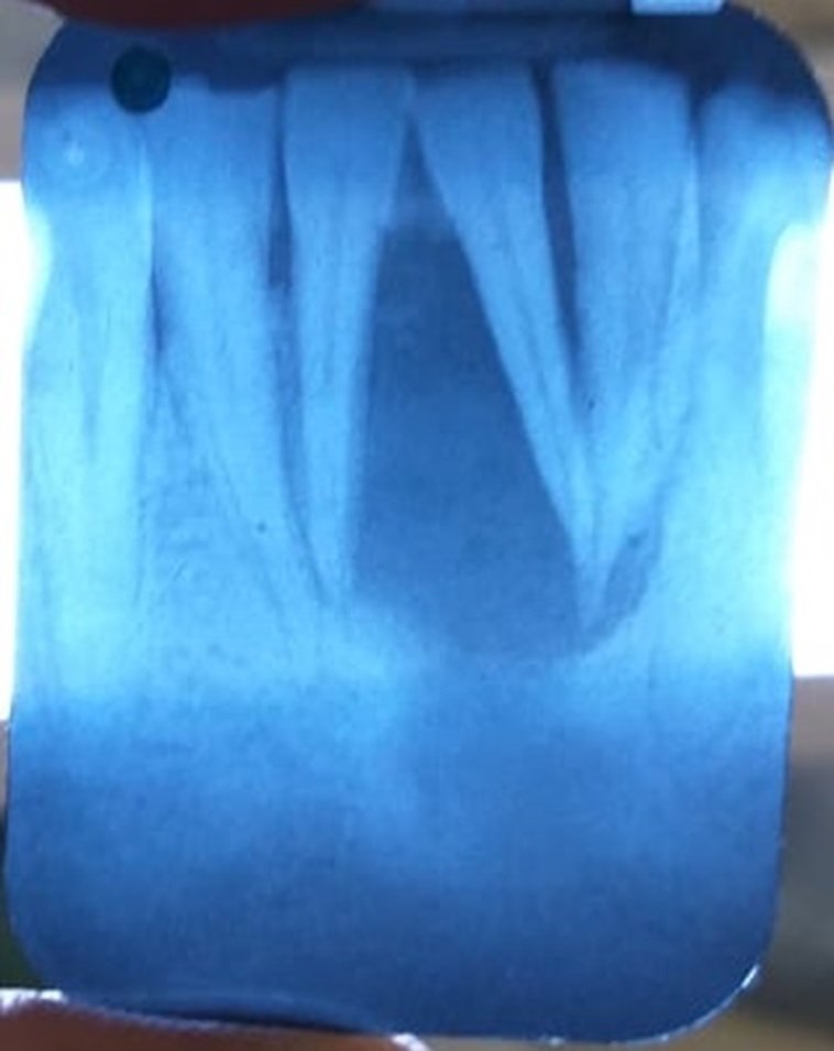
On examination, severe bilateral chemosis, blurred vision in relation to right eye and tenderness on palpation over right zygomatic buttress region were noted. Intraoral examination revealed palatal ecchymosis in relation to 14-16 teeth region. No signs of tooth mobility or pus discharge was noted.
Patient was planned for surgical debridement and endoscopic endonasal debridement of nose and paranasal sinuses under general anaesthesia.
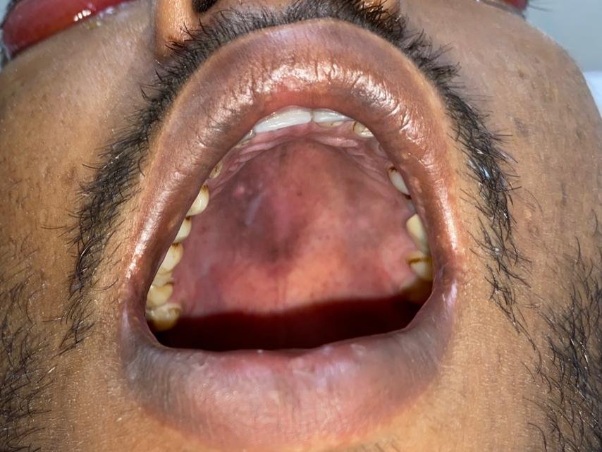
Fig 1
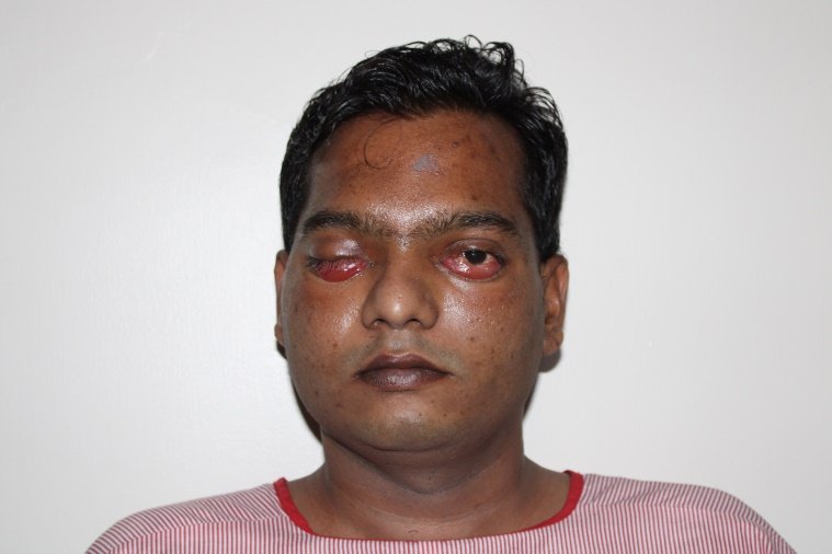
Fig 2
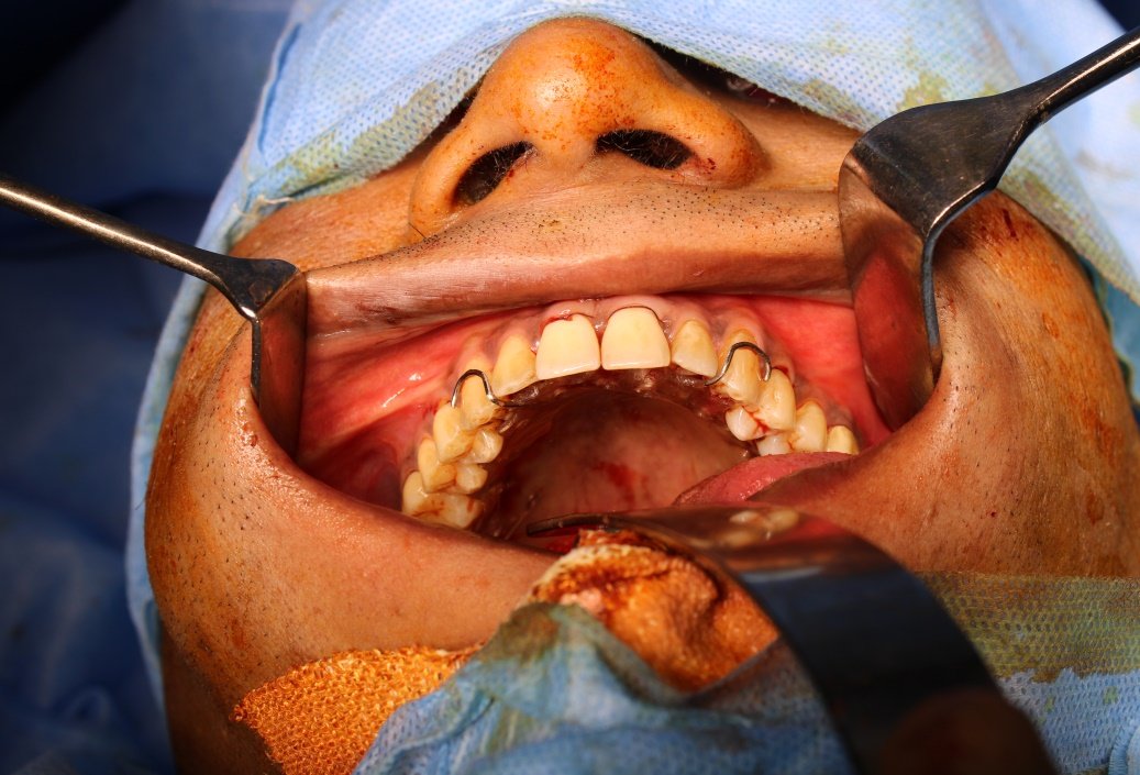
Fig 3
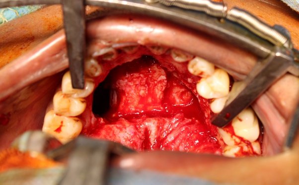
Fig 4
Crevicular incision was placed from 16-26 teeth and palatal flap elevated. Debridement of right maxilla was done until fresh bone bleed was noted. Gauze pack impregnated with conventional amphotericin was placed in to the defect. Palatal mucosa was then sutured in position using 4-0 vicryl .A palatal plate was then placed to counter any postoperative haematoma.
On postoperative day 1, patient was restarted on liposomal amphotericin B and was administered a cumulative dose of 7400 mg.
Postoperative MRI of bilateral orbit and paranasal sinus showed complete resolution of infection.
On one month follow-up, bilateral chemosis had resolved and surgical site healing was satisfactory.Patient still complained of blurred vision in relation to right eye.
Patient is currently on a three month follow up.
ONE MONTH FOLLOWUP:

Fig 5
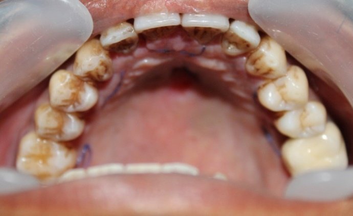
Fig 6
THREE MONTH FOLLOWUP:

Fig 7

Fig 8
DISCUSSION
Mucormycosis is an opportunistic fungal infection caused by Rhizopus oryzyae. It may be disseminated involving sinuses, nose, orbit, lung, central nervous system, jaw bones and gastrointestinal tract.
Globally incidence varies from 0.005 to 1.7 per million population, with the prevalence in India being 80 times higher than the other developed countries.
In a systemic review and meta-analysis published in 2018, death was reported in 46% of patients.
According to global epidemiologic studies of mucormycosis, it was found that India contributed to approx. 45% of new cases detected globally during the second wave of COVID 19.
Surge is seen higher in patients with pre-existing diabetes, those on systemic corticosteroids.
Early diagnosis and prompt initiation of appropriate antifungals are important for improving outcomes.
Biopsy and fungal staining (KOH mount) are the mainstay of laboratory diagnosis. Fungal culture and susceptibility testing help to confirm the diagnosis. Treatment should be initiated as soon as possible on diagnosis. Imaging tests such as a CT scan, may also be used to support the diagnosis.
Surgical management of mucormycosis varies from simple surgical debridement to aggressive resection of the involved bone.
Removal of tissue harboring the fungal elements halts the spread of infection. Being a relatively bloodless field, surgical debridement proceeds quickly.
Maxillary resection causes excessive mutiliation to the patient. Complete removal of the hard palate results in hyper nasal speech and masticatory difficulties. Oro antral communication can be a source of discomfort as food and fluids enter the sinus cavity thereby leading to sinusitis and otitis.
Preservation of palatal mucosa minimizes the unpleasant sequalae post-surgery thereby improving the psychological status of the patient. Postoperative nutrition, speech and healing are better when patient has a palatal mucosal cover.
CONCLUSION
Maintaining oronasal separation with the help of palatal mucoperiosteum following maxillary resection guarantees improved palatal function, adequate support for rehabilitation and minimizes complications with a good overall prognosis.
REFERENCES
- Leitner C, Hoffmann J, Zerfowski M, Reinert S. Mucormycosis: necrotizing soft tissue lesion of the face. J Oral Maxillofac Surg 2003;61(11):1354-1358.
- Pogrel MA, Miller CE. A case of maxillary necrosis. J Oral Maxillofac Surg 2003;61(4):489-493.
- Jones AC, Bentsen TY, Fredman PD. Mucormycosis of the oral cavity. Oral Surg Oral Med Oral Pathol 1993;75(4):455-460.
- Kofteridis DP, Karabekios S, Panagiotides JG, Bizakis J, Kyrmizakis D, Saridaki Z, Gikas A. Successful treatment of rhinocerebral mucormycosis with liposomal amphotericin B and surgery in two diabetic patients with renal dysfunction. J Chemother 2003;15(3):282-286.
- Chalian, VA, Drane JB, Standish SM. The evolution and scope of maxillofacial prosthetics. In: Chalian VA, Drane JB, Standers SM editors. Maxillofacial Prosthetics: Multidisciplinary Practice.Williams and Wilkins Company; Baltimore: USA, 1972.
- Beumer J, Curtis TA, Marunick MT. Maxillofacial rehabilitation: prosthodontic and surgical considerations. St Louis: Ishiyaku Euro America;1996. p. 225-229.
- Keyf F. Obturator prosthesis for hemimaxillectomy patients.J Oral Rehab 2001;28(9):821-829.
- Yoshida H, Furuya Y, Shimodaira K, Kanazawa T, Kataoka R, Takahashi K. Spectral characteristics of hypernasatity in maxillectomy patients. J Oral Rehab 2000;27(8):723-730.
- Sykes LM, Wolfaardt JF, Sukha A. Prosthodontic rehabilitation of a patient with total avulsion of the maxilla (a clinical report). J Prosthet Dent 2002;88(4):362-366.
- Haraguchi M, Mukohyama H, Taniguchi H. A simple method of fabricating an interim obturator prosthesis by duplicating the existing teeth and palatal form. J Prosthet Dent. 2006;95:469‑72.
- Jacob FJ. Clinical management of the edentulous maxillectomy patient. In Taylor TD. Clinical maxillofacial prosthetics. Chicago: Quintessence; 2000. pages 85‑7.
Disclaimer from Guident
The views and opinions expressed in articles published in Guident are solely those of the authors and do not necessarily reflect the views of the editors, the editorial board or the publisher, Ivory India. Guident does not endorse any product, technique or treatment described. Clinical content is intended for qualified dental and medical professionals and is not a substitute for individual clinical judgement. Guident, its editors and the publisher accept no liability for any consequence arising from the use of information published in the journal.
How to cite this article
Dr. A. Emmanuel D.S., Dr. C. Deepak, Dr. Rakhi R.. From Dependence to Independence-Management of Mucormycosis with Preservation of Palatal Mucosa: A Case Report. Guident. 2021 Dec;15(1-2). Available from: https://ivoryindia.in/articles/oral-surgery/from-dependence-to-independence-management-of-mucormycosis-with-preservation-of-palatal-mucosa-a-case-report.html