Giant Compound Odontoma-a Case Report
Department of Oral and Maxillofacial Surgery, , Annoor Dental College and Hospital, Muvattupuzha India
ABSTRACT
Odontomes are common benign tumors which arise from odontogenic tissues. They are classified into two types – Compound odotomes and Complex Odontomes absed on its morphology. Usually they are asymptomatic, but at later stages can present as a swelling or may become infected and cause pain. They are usually associated with unerupted permanent or primary teeth. Radiographic evaluation is diagnostic. Early surgical treatment is of paramount importance to minimize complications and further interventions. In this, we present a case report of a 19 years old male with Giant compound odontome in anterior maxilla.
INTRODUCTION
Broca first coined the term Odontoma in 1866.1 Odontomas are less aggressive, most prevalent benign tumors.1 They are developmental in origin and arise from odontogenic tissues.5 Morphologically they are subdivided into Complex and Compound odontomas.2 They are usually asymptomatic and are diagnosed on routine dental/radiographic examination as small, solitary, multiple radio-opaque lesions1,3 Rarely this lesions attain a larger size.5 They are radiographically and histologically characterized by the production of mature enamel, dentin, cementum as well as pulp.5 Early diagnosis and prompt treatment is adviced to prevent lengthy and expensive corrective procedures.4 Here we present a case of Giant Compound Odontoma in maxillary anterior region of a 19 years old male patient.
CASE REPORT
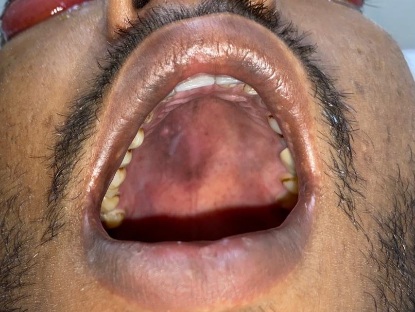
A 19-year-old male reported to the department of Oral and Maxillofacial Surgery, Annoor Dental College and Hospital, Muvattupuzha with Chief complaint of missing teeth and gap in upper left front tooth region. Patient gives no history of pain or swelling. There were no complaints in other facial issues, no history of trauma, and other health conditions were well-adjusted. On Extraoral examination, no abnormalities were detected. However on Intraoral examination, Permanent Maxillary left canine(23) was missing with a mild swelling above permanent maxillary first premolar (24) region .
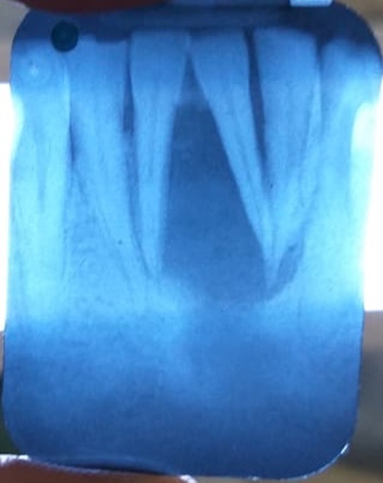
An orthopantamogram(OPG) was taken which shows multiple radio-opaque tooth like structures in the left maxilla. Also an impacted tooth was present above the lesion and associated mesiodens in relation to upper maxillary incisors.

It was provisionally diagnosed as Compound odontoma and was surgically removed under general anesthesia. The tumor was surgically removed using crestal incision extending from 21 to 25 region and a full thickness mucoperiosteal flap was elevated. Surgical site was exposed and 51 odontomes were removed along with the impacted left maxillary canine(23). Closure was done with 3-0 vicryl. Specimen sent for histopathological examination.

The histological examination was also done which revealed that the lesion was encapsulated with evidence of cementum and dentine like structures along with pulpal tissue and epithelial remnants.
Postoperatively follow up done after 1 week, 2 weeks, 1 month and 3 months clinically and radiographically(OPG). Healing is satisfactory and no evidence of recurrence noted.

DISCUSSION
Odontome’s are relatively common,hamartomous malformation usually associated with an impacted tooth.2 In 2005, WHO classified Odontomes into two types – Complex odontome and Compound Odontome.5
Compound odontoma usually contains multiple radiopaque, miniature tooth-like structures known as denticles of varying size and shape surrounded by a narrow radiolucent zone whereas complex odontoma consist of an irregular mass of hard and soft dental tissues surrounded by a narrow radiolucent band with a smooth outer periphery1,6
Compound odontomas are usually asymptomatic swelling which cause bony expansion.7 It may occur anywhere in the jaws but is commonly seen in maxillary canine/premolar region.5 They are usually detected before the age of 20 years and may be associated with a retained primary tooth or an unerupted primary/permanent tooth.3,9
Diagnosis is usually fone with the help of radiographic evaluation using orthopantomogram.8
The literature defines two treatment modalities one in which the impacted teeth is removed, the second option where the impacted teeth is orthodontically extruded into occlusion following removal of interference caused by odontome.8 In the case, we have removed the impacted tooth along with the lesion.
Surgical removal is the choice of treatment for this condition as its presence can cause problems like root resorptions of the neighboring teeth.10 Small, localized odontomas are easier to manage, but large odontomas require a more complex treatment approach which include osteoplasty, reconstruction of soft tissue and dental prosthesis.7
CONCLUSION
The present case report is of compound odontome of anterior maxilla associated with an impacted canine. As odontomes are encountered earlier in life mostly not after 2nd decade, clinical and radiographic evaluation of younger age group with missing permanent teeth is of paramount importance to minimize interventions as well as complications following enucleation.
REFERANCES
- Van HA, Nguyen TT, Vo NQ, Vo TH, Chansomphou V, Pham NT, Trinh CT, Aiuto R. Giant compound odontoma of the mandible in an adolescent. Journal of Pediatric Surgery Case Reports. 2021 Feb 1;65:101755.
- Rana V, Srivastava N, Kaushik N, Sharma V, Panthri P, Niranjan MM. Compound odontome: A case report. International journal of clinical pediatric dentistry. 2019 Jan;12(1):64.
- Eswara UM. Compound odontoma in anterior mandible—a case report. The Malaysian journal of medical sciences: MJMS. 2017 May;24(3):92.
- de Oliveira BH, Campos V, Marçal S. Compound odontoma-diagnosis and treatment: three case reports. Pediatric dentistry. 2001 Mar 1;23(2):151-7.
- Akerzoul N, Chbicheb S, El Wady W. Giant Complex Odontoma of Mandible: A Spectacular Case Report. Open Dent J 2017; 11: 413–9.
- Salgado H, Mesquita P. Odontoma Composto: Caso Clínico.
- Kunusoth R, Sahu V, Fathima A, Prakash R, Kumar S, Pala KN. Compound odontoma in the anterior maxilla. Oncology and Radiotherapy. 2019;1(46):43-5.
- Meneses-Santos D, Góis AS, de Souza Amorim K, Junior RL, de Almeida Souza LM. Compound odontoma associated to permanent teeth impaction in jaw: case report. Journal of Oral Diagnosis. 2018;3(1):1-6.
- Raval N, Mehta D, Vachhrajani K, Nimavat A. Erupted odontoma: a case report. Journal of clinical and diagnostic research: JCDR. 2014 Jul;8(7):ZD10.
- Souza EQ, Ganzaroli VF, de Almeida IR, Freire JD, de Abreu Costa L, Toro LF, Guiati IZ, Ponzoni D, Ervolino E, do Couto Filho CE. Compound odontoma in the anterior region of the mandible: clinical case report. Research, Society and Development. 2021 Oct 4;10(13):e50101320987-
Disclaimer from Guident
The views and opinions expressed in articles published in Guident are solely those of the authors and do not necessarily reflect the views of the editors, the editorial board or the publisher, Ivory India. Guident does not endorse any product, technique or treatment described. Clinical content is intended for qualified dental and medical professionals and is not a substitute for individual clinical judgement. Guident, its editors and the publisher accept no liability for any consequence arising from the use of information published in the journal.
How to cite this article
Dr. Eldhose K.G., Dr. Paul Steaphen, Dr. Anu Jose Senior. Giant Compound Odontoma-a Case Report. Guident. 2021 Oct;14(11). Available from: https://ivoryindia.in/articles/oral-surgery/giant-compound-odontoma-a-case-report.html