Ranula in a 7 Year Old Child : A Case Report and Review of Literature
Department of Oral Pathology and Microbiology, SRM Kattankulathur Dental College and Hospital, Potheri
ABSTRACT
Ranula is a mucus filled cavity, specifically occurring in the floor of the mouth that is related to the functioning of the ducts of the sublingual salivary gland or rarely the submandibular gland. The pathogenesis of ranula is mainly due to blockage of salivary gland duct after trauma. A variety of treatments for ranula has been suggested that includes aspiration of cystic fluid, sclerotherapy, marsupialisation, incision and drainage, and excision of the sublingual gland with or without ranula. These various treatments have shown diversified results. Most surgeons agree that removal of the sublingual gland is necessary in oral and plunging ranula. Herewith, we discuss a case of ranula with review of literature and emphasis on management of the lesion.
Keywords: Ranula, Sublingual gland, Plunging ranula
INTRODUCTION
The name “ranula” is used to describe an extravasation cyst which is found in the floor of the mouth. This lesion resembles the bulging underbelly of a frog and is derived from the Latin word “rana” meaning “underbelly of frog”. The other names for Ranula are Mucous Extravasation Cyst/ Retention Phenomenon/Sublingual Cyst/Cervical or Plunging Ranula/Mucocele. The most usual type which appears in the oral cavity is the mucous cyst.
Based on their site of occurrence, they can be classified into three groups namely , Sublingual, Sublingual-submandibular and Submandibular.1 Ranula is a mucus filled cavity, specifically occurring in the floor of the mouth that is related to the functioning of the ducts of the sublingual salivary gland or rarely the submandibular gland. The pathogenesis of Ranula is mainly due to blockage of salivary gland duct after trauma.2
CASE REPORT
A seven year old female child reported to my hospital with a complaint of swelling in the floor of the mouth for the past 1 month. Patient was apparently alright one month back since then she noticed swelling that started as a small one in the beginning and gradually increased to the present size. On examination, swelling was firm, non-tender with a bluish hue on the left side ventral aspect tongue and not associated with pain. The swelling has extended diffusely to the left submandibular region and is tender and fluctuant. Intra orally the floor of the mouth is raised from 41 to 36 region, surface mucosa. A provisional diagnosis of cyst was given.
The differential diagnosis includes Ranula, thyroglossal duct cyst, Sialolithiasis, cystic hygroma. MRI imaging was done which revealed a large multiseptated cystic lesion in the left sublingual space measuring about 43 mm x 33 mm x 34 mm extending up to the midline displacing the left side of the tongue and hilum of the submandibular gland. Based on the clinic-radiologic correlation, a diagnosis of Ranula was given. Patient was advised to do a biopsy for further confirmation.
Under General Anaesthesia, the cystic lesion along with sublingual gland is excised and sent for Histopathological examination. Excised soft tissue specimen received in formalin, measured about 3.5 x 3.0 cm brown in colour, firm in consistency with surface corrugations. Specimen was cut into 3 longitudinal sections. All the pieces were submitted for processing.
The histopathological examination of the soft tissue specimen shows several large collapsed empty spaces of presumed mucin accumulation surrounded by compressed connective tissue stroma predominantly thrown into folds and papillary projections. The compressed connective tissue appears edematous in few areas along with fibroblasts, chronic inflammatory cells, foamy macrophages, numerous engorged thin-walled blood vessels and several areas of haemorrhage. Several lobules of mucous gland acini and ductal elements along with a dilated feeder duct are also seen. Small nerve bundles along with an area showing neurovascular bundles are also evident.
Based on the correlation with clinical findings, imaging and histopathology, a final diagnosis of mucous retention cyst was given.
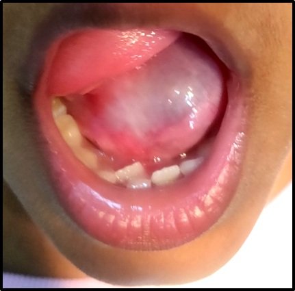
Figure 1 : The clinical picture shows a bluish hue with swelling in the floor of the mouth which is smooth and soft in consistency.

Figure 2 : The histopathological picture shows several large collapsed empty spaces of presumed mucin accumulation surrounded by compressed connective tissue stroma predominantly thrown into folds and papillary projections.
DISCUSSION
Ranula is an extravasation cyst and develops from extravasation of mucus after trauma to the sublingual gland and rarely submandibular gland or obstruction of the salivary ducts. Many theories for the origin and pathogenesis of these cysts have been postulated by Hippocrates, who described that ranulas are the effect of local inflammation of sublingual salivary gland.3 Ranulas can be classified into three groups. Oral ranulas present with intraoral swelling only, while a plunging ranula exhibits cervical swelling without swelling of the mouth floor. A mixed ranula has both intraoral and cervical swelling.4 Trauma, obstruction, or inflammation can damage one or more ducts, causing leakage of salivary contents into the surrounding tissue, inducing a fibroblastic reaction that seals the mucus in a connective tissue sac, referred to as an extravasation pseudocyst. This is the most common type of ranula. In less than 10% of all ranulas, congenital obstruction, by either an imperforate salivary duct or an ostial adhesion, leads to the formation of a retention cyst.5 The pathogenesis of plunging ranula may be as extension of a simple ranula, congenital in origin via ectopic cell rests or iatrogenic in nature due to transposition of tissue during surgery involving the salivary structures. Extensive extension has been reported in rare occurrences, producing pressure symptoms like dysphagia and airway obstruction. The current consensus is that submandibular glands are involved by extension of a plunging ranula into it and engulfing it, rather than being an intrinsic pathology of the gland.6 The typical clinical manifestation of a ranula is a slow-growing (usually unilateral but can be bilateral), pliable, painless mass in the floor of the mouth.5 Ranulas account for 6% of all salivary gland cysts.7 Clinically, it appears round or oval in shape, bluish in color, mobile and soft in consistency, and fluctuant upon palpation opposed to lymphomas and tumors of the salivary glands. Further enlargement of the swelling might lead to dysphagia, difficulty in speech, or even airway blockage.8There seems to be a higher frequency among young adults in their second and third decades as well as in children younger than 10 years, with a slight female preponderance.5
The most common technique for the management of ranula in earlier days was marsupialisation. Though marsupialisation was done, the cyst had a high recurrence rate of 61%-89% as the cyst is not completely removed. In some instances, it served as a precursor for plunging ranula. However, the packing of the ranula cavity with gauze after marsupialisation was found to be effective in reducing the recurrence rate which is generally recommended for cyst less than 2 cm in diameter.Ogita S et al., introduced OK-432 (lyophilized streptococcal preparation) for lymphangioma which was considered as an alternative method to surgical management.1
CONCLUSION
Ranula is an extravasation cyst and may develop from extravasation of mucus after trauma to the sublingual gland and rarely submandibular gland or obstruction of salivary ducts. Many theories for the origin and pathogenesis of these cysts have been postulated by Hippocrates, who described that ranulas are the effect of local inflammation of sublingual salivary glands .
REFERENCES
- Saraniya Packiri, Deepa Gurunathan, and Kathiravan Selvarasu. Management of Paediatric Oral Ranula: A Systematic Review. J Clin Diagn Res.2017 Sep; 11(9): ZE06–ZE09.
- Tanay Chaubal, Ranjeet Bapat. Ranula : Frog’s Belly.The American Journal of medicine 2021;23-03.
- Nikolaos Kolomvos , Evangelos Kalfarentzos, Nikolaos Papadogeorgakis. Surgical treatment of plunging ranula: Report of three cases and review of literature.2019; 2214-5419
- Moon-Gi Choi. Case report of the management of the ranula. (J Korean Assoc Oral Maxillofac Surg 2019;45:357-363).
- Elizabeth Ayers, MS, RDMS, RVT- Journal of Diagnostic Medical Sonography. Plunging Ranula: A Case Report . 2018, Vol. 34(4) 285–290
- Ramji AN. Int J Otorhinolaryngol Head Neck Surg. Sequestered plunging ranula: a case report and literature review.2019 Sep;5(5):1411-1415
- Arunkumar Kamalakaran ,Balaji Jayaraman , Saravanan Balasubramaniam ,Rohini Thirunavukkarasu ,Bharathi Ramakrishnan. Plunging Ranula in a 78- year- old Male – a Rare Case Report. 2018;10(1):e92-
Disclaimer from Guident
The views and opinions expressed in articles published in Guident are solely those of the authors and do not necessarily reflect the views of the editors, the editorial board or the publisher, Ivory India. Guident does not endorse any product, technique or treatment described. Clinical content is intended for qualified dental and medical professionals and is not a substitute for individual clinical judgement. Guident, its editors and the publisher accept no liability for any consequence arising from the use of information published in the journal.
How to cite this article
Dr. K.T. Magesh, Dr. R. Ramya, Dr. M. Nivedhidha. Ranula in a 7 Year Old Child : A Case Report and Review of Literature. Guident. 2021 Oct;14(11). Available from: https://ivoryindia.in/articles/oral-medicine/ranula-in-a-7-year-old-child-a-case-report-and-review-of-literature.html