Commonly Indicated Clinical Conditions for CBCT: A Factual Survey Based on a CBCT Centre
Department of Oral Medicine and Radiology, K D Dental College and Hospital Mathura, India
ABSTRACT
Introduction: Cone Beam Computed Tomography (CBCT) is a relatively new technique that was first used for angiography in the early 1980s and has since been used for a variety of other purposes. CBCT allows for 3D assessment of hard tissues in the maxillofacial area, ushering in a transition from a two-dimensional to a volumetric approach in maxillofacial imaging.
Objectives: To assess the commonly indicated clinical conditions for CBCT in a private CBCT Centre in two tier city, to evaluate the additional diagnostic tool and prevalence of additional findings in around the base of skull from large field of view CBCT as compared to conventional radiographs of the maxillofacial region and to determine their clinical importance.
Methods: The survey was based on 170 scans which were referred in the span of 3 months by dental practitioners of Mathura. The scans were taken from a private P square maxillofacial imaging CBCT center, Mathura. CARESTREAM Dental CS 9600 machine was used.
Result: The 170 scans were studied, analyzed and segregated according to their indications and were categorized according to their specialities. The findings which were beneficial over the conventional radiographs were noted. 61% of the scans were of implant dentistry, 17%scans of endodontics, 5.8%pathology and impaction, 4.11% scans of orthodontics, 2.3 % peditric dentistry, 1.7% scans of maxillofacial trauma and 0.5% scans were of TMJ disorder and ENT. Different FOV used for this study was also evaluated and recorded.
Conclusion: There are numerous types of CBCT machines available throughout the world with varying features, such as FOV 4x4 to 17x23. It is mandatory to do a market research according to the frequency and type of scans indicated and cost analysis according to the machine required or preferred.
Keywords: Cone Beam Computed Tomography (CBCT), 3D, FOV (field of view)
INTRODUCTION
Cone Beam Computed Tomography (CBCT) is a relatively new technique that was first used for angiography in the early 1980s and has since been used for a variety of other purposes. CBCT allows the 3dimensional assessment of hard tissues of the maxillofacial region which heralded a shift from a two-dimensional to a volumetric approach in maxillofacial imaging. The advantages of good image quality, volumetric analysis, fast scan times, and comparatively less radiation dose than traditional medical CT and better image quality than conventional radiographs have resulted in greater ubiquity as an imaging modality across all disciplines of dentistry. Fields including prosthodontics, orthodontics, oral surgery, periodontics, and endodontics find unique utility of the 3-dimensional reconstructions provided by CBCT. It has become an important adjunct in diagnosis due in part to the diverse image reconstructions available and the ability to visualize bony levels, and the sub-millimeter accuracy enabling linear measurements. The present study aimed to evaluate the commonly indicated clinical conditions for CBCT on a CBCT Centre and to evaluate the additional diagnostic tool and prevalence of functional findings in around the base of skull from large field of view CBCT as compared to conventional radiographs of the maxillofacial region and to determine their clinical importance. (Fig.1 & 2) Clinical benefits and limitations of CBCT. This study was to assess the commonly indicated clinical conditions for CBCT in a private CBCT Centre in two tier city, to evaluate the additional diagnostic tool and prevalence of additional findings in around the base of skull from large field of view CBCT as compared to conventional radiographs of the maxillofacial region and to determine their clinical importance. And to enlist the different types of FOV required for the indicated scans.
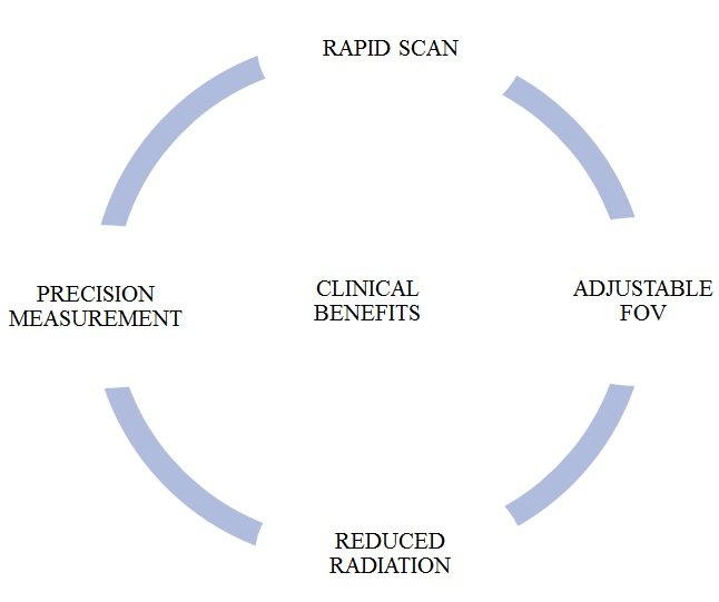
Fig. 1: Clinical benefits of CBCT

Fig. 2: Limitations of CBCT
MATERIALS AND METHODS
The survey was based on 170 scans which were referred in the span of 3 months by dental practitioners of Mathura. The scans were taken from a private P square maxillofacial imaging CBCT center, Mathura. CARESTREAM Dental CS 9600 machine was used.
RESULTS
Data were assessed according to frequency distribution. These scans were studied, analyzed and segregated according to their indications. They were categorized according to their specialities and the findings which were beneficial over the conventional radiographs were noted. Different FOV used for this study was also evaluated and recorded. (Fig.3) summary of indicated scans.

Fig. 3: summary of indicated scans.
61% of the scans were of implant dentistry, 17%scans of endodontics, 5.8%pathology and impaction, 4.11% scans of orthodontics, 2.3 % peditric dentistry, 1.7% scans of maxillofacial trauma and 0.5% scans were of TMJ disorder and ENT. The scans indicated:
IMPLANT DENTISTRY
- CBCT were advice for assessment of bone quality and bone density.
- Proximity to vital structure. (Fig.4)
- to assess the success of bone graft
- Virtual implant placement guidance.
- Out of 105 patients – there were 15 cases referred for full mouth implant (12x10 FOV) CARESTREAM Dental CS 9600

Fig. 4: Nerve proximity
ENDODONTIC ASSESSMENTS
- For diagnosing apical lesions.
- For detecting fractured roots. (Fig.5)
- To determine root morphology, the number of roots and accessory canals, and angulations of roots.
- For assessing root-canal fillings
- Calcified canal identification
- The use of CBCT in some of the cases significantly helped in additional diagnosis as compared to 2D imaging.
- There were evident additional findings of: (Fig.6)

Fig. 5: Fractured root in CBCT scan

Fig. 6: Additional findings in CBCT.

Fig. 7: comparision of C-shaped canal in conventional radiograph and CBCT.

Fig. 8: Comparision of Radix entomolaris in conventional radiograph and CBCT.
PATHOLOGY
- For diagnosis and for better treatment planning.
- For assessing architecture of the pathology in 3D imaging.
- To analyze localization of pathology in maxillofacial region.
- To assess the proximity to vital structures.
- Invasion to para oral structures.
ORTHODONTICS
- CBCT is a reliable tool in the assessment of the Proximity to vital structures that may interfere with orthodontic treatment.
- In detecting localization of impacted canine.
- Assessing palatal bony structures and suture closure.
IMPACTION
- CBCT were referred to assess types of impaction in relation to buccal/lingual direction.
- To determine the proximity to neurological involvement.
- For better treatment planning.

Fig. 9: Proximity of vital structure is detected better in CBCT
PEDIATRIC
- Jaw pathology
- maxillofacial fracture
- For detection in fibro-osseous ankylosis of condyle.
ORAL AND MAXILLOFACIAL TRAUMA
- For 3D reformatted images
- For detecting orbital floor fractures.
- For preoperative virtual planning.
- Analyzing and assessing paranasal sinuses
ENT
- CBCT was advised for sinus pathology.
- Temporomandibular joint disorder
- One of the most significant benefits of CBCT is its ability to define the true position of the condyle in the fossa, which frequently shows potential dislocation of the disc in the joint and the degree of translation of the condyle in the fossa.
- For detection in fibro-osseous ankylosis of condyle.
DISCUSSION
CBCT provides accurate, high resolution images in formats which permit 3 dimensional display of the complex anatomy of maxillofacial region. The clinical applications for CBCT imaging in dentistry are increasing. The results of this study demonstrate that 170 cases were clinically relevant and that the most common clinical applications are in the field of, implant dentistry, endodontics, orthodontics, oral and maxillofacial surgery, pediatric dentistry and ENT. The size of the scanned object volume is called the field of view. The size of the FOV significantly affects the resolution of the CBCT scan.
The FOV most typically included the jaws, midface, and skull base. Although FOVs as large as 17x23, 23x16.5 are available in the market, they are seldomly used. In this study the FOV required in 130 out of 170 cases fields were 4x4 and 5x5 and only 40 cases required 12x10 FOVs.(CARESTREAM Dental CS 9600).
FOV largely required in 170 cases

Fig. 10: Different sizes of FOV
Large FOV: covers most of the craniofacial skeleton, at least from below the soft tissue of the chin to nasion. Usually greater that 13 cm maximum scan height.
Medium FOV: Single or inter-arch ranging from 5 cm to 10 cm incorporating the maxilla and/or mandible.
Small FOV: approximately 5 cm or less vertical height covering localized regions such as a segment of the dental arch or temporomandibular joints.

Fig. 11
CONCLUSION
There are numerous types of CBCT machines available throughout the world with varying features, such as FOV 4x4 to 17x23. Every time you need not to go for higher FOV to get a wider scan, there is a possibility of reformation by STITCHING OR BLENDING where it involves obtaining data from two or more separate scans, superimposing and overlapping CBCT data volumes using corresponding fiducial reference landmarks (referred to as either ‘bio-image registration’ or mosaicking) and fusing adjacent image volumes (stitching or blending) to create larger volumetric dataset, either in the horizontal or in the vertical dimension.
Also, there is another possible method by increasing the height or width of the FOV using a smaller area detector to offset the position of the detector, collimate the beam asymmetrically and scan only half the patient’s ROI (region of interest) in each of the two offset positions.
REFERENCES
- S Do lekog lu et al. The usage of digital radiography and cone beam computed tomography among Turkish dentists. Dentomaxillofacial Radiology (2011) 40, 379–384. doi: 10.1259/dmfr/27837552.
- Alamri et al. applications of CBCT in dental practice: A review of literature. General dentistry. september 2012.60(5)390-400. www.agd.org
- Shah Akash. Implications of CBCT in Dentistry: A Review. Medical & Clinical Reviews. October 23,2017. Vol. 3 No. 3: 15. DOI: 10.21767/2471-299X.1000057
- Abramovitch & Rice. Basic Principles of Cone Beam Computed Tomography. dental.theclinics.com . 2014, 58 (2014) 463–484. http://dx.doi.org/10.1016/j.cden.2014.03.002
ACKNOWLEDGEMENTS
We would like to acknowledge Dr. PRAGYA GOYAL proprietary of P Square Advanced Maxillofacial Imaging centre, mathura for providing the data for the study.
We acknowledge the important contribution of Dr. VINAY MOHAN, Dr. ANUJ GAUR & Dr. NIHARIKA in the making of this review article.
Disclaimer from Guident
The views and opinions expressed in articles published in Guident are solely those of the authors and do not necessarily reflect the views of the editors, the editorial board or the publisher, Ivory India. Guident does not endorse any product, technique or treatment described. Clinical content is intended for qualified dental and medical professionals and is not a substitute for individual clinical judgement. Guident, its editors and the publisher accept no liability for any consequence arising from the use of information published in the journal.
How to cite this article
Dr. Ngurang Anam, Dr. Gopa Kumar Nair, Dr. Binish Khan. Commonly Indicated Clinical Conditions for CBCT: A Factual Survey Based on a CBCT Centre. Guident. 2021 Jul;14(8). Available from: https://ivoryindia.in/articles/oral-medicine/commonly-indicated-clinical-conditions-for-cbct-a-factual-survey-based-on-a-cbct-centre.html