
Peritonsillar Abscess Extending to Masticator Space Initially Diagnosed as Temporomandibular Joint Disorder- a Case Report
Department of Oral and Maxillofacial Surgery, H.P. Govt. Dental College and Hospital, Shimla, Yamunanagar Dental College, Yamunanagar, HAR
ABSTRACT
Background: To describe a novel case of peritonsillar abscess extending to the masticator space initially diagnosed as Temporomandibular Disorder (TMD).
Methods: The method includes clinical case records including orthopantomogram (OPG), Ultrasonography (USG), Contrast Enhanced Computed Tomography (CECT), Magnetic Resonance Imaging (MRI) and surgical approach.
Results: A 30-year-old male with preauricular pain and restricted jaw function was clinically evaluated, diagnosed using conventional radiographs and treated for temporomandibular joint disorders (TMD). However, the patient reported back with further worsening of pain, swelling and jaw function, so advanced imaging modalities were used to make a differential diagnosis. The case was diagnosed as peritonsillar abscess with secondary involvement of masticator space and drained intraorally.
Conclusion: it is crucial to use advanced diagnostic modalities while considering primary diagnosis of TMD as conservative treatment is the initial treatment for many TMD’s while other diagnosis has great potential for morbidity and the unusual route of spread for head and neck infections should always be kept in mind.
Key words: temporomandibular disorders, peritonsillar abscess, masticator space, lateral pharyngeal space.
INTRODUCTION
To determine the identity of a disease still remains the most important and crucial step for treating the patients. Patients with classic symptoms of restricted jaw movements and orofacial pain are often diagnosed with TMD without satisfactorily thinking about other causes like infection, trauma and neoplasms, hence it is essential to consider unusual causes while making an initial diagnosis as symptoms could hide an insidious lesion and lead to fatalities.1 Deep neck infection is a grave disorder that often spreads to other organs leading to significant morbidity and even prove fatal. The presence of cervical fascia leads to predictable and relatively constant trend in the distribution of infection along the anatomic spaces, however this pattern of spread could vary among patients. Peritonsillar abscess is the most common deep neck infection that sometimes spreads to the parapharyngeal space and involves the lower cervical space.2 An infection of lateral parapharyngeal space is one of the conditions which may present with symptoms similar to TMD.3 We report an unexpected case which initially was diagnosed as TMD but with advanced investigating modalities it was finally diagnosed as case of peritonsillar abscess taking unusual route of spread. The advanced imaging modalities are of great help to diagnose and treat such unusual and rare cases.1CASE REPORT
A male patient aged 30 years reported to the Department of Oral and Maxillofacial Surgery with chief complaint of pain for last 19 days and swelling for last 3 days in front of the left ear. He was already under medication for internal derangement of left TMJ for one week but symptoms deteriorated. Patient was afebrile with no associated comorbidities. Physical examination revealed interincisal opening-10mm with swelling: size 0.5×0.5 cm, non-fluctuant, normal surface temperature with tenderness on palpation over left preauricular region. Odontogenic cause of any infection was ruled out. Laboratory examinations were within normal range. On panoramic radiograph (Fig. 1) bony changes over left condyle were detected so, provisional diagnosis of left TMD was made. Analgesics with muscle relaxant, hot fermentations, soft diet and physiotherapy were prescribed.But after 2 days patient (Fig. 2) reported back with breathing difficulty, increased facial swelling: size 2.5×3.5 cm, fluctuant in nature with increased surface temperature, maximum interincisal opening-4mm and increased pain intensity. Thus, to arrive at the differential diagnosis USG guided Fine Needle Aspiration Cytology (FNAC) was performed. USG revealed bulky heterogeneous echotexture involving left temporalis and masseter muscle with questionable diagnosis of cervical lymphadenopathy due to infectious aetiology. Aspirate was sent for culture and sensitivity testing. The patient then received intravenous injection of amoxiclav 1.2g twice daily, infusion ornidazole 500 mg twice daily but to no avail. Culture and sensitivity report revealed staphylococcus aureus sensitive to piperacillin and tazobactam, so revised treatment with piperacillin and tazobactam 4.5 g thrice daily intravenous was ordered.

Preoperative Panoramic Radiograph demonstrating bony changes in Left condyle

Unilateral large swelling
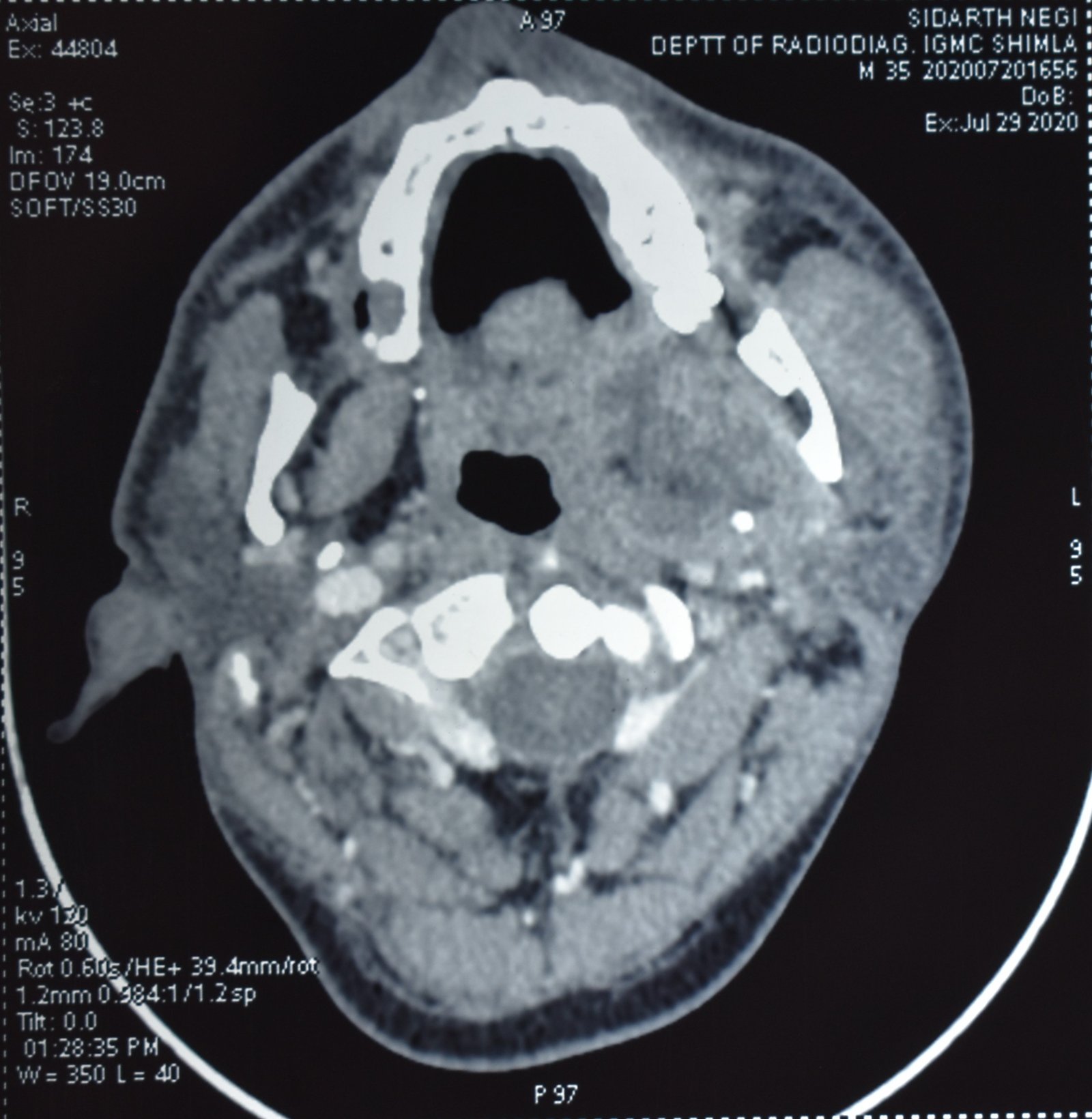
Preoperative CECT (axial view)

Preoperative MRI (axial view) with contrast

Mouth opening= 29 mm at 2 months follow up post operativ
DISCUSSION
Distinguishing particular condition from other that presents similar clinical features is an art. The initial treatment modalities of TMD’s are generally conservative and reversible but the therapy for other conditions within the differential diagnosis are not necessarily as conservative as it is for TMD’s. Eventually, inability to diagnose and treat deep neck infections may prove lethal.3Sometimes symptoms of infection such as swelling, warmth, and redness may develop later and the importance of parameters of infection like increased temperature and palpability of the lymph nodes in evaluating possible infection cannot be ignored.3 However, the patient in our case at the time of presentation was afebrile with no palpable lymphadenopathy. Therefore, even in the absence of specific findings, an infection or other pathology should be considered.4 Similarly, in our case we did not suspect any infection.
A survey of the writing uncovered 53 patients who were at first misdiagnosed as having TMD. So, the diagnosis ought not be founded exclusively on the clinical assessment yet additionally on advanced imaging modalities, for example, CECT and MRI.4 In our case also initially we suspected TMD but later on worsening of symptoms CECT and MRI were done which revealed infectious aetiology.
Peritonsillar abscess represents an infection between the tonsillar capsule and superior constrictor muscle within the crypts of tonsil. Infections in this region usually spread to parapharyngeal space with further downward extension via the submandibular space.2,5 The masticator space infections are usually odontogenic, arising in the mandible and spreading first into the masseter and/or medial pterygoid muscles. The parapharyngeal space is a secondary site of spread for these infections. That is, infections from the pterygoid muscles sometimes spread medially to the parapharyngeal space.2 Conversely, in the present patient there were no findings of an odontogenic infection.
Another hypothetical route of extension to the masticator space from the parapharyngeal space would be anteriorly from the parotid space.2 The present patient had involvement in the parotid space; as visualized both on CECT and MRI. We can thus conclude that in the current case, infection originating from the peritonsillar abscess, showed an unusual route of extension to the masticator space via the parotid space or along the surface of the mandibular ramus.
Surgical incision and drainage along with antimicrobial administration remains the primary treatment for deep neck abscess. Every patient should be given initial empiric antibiotic therapy until culture and sensitivity results are available. Earlier external approaches for incision and drainage of masticator abscess was described but recent literature supports an intraoral approach. Intra-oral approach provides safety with regards to nerve injury, better cosmetic outcome and short anaesthesia time, however consequences of pus aspiration from intraoral drainage cannot be ignored.2,5 The collected pus with swabs was immediately sent for microbiological analysis but culture was reported as sterile because antibiotics were already escalated to piperacillin combined with tazobactam based on pre-operative culture and sensitivity report. Antibiotic therapy for the deep space infections has been reported to range from 10 to 30 days, consisting of combined intravenous and oral routes.
CONCLUSION
Trismus, preauricular pain and deviation of mouth opening are not specific for TMD alone. The possibility of other causes should be carefully considered. Differential diagnosis should be aided by advanced imaging modalities.Conflict of interest statement (None).
The author has not received any research grant, remuneration, or speaker honorarium from any company or committee whatsoever.
Funding
This research did not receive any specific grant from funding agencies in the public, commercial, or not-for-profit sectors.
REFERENCES
- Ogi N, et. al. Chronic dental infections mimicking temporomandibular disorders. Australian Dental Journal 2002;47:(1):63-65
- Hidaka H, et. al. Unusual Parapharyngeal Extension of Peritonsillar Abscess to the Masticator Space: Successfully Drained by Extraoral and Intraoral Endoscopic Approaches. Annals of Otology, Rhinology & Laryngology 2014, Vol. 123(5) 333-337.
- Brown RS, Johnson CD, Fay RM. The Misdiagnosis of Temporomandibular Disorders in Lateral Pharyngeal Space Infections-Two Case Reports. 1994; 12:3, 194-198.
- Heo MS, An BM, Lee SS, Choi SC. Use of advanced imaging modalities for the differential diagnosis of pathoses mimicking temporomandibular disorders. Oral Surg Oral Med Oral Pathol Oral Radiol Endod. 2003; 96: 630-638.
- Hidaka H, Suzuki T, Ishida E, RisakoKakuta, Yano H. Unique Extension of Peritonsillar Abscess to the Masticator Space Encountered in the Early Phase of Antiviral Therapy for Hepatitis C. Clin Microbial. 2013; 2: 117.
Disclaimer from Guident
The views and opinions expressed in articles published in Guident are solely those of the authors and do not necessarily reflect the views of the editors, the editorial board or the publisher, Ivory India. Guident does not endorse any product, technique or treatment described. Clinical content is intended for qualified dental and medical professionals and is not a substitute for individual clinical judgement. Guident, its editors and the publisher accept no liability for any consequence arising from the use of information published in the journal.
How to cite this article
Dr. Monika Parmar, Dr. Manish Sahore, Dr. Abhishek Soni. Peritonsillar Abscess Extending to Masticator Space Initially Diagnosed as Temporomandibular Joint Disorder- a Case Report. Guident. 2020 Nov;13(12). Available from: https://ivoryindia.in/articles/oral-surgery/peritonsillar-abscess-extending-to-masticator-space-initially-diagnosed-as-temporomandibular-joint-disorder-a.html


