
Transmigrated Mandibular Canine and its Surgical Management-a Rare Case Report
Department of Oral and Maxillofacial Surgery, Manipal College of Dental Sciences, Mangalore
Abstract
Transmigration of mandibular canine is a rare phenomenon. Transmigration and impaction appear to be more common. Most of the patients do not have any symptoms, and these canines are often discovered at the radiological examination before orthodontic treatment. The aetiology is obscure, the treatment of impacted transmigrated canine is very complicated if it is diagnosed at a later stage. Early diagnosis of impacted canines is mandatory for timely treatment to ensure facial harmony and improved function. Here we present one such rare case of impacted mandibular canine which has transmigrated to opposite side and was accidentally found on radiological examination before orthodontic treatment and its surgical management.
Key words: Impaction, Mandibular canine, Surgical Extraction, Transmigration
Introduction
Failure to erupt tooth in an appropriate location in the dental arch may lead to numerous clinical conditions such as tooth impaction, translocation or even transmigration.1 Such clinical entities are often frequently encountered to oral and maxillofacial surgeons in their clinical practice. In the human dentition, Canines are the most crucial and cornerstone teeth of the dental jaws. They play a vital part in retaining the facial aesthetics and functional efficacy and any alteration in its eruption makes it a significant concern to the patient.2
Impacted canines in the mandible are rare as compared to maxilla. It is an even unusual incidence when an impacted mandibular canine crosses the midline and migrates to the opposing side of the mandible. This unusual phenomenon has been named as transmigration.3 Ando et al. first described this term and first case of transmigration of canine was reported in year 1951.4
Transmigration is a clinical phenomenon of yet unfamiliar aetiology. Transmigrated canines may persist as impacted and may remain asymptomatic or else while they can cause root resorption of neighbouring teeth, with other symptoms such as discomfort, pain and neuralgic symptoms to the patient.3,5 Incidence of impacted mandibular canine is recorded to be 20 times lesser in comparison to maxillary canines. Overall, the prevalence of transmigrated canines in mandible varies as 0.14% to 0.31%.6 Clinical outcomes related with transmigration of the canines comprises of atypical retention of mandibular deciduous canine or absence of mandibular permanent canines in the dental arch. Majority of times the transmigrated canines, were lying below the apices of the anterior teeth in a horizontal situation.7 Here we report such a rare case of transmigrated mandibular canine and its surgical management
Case Report
A 20 year-old female was referred from the department of orthodontia to our department in view of impacted canine in both upper and lower jaw seeking orthodontic treatment, on clinical examination , no gross facial asymmetry noted, TMJ movements were within normal limits with adequate mouth opening, intraoral examination revealed retained deciduous canine on both upper and lower dentition on left side of the jaw with all four third molar impacted showing class I molar relationship, on palpation mild tenderness was elicited on lower jaw with no swelling or pus discharge (Figure 1). Dental and medical histories revealed no significant findings.

Figure 1: Pre-operative Intraoral view
Panoramic radiograph revealed impacted 23, 33, 18,28,38,48 and 43 was infra erupted (Figure 2). Cone beam computed tomopraghy (CBCT) was advised to find the precise location of the mandibular canine relative to its neighbouring tooth and to plan the further treatment. CBCT revealed impacted maxillary left canine and horizontally transmigrated mandibular left canine with the crown of the tooth passing the midline and approaching the opposite side canine and lying horizontally below the apices of the mandibular anterior teeth labially, approaching the inferior border of mandible. (Figure 3, 4)
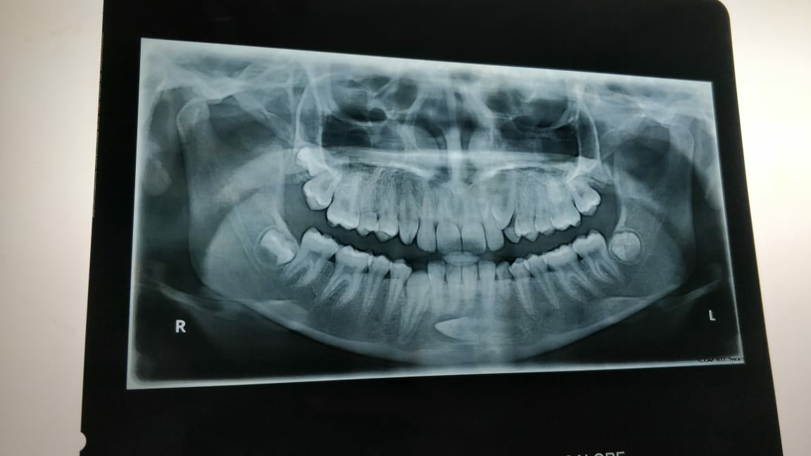
Figure 2: OPG showing Transmigrated canine

Figure 3: CBCT showing Impacted canine lying at inferior border of mandible

Figure 4: CBCT showing horizontal position of impacted canine
Two surgical approaches were possible: an extra-oral approach through the lower border of the mandible, or an intraoral labial approach through the mucobuccal fold. The intraoral labial approach was decided on because an extra-oral approach would necessitate a sub-mental incision at least an inch in length. This approach seemed contraindicated for aesthetic reasons. The intraoral approach seemed more conservative. Prophylactic 1 g Amoxicillin orally was given to patient before the start of the procedure. Using sterile protocols, local anaesthesia containing 2% lignocaine with 1:200000 adrenaline was injected as Left inferior alveolar nerve block (IANB). Incision was given with no. 15 blade extending from premolar to premolar on both the sides and a full thickness mucoperiosteal flap was reflected. Symphysis region was exposed up to the lower border of mandible (Figure 5).
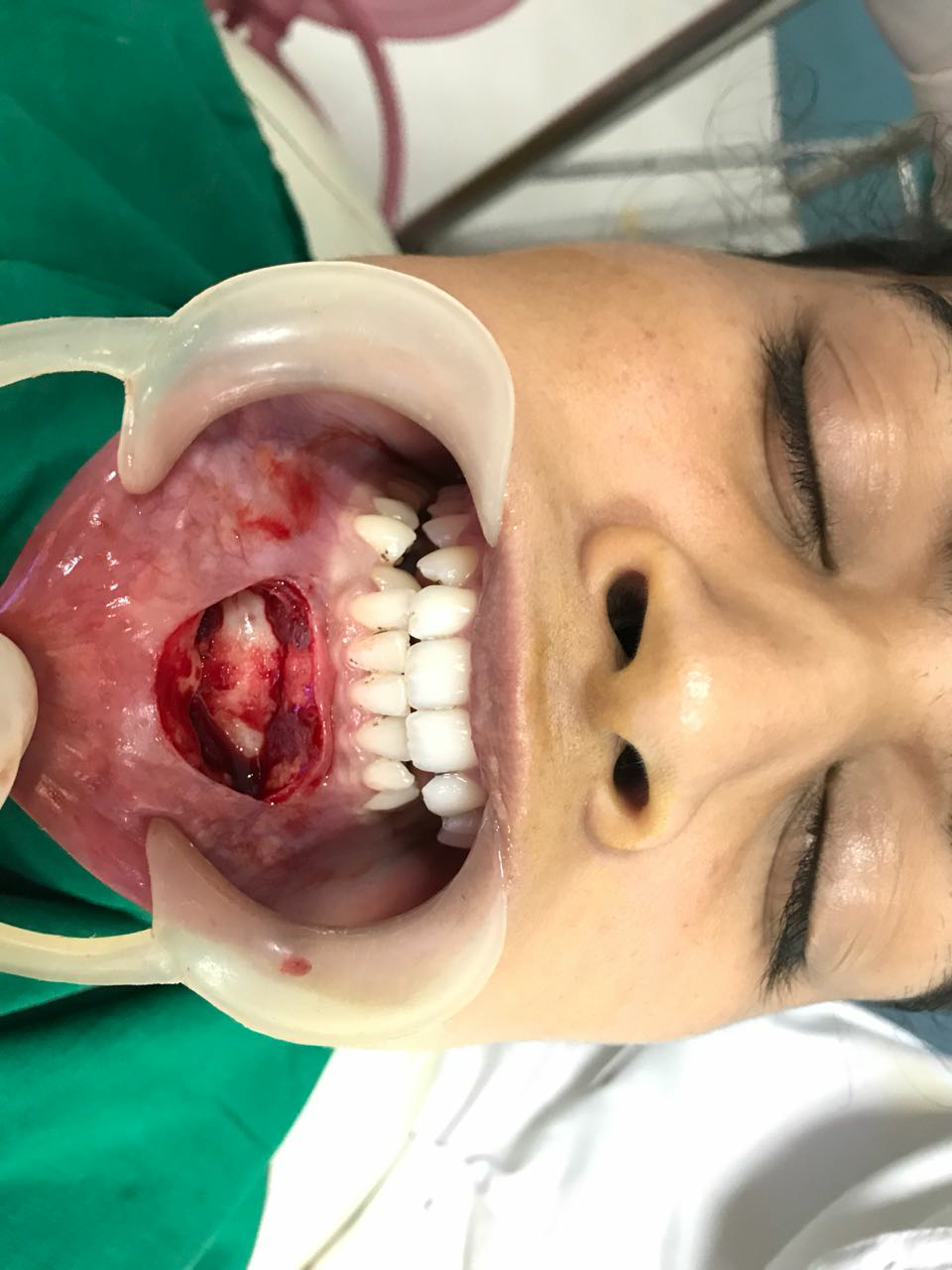
Figure 5: Intraoral exposure of canine
Bone cutting was done using round bur no 4 and window was created exposing the crown of the canine, followed by sectioning of the tooth beneath the cemento-enamel junction (Figure 6). The crown portion was divided and removed first followed by removal of root with slight luxation (Figure 7). Thorough irrigation was done with betadine and normal saline.

Figure 6: Sectioning of the tooth

Figure 7: Tooth specimen
Meanwhile patient blood was withdrawn from right ante-cubital vein and centrifuged at 10,000 rpm for 10 minutes to make Platelet rich fibrin (PRF) ( Figure 8) and infused into the defect created (Figure 9) and surgical closure was done layerwise using 3-0 silk (Figure 10) and pressure dressing was given extra-orally. Patient was advised analgesics for 3 days with intermittent ice application for first 24 hours. Suture removal was done on 7th post-operative day revealed good healing with no post-operative complications.

Figure 8: Platelet Rich Fibrin (PRF)
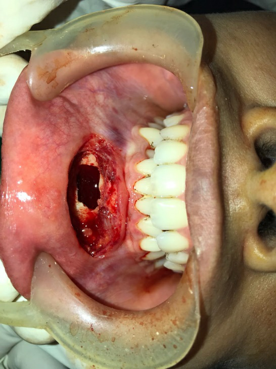
Figure 9: Defect packed with PRF

Figure 10: Surgical Closure
Patient underwent orthodontic treatment and 9 months Post-op orthopantomogram showed good amount of bone at the defect site with all the teeth in alignment and good healthy tissue (Figure 11, 12).

Figure 11: Post-op intraoral view

Figure 12: Post-op OPG
Discussion
The canines are considered to play significant aesthetic and functional roles in human dentition. Conditions such as impaction, translocation and, in particular, transmigration related to mandibular canines are quite uncommon.2,8 the literature on mandibular canine impactions is sparse. Few references have been made to diagnostic criteria or surgical management of these teeth. The lack of material in this area is probably attributable to the rarity of these impactions.
Panoramic radiographs are routinely advised along with the occlusal radiographs to detect the transmigrated canines. Other method of investigations includes lateral cephalogram, CBCT and very rarely computed tomography (CT). With the advent of CBCT in the field of dentistry it becomes possible to accurately localize the impacted canines and associated vital structures or any pathology and also to detect root resorption of the adjacent teeth.9
Mupparapu has suggested a classification system for transmigrated teeth and he categorized it into 5 types based on position the teeth in jaw and its pattern of migration.10 following this classification, our case represents type 2 category.
According to Thoma11, transmigration of the mandibular canines is quite rare scenario and it is very uncommon to find it in a horizontal position in the lower jaw. Various theories describing the aetiology of the process of transmigration have been proposed. Displacement of the dental lamina to an abnormal position in early life may result in abnormal eruptive path. Distant migration is likely in the developmental stage of the tooth apex because of rich blood circulation and active alveolar bone expansion of alveolar bone. In addition to these, other factors such as conical shape of the canine, congenitally missing lateral incisors, cystic lesions of the canine, prematurely lost or over retained primary teeth, tooth size arch size discrepancies, and pathological conditions can result in the diversion of canine from its line of eruption.4,12
Majority of times transmigrated canines are usually asymptomatic, although follicular cyst development surrounding the impacted tooth and chronic infection along with fistula creation have been recorded.13 Nodine5, stated that migrated mandibular canines are frequently revealed without producing any obvious symptoms indicative of their existence. Ando et al.4 also described that they had not found any symptoms like pain or compression of mandibular nerve due to the transmigrated canines in their patient. As in our case, patient was accidentally diagnosed with impacted canine with absence of clinical symptoms.
The persistent retention of the primary canine to a certain extent is a consistent sign resulting in the detection of its impacted permanent successor. The left canine is more affected as compared to right canine, and females have a tendency to affect more commonly than males14, which completely fits as in our case. Observation by Joshi et al.15 shows that, due to lack of the developing permanent mandibular canine beneath the primary canine, the root resorption of the primary canine is comparatively slow, as our patient showed retained deciduous canine on the left side of the mandible.
Caldwell and Bruszt16 in their study stated that the transmigrated canine retains its nerve supply from its area of origin. Therefore, it is obligatory to anesthetize the nerve of the involved side to where the impacted canine is located, especially under local anaesthesia. Henceforth this justifies administration of left inferior alveolar nerve block in our patient.
The management of transmigrated canine is becomes quite difficult if it is detected at a late stage. Treatment options includes orthodontic repositioning, extraction of the abnormally positioned canine surgically, transplantation of the tooth and the surgically exposing the canine with orthodontic alignment.17,18 As in our case the mandibular arch was crowded and position of transmigrated canine is near lower border of mandible patient so orthodontic repositioning was not promising, surgical removal of the transmigrated teeth is the treatment of choice. Henceforth Surgical removal was done and defect site was infused with PRF for superior healing. PRF contains various growth factors which promotes early healing with rapid bone formation as our patient was young and warrants urgent treatment we provided this treatment choice as best available option. However, in remarkable circumstances when all other teeth are in usual place and the space exists for the migrated canine, transplantation technique is one mode of managing such cases.
To best of our knowledge very few cases in literature are managed with surgical extraction along with PRF infusion therapy. Hence our case present as a unique case report.
Conclusion
Transmigration of the mandibular canine crossing the mandibular midline is an erratic and elusive phenomenon defined in the dental literature. Radiographic investigation through panoramic radiographs is indispensible to diagnose impacted canines and with newer technologies such as CBCT, it becomes very effective to exactly diagnose transmigrated canines. Well-timed recognition and well-designed treatment plan aids to preserve such canines, adjoining tissues and dentition, ensuing in enhanced aesthetic and function.
Furthermost, the canine sometimes migrates devoid of any pathology, but in a rare case a cyst or odontome supplements with such teeth. Aetiology of this phenomenon is obscure. Surgical removal of the transmigrated canine under local anaesthesia appears to be the best form of treatment in majority of the cases. A comprehensive diagnostic and therapeutic method, along with sound information of all prognostic consequences, is obligatory for appropriate diagnosis and accurate treatment planning.
Financial support and sponsorship
Nil
Conflicts of interest
There are no conflicts of interest.
References
- Camilleri S, Scerri E. Transmigration of mandibular canines—a review of the literature and a report of five cases. The Angle Orthodontist. 2003 Dec;73(6):753-62.
- Kharbanda OP, Choudhury AR. Extreme transmigration of mandibular cuspid: report of two cases. The Journal of clinical pediatric dentistry. 1994;18(4):307-8.
- Joshi MR. Transmigrant mandibular canines: a record of 28 cases and a retrospective review of the literature. The Angle Orthodontist. 2001 Feb;71(1):12-22.
- Ando s, Aizawa k, Nakashima t, Sanka y, Shimbo k, Kiyokawa k. Transmigration process of the impacted mandibular cuspid. The Journal of Nihon University School of Dentistry. 1964;6(2):66-71.
- Nodine AM. Aberrant teeth, their history, causes and treatment. Dent Items Interest. 1943 Sep;65(2):440-51.
- Aydin U, Yilmaz HH, Yildirim D. Incidence of canine impaction and transmigration in a patient population. Dentomaxillofacial Radiology. 2004 May;33(3):164-9.
- Miranti R, Levbarg M. Extraction of a horizontally transmigrated impacted mandibular canine: report of case. The Journal of the American Dental Association. 1974 Mar 1;88(3):607-10.
- Bhullar MK, Aggarwal I, Verma R, Uppal AS. Mandibular canine transmigration: Report of three cases and literature review. Journal of International Society of Preventive & Community Dentistry. 2017 Jan;7(1):8.
- Laffranchi L, Dalessandri D, Fontana P, Visconti L, Sapelli P. Cone beam computed tomography role in diagnosis and treatment of impacted canine patient's: a case report. Minerva stomatologica. 2010 Jun;59(6):363-76.
- Mupparapu M, Auluck A, Suhas S, Pai KM, Nagpal A. Patterns of intraosseous transmigration and ectopic eruption of bilaterally transmigrating mandibular canines: radiographic study and proposed classification. Quintessence international. 2007 Nov 1;38(10).
- Thoma KH. Oral Surgery. 2nd ed. St. Louis, Missouri: CV Mosby; 1952.
- Shapira Y, Mischler WA, Kuftinec MM. The displaced mandibular canine. ASDC J Dent Child. 1982;49:362–364.
- Rebellato J, Schabel B. Treatment of a patient with an impacted transmigrant mandibular canine and a palatally impacted maxillary canine. The Angle Orthodontist. 2003 Jun;73(3):328-36.
- Javid BR. Transmigration of impacted mandibular cuspids. International journal of oral surgery. 1985 Dec 1;14(6):547-9.
- Joshi MR, Shetye SB. Transmigration of mandibular canines: a review of the literature and report of two cases. Quintessence international. 1994 Apr 1;25(4).
- Bruszt P. Neurological anomaly associated with extreme malposition of a mandibular canine. Oral Surg Oral Med Oral Pathol.1955;8:484–487.
- Díaz-Sánchez RM, Castillo-de-Oyagüe R, Serrera-Figallo MÁ, Hita-Iglesias P, Gutiérrez-Pérez JL, Torres-Lagares D. Transmigration of mandibular cuspids: review of published reports and description of nine new cases. British Journal of Oral and Maxillofacial Surgery. 2016 Apr 1;54(3):241-7.
- González-Sánchez MA, Berini-Aytés L, Gay-Escoda C. Transmigrant impacted mandibular canines: a retrospective study of 15 cases. The Journal of the American Dental Association. 2007 Nov 1;138(11):1450-5.
Disclaimer from Guident
The views and opinions expressed in articles published in Guident are solely those of the authors and do not necessarily reflect the views of the editors, the editorial board or the publisher, Ivory India. Guident does not endorse any product, technique or treatment described. Clinical content is intended for qualified dental and medical professionals and is not a substitute for individual clinical judgement. Guident, its editors and the publisher accept no liability for any consequence arising from the use of information published in the journal.
How to cite this article
Dr. Mihir Dani, Dr. Mohan Baliga, Dr. Aananya Mishra. Transmigrated Mandibular Canine and its Surgical Management-a Rare Case Report. Guident. 2020 Oct;13(11). Available from: https://ivoryindia.in/articles/oral-surgery/transmigrated-mandibular-canine-and-its-surgical-management-a-rare-case-report.html


