Neutral Zone Approach for Patients with Severely Resorbed Mandibular Ridge
Department of Prosthodontics & Implantology K.D. Dental College & Hospital, Mathura (U.P) India
ABSTRACT
Resorption of mandibular ridges is a multifactorial and biomechanical disease which is chronic, progressive, irreversible, and cumulative leading to loss of sulcular depth, vertical dimension loss, and decreased lower facial height. The denture becomes passive due to complex neuromuscular control and causes difficulties in impression-making, mastication, and swallowing, which in turn leads to loss of retention and stability in complete dentures. The neutral zone concept plays a significant role in overcoming these challenges. The neutral zone is the area where the outward forces from the tongue are neutralized or nullified by the inward forces of the lips and cheeks acting during functional movements. The neutral zone technique is an alternative approach for the construction of lower complete dentures. Various materials and techniques are used in recording neutral zone, which have their own advantages and disadvantages. This article describes two different techniques for recording neutral zone.
INTRODUCTION
The Neutral Zone is defined as “the potential space between the lips and cheeks on one side, and the tongue on the other; that area or position where the forces between the tongue and cheeks or lips are equal”. It is also referred to as the dead space and the zone of minimal conflict. This concept considers the actions of the tongue, lips, cheeks and floor of the mouth on a recording material during a specific oral function. It was first described by Wilfred Fish who reported the influence of the polished surfaces on retention and stability of complete dentures in 1931. He stated that the polishing surface contour should conform to the shape of the tongue, lips, and cheeks. These tissues, in function or at rest, would exert an elastic pressure on the dentures, and retain them in place rather than dislodge them. Various movements including sucking, grinning, whistling and pursing the lips push the material into a position where the bucco-lingual forces are neutralized.
Swallowing and Speech are used as the principle modeling function for obtaining the neutral zone. Swallowing is used in the traditional ‘modeling compound-swallowing impression technique’ and Speech is utilized in a technique called piezography. Piezography records the mandibular denture space by means of pressure developed during oral functions, mainly speech . This technique customizes the contours of the lingual surface and precludes over-extension.
TECHNIQUE 1
- Maxillary and Mandibular primary impressions were made in stock trays using Impression compound (DPI - Pinnacle Impression Compound and Tracing Sticks, The Bombay Burmah Trading Corporation, Ltd., Mumbai, India). Custom trays were fabricated in autopolymerizing resin (DPI RR Cold Cure, The Bombay Burmah Trading Corporation, Mumbai).
- Border moulding was done using tracing compound (DPI -Tracing Sticks, The Bombay Burmah Trading Corporation, Ltd., Mumbai, India) and then, tentative jaw relation was recorded using impression compound (DPI - Pinnacle Impression Compound and Tracing Sticks, The Bombay Burmah Trading Corporation, Ltd., Mumbai, India). The impression compound was softened in hot water; the softened compound was kneaded and placed as roll on the maxillary and mandibular special tray. It was reheated and placed in patients mouth and the functional and physiological movements like swallowing, sucking, pursing lips, sipping water, pronouncing vowels and protruding tongue several times. Once the neutral zone was recorded, it was removed from mouth and placed in cool water. Final impression was taken in zinc oxide eugenol impression paste (DPI Impression Paste, The Bombay Burmah Trading Corporation, Mumbai) using closed mouth approach (Fig. 1).
- The master casts were poured in dental stone (Zhermack Elite Model Stone, Zhermack SpA, Badia Polesine [RO], Italy) and record bases were constructed with self-cure acrylic resin. Jaw relation records were then recorded using conventional occlusal rims made of modelling wax (HIFLEX-Modelling Wax, Prevest Denpro Limited, Jammu, India) and occlusal blocks were mounted on articulator (Fig. 2).
- Mandibular occlusal rim was removed and neutral zone record obtained earlier was placed. Height of compound rim was adjusted to the set vertical dimension (Fig. 3). The base of the cast was indexed by making notches. Addition silicone Putty index of this recorded zone was made (Fig. 4).
- The neutral zone record was then removed, and the denture base was kept in place (Fig.5). Modelling wax was poured in the space representing the neutral zone, forming the new occlusal rim on the mandibular record base (Fig.6).
- Teeth setting was done in a conventional matter and a wax try-in was performed to evaluate mandibular record base stability, aesthetics, and intraoral occlusion (Fig.7). A tertiary impression was made using zinc oxide eugenol impression paste (DPI Impression Paste, The Bombay Burmah Trading Corporation, Mumbai) in closed mouth approach (Fig.8). Casts were poured and the trial dentures were processed with heat-cure acrylic resin. The denture was inserted and verified for retention, stability, and occlusion (Fig.9).
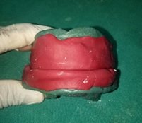
Fig. 1
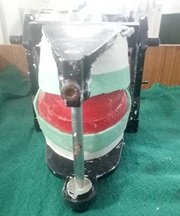
Fig. 2

Fig. 3

Fig. 4
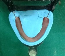
Fig. 5
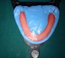
Fig. 6
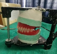
Fig. 7

Fig. 8

Fig. 9
TECHNIQUE 2
- Maxillary impression was made using irreversible hydrocolloid (Vignette Chromatic, Dentsply, Gurgaon, India) and mandibular impression was made using admix material – three parts by weight of impression compound and seven parts by weight of tracing compound (DPI - Pinnacle Impression Compound and Tracing Sticks, The Bombay Burmah Trading Corporation, Ltd., Mumbai, India) (Fig.10).
- Customized tray (Fig.11) is fabricated with autopolymerising acrylic resin (Rapid Repair, Dentsply, Gurgaon, India) according to Dynamic Impression Technique. Tray with 1 mm wax spacer and cylindrical mandibular rests in the posterior region are made at increased vertical height. High-fusing impression compound is softened, placed on top of the mandibular rests and inserted in the patient’s mouth (Fig.12). Patient is advised to close his mouth so that the mandibular rests fit against the maxillary alveolar ridge.
- McCord and Tyson’s technique for flat mandibular ridges is followed for definitive impression. Wax spacer is removed from mandibular customized tray, admix material is loaded and patient is guided to close his mouth on the mandibular rests.
- For recording the functional state, patient is instructed to run his tongue along his lips, suck in his cheeks, pull in his lips and swallow by keeping his mouth closed, as in closed mouth impression technique, till the impression material hardens (Fig.13). Maxillary secondary impression was made in a conventional manner. Master casts were poured and record bases were fabricated using autopolymerizing resin (DPI-RR Cold Cure, The Bombay Burmah Trading Corporation, Ltd., Mumbai, India)
- An additional autopolymerizing resin mandibular denture base is fabricated and is attached with retentive loops made of thin orthodontic wire in the centre. Two vertical pillars made of low fusing compound (Tracing Sticks, DPI PINNACLE, The Bombay Burmah Trading Corporation, Mumbai.) are placed in First molar region (Fig.14).
- Maxillary occlusal rim was placed in the mouth and parallelism was verified using the Fox occlusal plane. The horizontal jaw relation was recorded using the admix material. The admix material was manipulated, placed over the mandibular base and inserted in the mouth. Patient was instructed to perform routine mandibular movements (including swallowing, sucking of the lips and exaggerated ‘EEE’ and ‘OOO’ sounds), which aided in molding the neutral zone space (Fig15).
- The maxillary and mandibular occlusal rims were articulated in a mean value articulator to fabricate indices surrounding the neutral zone plaster impression on the mandibular cast (Fig.16).
- Three notches were made on the cast: one in the anterior and two in the posterior regions. This was followed by applying separating medium on the cast, the record base, and over the neutral zone record. Boxing was done with modeling wax, and plaster of Paris was poured into the boxing up to the upper surface. The plaster indices were sectioned into a labial and buccal index and a lingual index in order to guide the removal and placement of these indices (Fig.17).
- The neutral zone record was then removed (Fig.18) and wax was poured in the space representing the neutral zone, forming the new occlusal rim on the mandibular record base (Fig.19).
- The mandibular teeth were arranged following the index, and the maxillary teeth were arranged following the mandibular teeth arrangement (Fig.20). A wax try-in was performed to evaluate mandibular record base stability, aesthetics, and intraoral occlusion. The trial dentures were processed with heat-cure acrylic resin. The denture was inserted and verified for retention, stability, and occlusion (Fig.21).

Fig. 10

Fig. 11

Fig. 12

Fig. 13

Fig. 14

Fig. 15

Fig. 16
Fig. 17

Fig. 18

Fig. 19

Fig. 20

Fig. 21
CONCLUSION
The main goal of prosthodontic treatment is to restore the function and aesthetics. Complete denture is influenced by not only the ridge but also by the surrounding oral cavity, occlusion, biting forces and many others. In the atrophic mandible, one of the principal functional problems, other than instability, arises from the inability of the residual ridge and its overlying tissues to withstand masticatory forces. Furthermore, the muscle attachments are located near the crest of the ridge, with greater dislocating effect of the muscles. For these reasons, the range of muscle action, as well as spaces into which the denture can be extended without dislocation, must be accurately recorded in the impression.
A denture shaped by neutral zone technique will ensure that the muscular forces are working more effectively in harmony and gives advantage of stabilizing potential of oral and perioral musculature. Admix material was used for recording the neutral zone. It's a combination of impression compound and green stick (low-fusing) compound in the ratio of 3:7. The mixing of a low-fusing compound with the impression compound results in a low viscosity material allowing for ease in manipulation of the oral musculature. The admix material allowed better flow and an accurate impression. The techniques presented in this article have proved to be efficient for patients who are not satisfied with mandibular dentures.
REFERENCES
- Beresin VE, Schiesser FJ. The neutral zone in complete dentures. J Prosthet Dent. 1976;36(4):356–367. doi: 10.1016/0022-3913(76)90155-4.
- McCord JF, Tyson KW. A conservative prosthodontic option for the treatment of edentulous patients with atrophic (flat) mandibular ridges. Br Dent J. 1997;182:469–472. doi: 10.1038/sj.bdj.4809415.
- Saravanakumar, Prathibha et al. “Improvised Neutral Zone Technique in a Completely Edentulous Patient with an Atrophic Mandibular Ridge and Neuromuscular Incoordination: A Clinical Tip.” Cureus vol. 9,4 e1189. 24 Apr. 2017, doi:10.7759/cureus.1189.
- Agarwal S, Gangadhar P, Ahmad N, Bhardwaj A. A simplified approach for recording neutral zone. J Indian Prosthodont Soc. 2010;10(2):102-104. doi:10.1007/s13191-010-0019-8
- Praveen G, Gupta S, Agarwal S, Agarwal SK. Cocktail Impression Technique: A New Approach to Atwood's Order VI Mandibular Ridge Deformity. J Indian Prosthodont Soc. 2011;11(1):32-35. doi:10.1007/s13191-011-0055-z
- Ladha K, Gupta R, Gill S, Verma M. Patient satisfaction with complete dentures fabricated using two neutral zone techniques: a within-subject cross-over pilot study. J Indian Prosthodont Soc. 2014;14(2):161-168. doi:10.1007/s13191-013-0297-z
- Asnani P , Chandu GS , Mishra SK Neutral zone technique in complete denture –Case report..J App. Dent. Med. Sci. 2015; 1(1):15-18.
Disclaimer from Guident
The views and opinions expressed in articles published in Guident are solely those of the authors and do not necessarily reflect the views of the editors, the editorial board or the publisher, Ivory India. Guident does not endorse any product, technique or treatment described. Clinical content is intended for qualified dental and medical professionals and is not a substitute for individual clinical judgement. Guident, its editors and the publisher accept no liability for any consequence arising from the use of information published in the journal.
How to cite this article
Dr. Yashi Garg, Dr. Rahul Nagrath, Dr. Manesh Lahori. Neutral Zone Approach for Patients with Severely Resorbed Mandibular Ridge. Guident. 2020 Oct;13(11). Available from: https://ivoryindia.in/articles/prosthodontics/neutral-zone-approach-for-patients-with-severely-resorbed-mandibular-ridge.html