Odontogenic Keratocyst Associated with Impacted Maxillary Third Molar
Department of Maxillofacial & Oral Surgery, Department of Maxillofacial & Oral Surgery, Department of Periodontology, Government Dental College & Hospital, Mumbai, India
ABSTRACT
Odontogenic keratocyst is a specific histological type of developmental odontogenic cyst. OKC is a commonly occurring odontogenic cyst, first described by Philipsen in 1956. In 60% - 80% of all cases mandible is involved and men in younger age group are more affected than women. In 10% of patients it occurs as one of the signs associated with Gorlin-Goltz syndrome as recurrence rate of OKC is very high, treatment ranges from simple enucleation to resection of the jaw. In this paper, we present a case report of a keratocyst, which originated probably from epithelial remnants distal to an impacted maxillary third molar, and which expanded to include the lateral and orbital walls of the right maxillary sinus.
Keywords: Keratocyst, Enucleation, Maxillary antrum, impacted maxillary third molar.
Abbreviations: OKC - Odontogenic Keratocyst
INTRODUCTION
Odontogenic keratocyst (OKC) have the potential to recur and occasionally to undergo malignant transformation.1 If any remnants are left in the bone postoperatively they will soon recur, as they have a high potential for growth.2 OKCs though having multilocular appearance on radiograph may mimic a unilocular radiolucency similar to a dentigerous cyst involving an unerupted tooth,3 therefore treatment of OKC varies from conservative simple enucleation of the cyst to radical surgery i.e. resection of the jaw so as to keep the margins free of any remnant cystic tissue.4, 5, 6 The incidence of keratocysts varied from 3%-12% (mean 7%) of all odontogenic cysts.7 The most common important features of OKC are unusually high recurrence rate that ranges from 5% to 62.5%8, 9, 10 and its accidental discovery on the radiograph due to its insidious growth.
CASE REPORT
A 19-year-old male patient reported to the Department of Oral and Maxillofacial Surgery complaining of intermittent pain and swelling of the upper right side of face since 3-4 years. Patient gave history of intermittent painless swelling over right cheek followed by pus discharge in right buccal vestibule near maxillary molar. Then patient was advised analgesic and antibiotic by his general physician, which relieved both pain and swelling with persistence of pus discharge. However as the symptoms kept moving, the patient finally was referred for definitive diagnosis and management. History for major illness/ medical disorders/ allergies were negative.
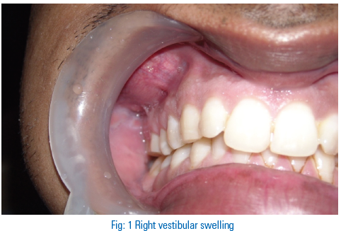
Extra-oral examination revealed asymmetrical face due to diffuse swelling over the right cheek. Intraoral Examination revealed a localised swelling in right maxillary buccal vestibule in relation to premolar and molar region (Fig 1). All the permanent teeth were present except the 18, and the 15, 16 and 17 were found to give a negative response to electrical stimulation. Orthopantomogram and computerized tomography scans of the maxilla confirmed an impacted tooth that looked like the upper third molar at the posterior lateral wall of the maxillary antrum (Fig 2, 3). There was a well demarcated unilocular radiolucency related to the impacted tooth and involving the right maxillary sinus.

All routine investigation was done and root canal treatment of 15, 16 and 17 was completed prior to surgery. Extraction of impacted teeth with excision or enucleation of associated lesion and curettage of right maxillary sinus to cease pus discharge was decided as treatment objectives so Caldwell-Luc surgery via intraoral approach under general anaesthesia was planned.
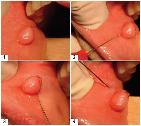
On the operation table, tooth was extracted. The cyst seem to approach the sinus roof i.e. orbital floor & posterior surface i.e. infratemporal fossa along with the lateral nasal wall which was eroded by the chronic lesion. During enucleation of cystic lesion copious amount of cheesy keratin like foul smelling material was obtained (Fig 4, 5 and 6).
Post enucleation, ribbon gauze impregnated with petroleum jelly & povidone-iodine was packed into the empty antral cavity and removed in stages over the next 72 hrs.
Patient was maintained under intravenous antibiotics & oral analgesics for three days post operatively. As recovery was uneventful patient was discharged on 4th post-operative day asked to continue antibiotics and analgesics for three more days along with tincture benzoin inhalation & local nasal decongestants drops.
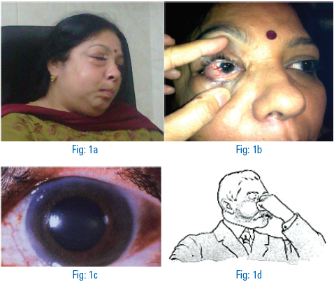
Two days after discharge however patient reported with peri orbital swelling involving the right eye & complained of diminished vision in same eye. The patient was readmitted & referred to ophthalmology OPD for opinion owing to the diminished vision & associated swelling. The ophthalmologist advised CT brain to rule out cavernous sinus thrombosis (CST). CT brain reports were negative for any pathological/abnormal changes. Patient was therefore kept under I.V. antibiotics and observation for three days. Subsequently recovery was uneventful & patients remain asymptomatic for the past one year when he has been followed up. (Fig 7 and 8)


Fig 8: 1 year postop intraoral view
DISCUSSION
OKC was first described by Phillipsen in 1956; the database continues to increase with influence to the pathogenesis, behaviour and treatment of cyst.11 Its origin is attributed to dental lamina remnants in the teeth bearing areas of mandible and maxilla.12, 13 Remnants of odontogenic epithelium persist in oral tissues after odontogenesis is complete, and from these remnants a variety of tumours and cysts might possibly arise.14 The most common location of occurrence is the posterior portion of the mandible. Other locations have also been reported, such as the anterior portion of the maxilla, the maxillary sinus and the maxillary third molar area.15 Apart from the Gorlin-Goltz syndrome, keratocysts cannot be diagnosed from clinical features alone, and misdiagnosis is common.16, 17 The main difference between OKCs and other jaw cysts are insidious growth of the OKC. OKCs recur more often than any other type of jaw cyst. The recurrence rate is almost comparable to that of the ameloblastoma.18, 19 The ideal treatment for the odontogenic keratocyst would be enucleation or curettage followed by treatment of the cavity with an agent such as liquid nitrogen or Carnoys solution to ensure total destruction of the epithelial remnants or satellite cysts.20 Some investigators claimed good success with decompression and enucleation only while other advocate enucleation, peripheral osteotomy, and chemical curettage.21, 22 High recurrence rate raises the question of the adequacy of treatment. There is no doubt that the entire cyst must be completely removed, but this is not always possible because of the size of the cyst, the fragility of the cyst wall, and the presence of satellite cysts. Nevertheless many authors agree that simple enucleation is enough, provided that patients are closely followed up both clinically and radiologically for several years.23-28 Many authors recommend the application of Carnoy’s solution, a tissue fixative to bony cavity following enucleation and curettage of cyst. Voorsmit demonstrated that a 5 minute application of the Carnoy’s solution allow penetrate to depth of 1.54mm and tissue fixation.29, 30
Post-operative follow-up with annual radiological examinations is essential for at least five years following surgery. Recurrence is documented even after 10 years of follow-up and treatment. However, it is difficult to diagnose recurrence with regard to cysts in the maxillary sinus after the surgical removal of the initial lesion. Therefore computerised tomogram is important in assessing the full extent of the recurrent lesion preoperatively during follow up period.
In this case, the fact to be highlighted is its occurrence in maxilla which is rare. We had success by means of enucleation and aggressive curettage. There was no sign of recurrence on follow-up for one year. The patient has been symptom free up till date and he is under long-term follow up. The potential for recurrence of the OKC relates to the high proliferation activity of the keratocyst epithelium. Due to the high recurrence rate and aggressive behaviour of OKCs, different treatment techniques can be chosen. Enucleation or curettage alone is the mostly preferred treatments in the management of OKCs. However, according to various authors, it is better to use Carnoys solution together with enucleation or curettage, since it may provide less chances of recurrence.
REFERENCES
- Klammt J. Zysten des Kieferknochens. Leipzig: Barth, 1976.
- Horch HH. Zahnlrztliche Chirurgie, Praxis der Zahnheilkunde. Mtinchen- Wien-Baltimore: Urban u. Schwarzenberg. 1989: Vol 9, 281-346.
- Woo Sook-Bin et al.: Odontogenic keratocyst in the anterior maxilla: Report of two cases, one simulating a nasopalatine cyst. Oral Surg Oral Oral Med Pathol 64:463-465,1987.
- der Keratozysten. Dtsch zahnarztl Z 1985; 40: 645-647.
- Lendrodt J, Gundlach KKH. Zur chirurgischen Therapie der gutartigen odontogenen Tumoren. Dtsch Z Mund-Kiefer-Gesichtschir 1978; 2. 3312.
- Wimmer L. 1st dies Unterkieferresektion bei Keratozysten indiziert? Dissertation Dusseldorf, 1989.
- Franscini M. Langzeit-Beobachtung von Keratozysten: Dissertation Erlangen. 1989.
- Absi EG, Sim RL. Odontogenic keratocyst involving the maxillary sinus: report of two cases. Dentomaxillofac Radiol 23:226-229, 1994.
- Brannon RB. The odontogenic keratocyst. A clinicopathologic study of 312 cases. Part I Clinical Features. Oral Surg Oral Oral Med Pathol 42:54-72, 1976.
- Voorsmit RA: The incredible keratocyst (thesis). University of Nijmegen, the Netherlands 1984.
- Philipsen H: Om keratocyster (kolesteatomer) I kaeberne (On keratocysts in jaws). Tandlaegabladet 1956;60:963.
- Regezi JA, Sciubba J: Oral Pathology. Clinical-Pathologic Correlations. 2nd ed. Philadelphia, Pennsylvania: W.B. Saunders Company. 1993, pp: 337-341.
- Brannon RB. The odontogenic keratocyst. A clinicopathologic study of 312 cases.
Disclaimer from Guident
The views and opinions expressed in articles published in Guident are solely those of the authors and do not necessarily reflect the views of the editors, the editorial board or the publisher, Ivory India. Guident does not endorse any product, technique or treatment described. Clinical content is intended for qualified dental and medical professionals and is not a substitute for individual clinical judgement. Guident, its editors and the publisher accept no liability for any consequence arising from the use of information published in the journal.
How to cite this article
Dr. Ankush Janardhan, Dr. Prajwalit Prakash, Dr. Rajesh Prabhakar. Odontogenic Keratocyst Associated with Impacted Maxillary Third Molar. Guident. 2013 Jan;6(3). Available from: https://ivoryindia.in/articles/oral-surgery/odontogenic-keratocyst-associated-with-impacted-maxillary-third-molar.html