Aesthetic Rehabilitation of Anterior Missing Teeth with Sieberts Ridge Defects: A Case Report
Department of Prosthodontics, Crown & Bridge and Implantology, K.D Dental College & Hospital, Mathura India
ABSTRACT
Prosthodontists may face challenges in treating patients with alveolar ridge defect in partially edentulous patients. The edentulous space may be due to loss of tooth, due to trauma during extraction or congenital defects which eventually leads to alveolar bone loss. This loss in the alveolar bone causes the intrusion of the overlying soft tissue during healing which ultimately creates contours.
The replacement/regeneration of lost tissues along with tooth replacement needs multiple visits of surgeries. In our report, we present three cases highlighting existing sieberts classification ridge defects around teeth and their correction with PFM crowns with gingival shaded porcelain . Loss of residual ridge contour may lead to unesthetic open gingival embrasures ("black triangles") ,food impaction and percolation of saliva during speech.
Use of gingival restoration can reduce the necessity for a surgical procedure to restore missing soft and hard tissues, thereby simplifying and reducing the time and cost of treatment.
KEYWORDS - SIEBERTS CLASSIFICATION, PFM CROWNS, GINGIVAL SHADED PORCELAN
INTRODUCTION
Pontic or artificial tooth, its name is derived from the Latin word- pons, meaning bridge.
Pontics are the artificial teeth of a fixed partial denture that replace missing natural teeth, restoring function and appearance.
They must be compatible with continued oral health and comfort. The design of the prosthetic tooth will be dictated by esthetics, function, ease of cleaning, maintenance of healthy tissue on the edentulous ridge, and patient comfort.
An ideally shaped ridge has a smooth, regular surface of attached gingiva, which facilitates maintenance of a plaque-free environment. Its height and width should allow placement of a pontic that appears to emerge from the ridge and mimics the appearance of the neighboring teeth.
Facially, it must be free of frenum attachment and of adequate facial height to sustain the appearance of interdental papillae. Loss of residual ridge contour may lead to unesthetic open gingival embrasures ("black triangles") ,food impaction and percolation of saliva during speech.
Use of gingival restoration can reduce the necessity for a surgical procedure to restore missing soft and hard tissues, thereby simplifying and reducing the time and cost of treatment.
SIEBERT’S CLASSIFICATION OF RESIDUAL RIDGE DEFORMITIES
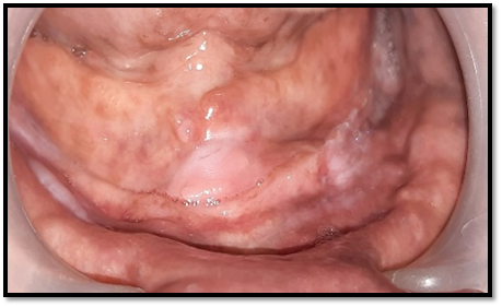
Class I defects: Faciolingual loss of tissue width with normal ridge height.
Class II defects: Loss of ridge height with normal ridge width.
Class III defects: a combination of loss in both dimensions.
This case report describes the treatment planning for a patient suffering from loss of soft and hard tissues using artificial gingiva in a tooth-supported fixed prosthesis.
CASE REPORT
A 45 year-old woman, 33 and 48 year old men presented as a new patient to the Department of Prosthodontics, KD Dental college , their chief complaint was loss of the upper anterior teeth with a request to have these replaced to improve the appearance of the anterior part of their mouth . Written informed consent was obtained from the patient for treatment and publication of this case report.
A thorough medical and dental history was taken at the first appointment.. A comprehensive clinical and radiographic examination was then undertaken. Primary impressions using alginate
An initial discussion with the patient about the feasible treatment options, from the ideal to the least ideal choice, along with an explanation of the advantages and disadvantages of each option.
Because the patient did not want to undergo any periodontal treatment due to financial and time constraints, they preferred fixed prosthesis (PFM crown ) as a treatment option, excluding other modalities.
Porcelain fused to metal (PFM) crown is the most widely used fixed restoration. It is a full metal crown having a facial surface (or all surfaces) covered by ceramic material. It consists of a ceramic layer bonded to a thin cast metal coping. It combines the strength and accurate fit of cast metal coping with the cosmetic of ceramic, So, this type combines the advantages of the strength of full metal crown and esthetic of all ceramic crown.
DIAGNOSTIC IMPRESSION
Primary impressions using alginate and wax occlusal records were taken to make articulated study models. Digital photographs were also taken. Waxup done and putty index were made for immediate temporisation. The waxup provided a matrix for fabrication of provisional restoration and also a blueprint for dental positioning and adjustment of the axes of the teeth.
TOOTH PREPERATION
Labial reduction PFM crown preparation requires deep facial reduction to give enough space for metal and porcelain, and thus avoiding over contouring and poor esthetic which would inevitably occur when no enough tooth structure is removed. The amount of labial reduction is 1.5-2 mm. Advantages of adequate reduction (deep facial reduction) 1. The restoration will properly contour (effect on esthetic & gingival health). 2. The shade & translucency of the restoration will match that of the adjacent natural tooth. Ø 0.5 mm for the metal coping. Ø 1 mm for porcelain (0.2 mm opaque layer, 0.5 mm body “dentin” layer, and 0.3 mm incisal “enamel” layer).
Because of the anatomy of the tooth labially, it should be reduced in two planes corresponding to the two geometric planes of the labial surface: a gingival plane and an incisal plane. Advantages of two plane reduction 1. To follow the anatomy of the surface. 2. To avoid hitting the pulp. 3. To give enough space for the metal and porcelain layers, so that avoiding poor esthetic or over contour.
Proximal reduction A pointed tapered fissure bur (long needle) is used to break the contact with the adjacent tooth, moving the bur up and down from the palatal to the labial. A round-end tapered fissure bur is then used to create a chamfer finishing line continuous with the chamfer finishing line of the palatal surface and joining the shoulder finishing line of the labial surface at a line angle called "wing".
Palatal (lingual) reduction
a. Cingulum area reduction D.O.G. of 1mm in depth is placed in the center using a round bur 1 mm in diameter. A small wheel diamond bur is then used to reduce this area following the concavity of this part of tooth surface.
b. Lingual axial reduction D.O.G. of 1mm in depth is placed parallel to the long axis of the tooth. A round- end tapered fissure bur is then used to reduce this area parallel to the long axis of the tooth to create chamfer finishing line.
The final impression of the prepared abutments was made using a double-stage technique with heavy and light body polyvinylsiloxane impression material (Hydrorise Heavy and Wash, Zhermack Inc) sent to lab along with selected shade for both pfm crowns and gingival shade for rehabilitation of soft tissue . The temporary bridge was then cemented with a zinc oxide eugenol-based temporary cement (TempBond®, Kerr Corporation, Orange, CA).
Final crown with gingival shaded porcelain is cemented.
1. SIEBERT’S CLASSIFICATION - Class-1

PRE OPERATIVE

TOOTH PREPERATION

PFM CROWNS

CROWN WITH GINGIVAL SHADED PORCELAIN

POST OPERATIVE
2. SIEBERT’S CLASSIFICATION - Class-2

PRE OPERATIVE

CLASS 2 DEFECT SIEBERTS CLASSIFICATION & TOOTH PREPERATION IRT 11,23

PFM CROWN WITH GINGIVAL PORCELAIN

FINAL CEMENTATION 11,21,22,23

BEFORE

AFTER

SIDE PROFILE
3. SIEBERT’S CLASSIFICATION - Class-3

PRE OPERATIVE

TOOTH PREPERATION

IMPRESSION

PFM CROWN WITH GINGIVAL PORCELAIN

BEFORE

AFTER
DISCUSSION
No matter how well biologic and mechanical principles have been followed during fabrication, the patient will evaluate the result by how it looks, especially when anterior teeth have been replaced.
Many esthetic considerations that pertain to single crowns also apply to the pontic.
Several problems unique to the pontic may be encountered when attempting to achieve a natural appearance.
An esthetically successful pontic will replicate the form, contours, incisal edge, gingival and incisal embrasures, and color of adjacent teeth. The pontic's simulation of a natural tooth is most often betrayed at the tissue-pontic interface.
The greatest challenge here is to compensate for anatomic changes that occur after extraction. Special attention should be paid to the contour of the labial surface as it approaches the pontic-tissue junction to achieve a "natural" appearance.
To re-establish natural crown ratios and natural gingival profiles in complex situations, artificial gingival restoration can reduce the necessity for technique sensitive surgical procedures. It also increases intraoral comfort because of the smooth uniform interface between the prosthetic gingiva and the remaining tissues, thereby simplifying and reducing the cost and duration of treatment. The diagnostic waxup is critical when the replacement of missing teeth is esthetically challenging.
Gingival defects may be treated with surgical or prosthetic approaches. With successful surgical treatment, the result mimics the original tissue contours. But the surgical costs, healing time, discomfort and unpredictability make this choice unpopular.
Prosthetic replacement, with acrylics, composite resins, porcelains and silicones, is a more predictable approach to replacing lost tissue architecture. It is especially useful when a larger amount of tissue needs replacement. Ideal tissue contours can be waxed, processed and then colored to match the surrounding tissue.
The patient need not undergo any additional surgical procedures and receives an esthetically pleasing, functional restoration. It is possible to show the patient a waxed-up result or even take a try-in prosthesis directly to the mouth for evaluation before significant treatment is initiated.
REFERENCES
- Coachman C, Salama M, Garber D, Calamita M, Salama H, Cabral G. Prosthetic gingival reconstruction in a fixed partial restoration. Part 1: Introduction to artificial gingival as an alternative therapy. Int J Periodontics Restorative Dent. 2009;29:471–477.
- Hannon SM, Colvin CJ, Zurek DJ. Selective use of gingival-toned ceramics: case reports. Quintessence Int. 1994;25:233–238.
- Kay HB. Esthetic considerations in the definitive periodontal prosthetic management of the maxillary anterior segment. Int J Periodontics Restorative Dent. 1982;2:45–49.
- Fondriest J. Shade matching in restorative dentistry: the science and strategies. Int J Periodontics Restorative Dent. 2003;23(5):467-79.
- Cal E, Guneri P, Kose T. Comparison of digital and spectrophotometric measurement of colour shade guides. J Oral Rehabil. 2006;33:221-8.
- Tung FF, Goldstein GR, Jang S, Hittelman E. The repeatability of an intraoral dental colorimeter. J Prosthet Dent. 2002;88(6):585-90.
- McDermott KC, Webster MA. Uniform color spaces and natural image statistics. J Opt Soc Am A Opt Image Sci Vis. 2012 Feb 1;29(2):A182-7.
- Dozic A, Kleverlaan CJ, meegdees M, Vander Zel J, Feilzer AJ. The influence of porcelain layer thickness on the final shade of ceramic restorations. J Prosthet Dent. 2003 Dec;90(6):563-70.
- Gokce HS, Piskin B, Ceyhan D, Gokce SM, Arisan V. Shade matching performance of normal and color vision – deficient dental professional with standard daylight and tungsten illuminants. J Prosthet Dent. 2010; 103(3)139-147.
- LeeYK, Yu B, Lim HN. Lightness, chroma and hue distribution of a shade guide as measured by a spectroradiometer. J Prosthet Dent. 2010 Sep; 104 (3): 173-181.
- Chaiyabutr Y, Kois JC, Lebeau D, Nunokawa G. Effect of abutment tooth color, cement color, and ceramic thickness on the resulting optical color of a CAD/CAM glass – ceramic lithium disilicate – reinforced crown. J Prosthet Dent.2011; 105(2)83-90
- Lee WZ, Ong MM, Yeo AB. Gingival profiles in a select Asian cohort: a pilot study. J Investig Clin Dent 2018;9(1).
- Rathee M, Rao LP, Bhoria M. Prevalence of gingival biotypes among young dentate North Indian population: a biometric approach. Int J Clin Pediatr Dent 2016;9(2):104–108. DOI: 10.5005/ jp-journals-10005-1343.
- Shah R, Sowmya NK, Thomas R, et al. Periodontal biotype: basics and clinical considerations. J Interdiscip Dent 2016;6:44–49.
- Chu SJ, Tarnow DP, Tan JH, et al. Papilla proportions in the maxillary anterior dentition. Int J Periodontics Restorative Dent 2009;29(4): 385–393.
- Crigger BJ. Declaration of Helsinki revised. IRB 2000;22
Disclaimer from Guident
The views and opinions expressed in articles published in Guident are solely those of the authors and do not necessarily reflect the views of the editors, the editorial board or the publisher, Ivory India. Guident does not endorse any product, technique or treatment described. Clinical content is intended for qualified dental and medical professionals and is not a substitute for individual clinical judgement. Guident, its editors and the publisher accept no liability for any consequence arising from the use of information published in the journal.
How to cite this article
Dr. C Pradeep, Dr. Siddharth Sisodia. Aesthetic Rehabilitation of Anterior Missing Teeth with Sieberts Ridge Defects: A Case Report. Guident. 2022 Dec;16(1). Available from: https://ivoryindia.in/articles/prosthodontics/aesthetic-rehabilitation-of-anterior-missing-teeth-with-sieberts-ridge-defects-a-case-report.html