An Accustomed Breakthrough: Rehabilitation of Bilaterally Distal Extension Maxillary Arch with Digital Cast Partial Denture: A Case Report
Department of Prosthodontics, Career Post Graduate Institute of Dental Sciences and Hospital, Lucknow
ABSTRACT
The main goal of prosthodontic rehabilitation in any patient is to restore form, function, esthetics and phonetics. Numerous treatment modalities like removable partial dentures (RPD), overdentures, conventional fixed partial dentures or implant supported prosthesis are available to rehabilitate a partially edentulous arch. Replacement of any tooth requires meticulous intraoral examination of the present clinical condition of the patient along with analysis of the associated risks, benefits and estimated expenditure of the selected treatment option after thorough discussion with the patient.
Replacement of single or multiple missing teeth with removable partial dentures in distal extension cases poses a great challenge to the dentist owing to the displacement of mucosa due to occlusal loads. Cast partial dentures have in the recent times, thus, become the most feasible treatment modality in cases where fixed treatment is contraindicated either due to patient’s intraoral condition or economic status of the patient. The laboratory procedures involved in fabrication of conventional CPD framework are quite complicated, time-consuming and require precision of the dentist which is made more simpler with the aid of digitization in the present dentistry.
This clinical report, therefore, emphasizes on the prosthetic rehabilitation of a partially edentulous patient maxillary arch with DMLS Co-Cr cast partial denture (CPD).
Key words: Removable partial dentures, financial status, distal extension, cast partial dentures, DMLS.
INTRODUCTION
In the present-day situation, edentulism has turned out to be one of the most depleting and irreparable conditions affecting the oral health of a person in totality. The most common population to be affected by this, is the older generation and thus it has become a major cause of concern worldwide. In a patient who is partially edentulous, it becomes extremely difficult to achieve an adequate prosthetic restoration due to the loss and constant degeneration of the alveolar bone, adjacent teeth, as well as the supporting structures. There are a wide variety of treatment options that are available for a partially edentulous patient with single or multiple missing teeth that may include; an interim removable partial denture, a definitive cast partial denture, tooth supported fixed partial denture or an implant supported prosthesis depending on the clinical condition.1,2
Removable partial denture is contemplated to be one of the most widely accepted and most cost-effective treatment option for patients with partially edentulous arch. The prosthetic rehabilitation of a distal extension RPD plays a very crucial role in order to achieve a successful long- term prognosis and also to preserve the remaining natural teeth and their supporting structures. The difficulties encountered in bilateral distal extension RPDs are mainly the cause of dissimilarity in the support between the abutment teeth and the residual ridges. Hence, it is extremely essential to ensure that an RPD is accurately designed for the preservation of the remaining natural teeth.3,4
Several clinical studies have come to the conclusion; that on use of an acrylic removable prosthesis, significant damage is observed in the periodontium of the abutment teeth more than that seen in case of cast metal removable partial dentures.3,5 On comparison of cast partial denture with acrylic partial denture, factors like retention, stability, comfort and periodontal health of abutments are greatly enhanced in a cast partial denture.3 The metal denture base in cast partial dentures is more accurate, durable, resistant to distortion, easily cleansable, light weight and is less bulky whereas the full acrylic partial dentures have various disadvantages which include an increase in thickness of the denture base affecting the phonetics, altered taste sensation, reduced tongue space, and dentures being more prone to fracture at their thinnest portions.6,7 The most commonly used metal in the framework for a Cast partial denture is made of cobalt-chromium which is biocompatible.3
Digital dentistry has gained popularity in the current scenario and is thus being substituted for traditional fabrication methods. Digital technology has enhanced digital surveying, designing of the component-based framework as well as 3D printing of the resin. Rapid prototyping (RP), also known as additive manufacturing, is mainly involved in the fabrication of accurate 3D models directly from the electronic data.8
Every clinical situation involving multiple missing teeth cannot always be rehabilitated with the help of a cast partial denture. Thus, it is the responsibility of the dentist to decide the type of prosthesis which would be most appropriate for the patient.3 The present case report emphasizes mainly, the fabrication of a cast partial denture for a bilateral distal extension maxillary arch to enhance retention, stability, support, aesthetics and provide an improved masticatory function to the patient.
CASE REPORT
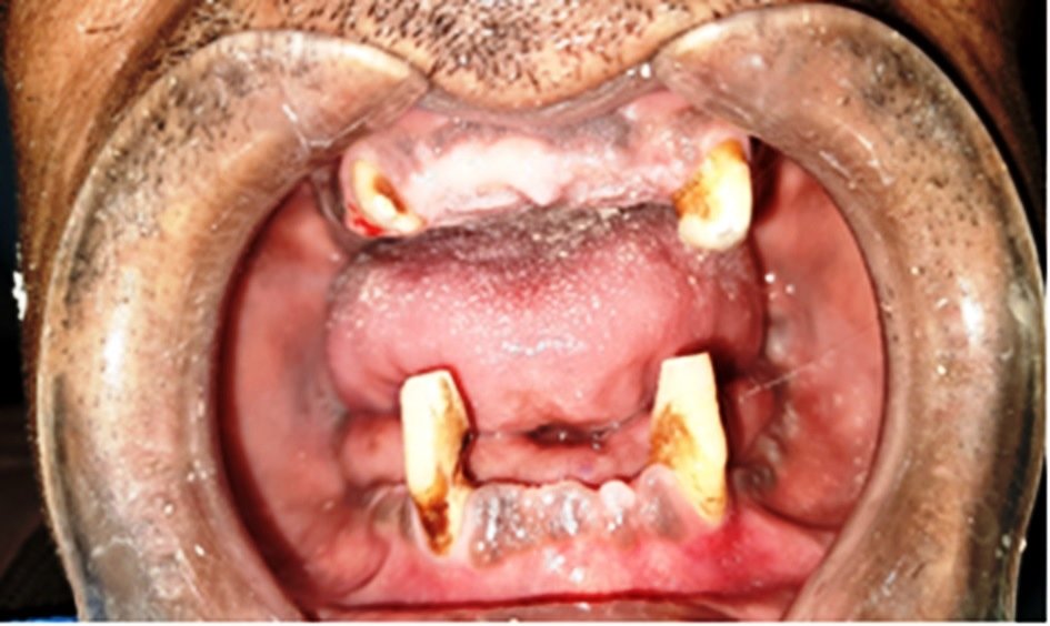
A 60-year-old male patient reported to the Department of Prosthodontics, Career Postgraduate Institute of Dental Sciences and Hospital, Lucknow with a chief complaint of missing teeth and inability to masticate. The patient had a history of multiple extractions which were done 2 years back due to grossly decayed posterior teeth in both maxillary and mandibular arches. Extra oral examination showed no evidence of facial asymmetry or TMJ abnormalities. On intra-oral examination, a dentate situation of Kennedy’s Class I in maxillary arch and Kennedy’s class III in mandibular arch was observed. Teeth 11,12,13,21,22 and 23 were present in the maxillary dentition with spacing present in both upper and lower anterior teeth. (Fig 1) Patient was already wearing an interim removable partial denture in the mandibular arch and was satisfied with the same so rehabilitation of only the maxillary edentulous area was planned. The teeth present in the maxilla were periodontally healthy to be considered as abutments for the removable prosthesis. Radiographic evaluation showed satisfactory bone support around the remaining teeth and no signs of periodontal and periapical lesions were seen.

Fig 1: Intraoral Preoperative view
Before treatment planning, complete oral prophylaxis was done and the patient was recalled for diagnostic impressions which were made with irreversible hydrocolloid impression material (Imprint, DPI, India) and poured in type III dental stone (Kalabhai Karson Pvt. Ltd). (Fig 2) The diagnostic casts were then studied and different treatment plans were proposed to rehabilitate and restore function and aesthetics, ranging from conventional removable partial dentures to implant supported fixed prosthesis. Prosthetic rehabilitation with dental implants was not a feasible option in this particular case due to presence of inadequate bone height and width and also due to the financial constraints of the patient. On, the other hand, the option of conventional fixed partial denture was also not possible due to absence of posterior abutments. Thus, after a detailed clinical examination and taking into consideration the patient’s age, profession, aesthetic and functional demands, a conventional cast partial denture in the maxillary arch would be an appropriate treatment option. The treatment steps were then explained to the patient and on his consent for the same, the rehabilitation was then carried out.

Fig 2: Diagnostic Impressions
With the help of diagnostic casts, Facebow transfer and interocclusal record was taken and mounted on Hanau semi adjustable articulator. Surveying of the maxillary cast was then done followed by designing of cast partial denture for the maxillary arch. In the maxillary distal extension base, the retentive meshwork must be extended to the hamular notch and the type of major connector should be selected based on the support and rigidity needed for preservation of tissues, and anatomic limitations. The maxillary major connector design that was planned in this case was antero-posterior palatal strap that had metal extension extending from the anterior strap and resting on the palatal surface of incisors for indirect retention. Also, cingulum rest seats were incorporated w.r.t 13 and 23 and I bar type of direct retainer was adapted over teeth 13 and 23 for better retention. The rest seat and the guide plane mock preparations in relation to 13 and 23 were done on the cast and required mouth preparations were carried out accordingly. Once the preparations were evaluated, border moulding was done followed by final impression made using polyvinyl siloxane addition silicone material (Aquasil Soft Putty, Aquasil Light Body, Densply, Germany), poured and retrieved. (Fig 3) Surveying was repeated and unfavourable undercuts were blocked out. The master cast was then scanned and the DMLS framework was digitally designed by specialized software (ExoCad) through a series of digital steps which replicate the conventional laboratory procedures. (Fig 4) The denture base framework was then tried intraorally for checking of accurate fit and adaptation. (Fig 5) The maxillary occlusal rim was then fabricated (Fig 6) and the maxillo-mandibular relation was recorded with the help of facebow transfer (Fig 7) and mounted on semi-adjustable articulator. The patient was recalled for the try-in (Fig 8) and necessary occlusal adjustments, followed by the conventional processing of the denture and the final insertion. (Fig 9)

Fig 3: Border moulding and final impression

Fig 4: Digital CPD Designing

Fig 5: Framework seated on master cast & intraoral tryin of framework

Fig 6: Fabrication of occlusal rim

Fig 7: Facebow transfer

Fig 8: Tryin

Fig 9: Final CPD Prosthesis
The patient was instructed to maintain proper oral hygiene and was reviewed after 1 week and minor adjustments were done. He was scheduled for follow-up after 1-month and 3-months and reported with no discomfort and was highly satisfied with the treatment outcome in terms of comfort and function.
DISCUSSION
Prosthodontic rehabilitation of partially edentulous patients can now be accomplished with the help of various treatment options. With the aid of recent advancements that include either CAD CAM, precision milled attachments, implant supported prosthesis or impression materials, the quality of RPD has gained a drastic improvement and acceptance by the patient as well.1
The treatment of choice is primarily dependent on the condition of the abutment teeth, which are likely found to be periodontally compromised.1 Cast partial dentures have become the treatment of choice over conventional fixed prosthesis in Kennedy class I situations due to absence of posterior abutments. Prosthodontic rehabilitation of bilateral distal extension maxillary arch with a conventional cast partial denture proved to be appropriate in this particular clinical case, since the patient insisted for an economic treatment modality. Implant supported fixed prosthesis; on the other hand; was avoided since it was a time taking procedure and expensive compared to the removable prosthesis. Another reason for not proceeding with implant prosthesis was the presence of insufficient bone height and width and most importantly, the patient’s denial to undergo any surgical procedures due to his economic status.3
The longevity of a RPD is influenced by the forces acting on the abutment teeth due to the dissimilarity in the nature and behaviour of tissues that support the prosthesis. In this particular case, the control over the forces acting on the abutment teeth was achieved through the metal extensions extending from the antero-posterior palatal strap major connector resting on the palatal surfaces of incisors along with cingulum rests as indirect retainers and I bar type of direct retainer on the posterior most abutment teeth with a stable denture base.1 Also, in the present case , CPD without a precision attachment was opted for maxillary arch rehabilitation taking into account the complex , time consuming treatment procedure and the poor financial status of the patient.
In the designing of a cast partial denture, the use of cobalt-chromium metal frameworks having higher strength and good thermal conductivity, incorporates consistent stability between deformation, stiffness, and elasticity of the prosthesis, acrylic dentures, on the other hand, with a stainless steel clasp are rigid and tend to deform very quickly.3 The credit for the manufacture of first Co-Cr RPD framework directly by SLM for a patient was attributed to Williams et al. Digital designing with help of using a 3D E-model has made it convenient for evaluation and surveying of the cast, blocking out undercuts, performing laboratory procedures, and increasing better reproducibility.8 With the emergence of newer techniques, the conventional method of replacing missing teeth in partially edentulous individuals still remains in the forefront, if proper diagnosis, treatment planning and execution has been carried out for a particular clinical situation.
CONCLUSION
Cast partial dentures prove to be an appropriate treatment modality for a partially edentulous patient, in clinical situations where implant supported prosthesis and conventional fixed partial dentures are contraindicated. The distal extension removable dentures should be designed in an accurate manner and proper selection of design considerations needs to be done after clinical evaluation of each patient also taking into account the merits and demerits of each component being used for the same. Cast partial dentures, thereby help in restoring the masticatory efficiency as well as improve the esthetics, phonetics, function of the patient without involvement of any complex treatment procedures.
REFERENCES
- Pradhan B, Joshi SP, Verma A. A Viable Rehabilitation Approach with Maxillary Cast Partial Denture and Mandibular Over Denture-A Case Report. Journal of Nepalese Prosthodontic Society. 2019 Dec 24;2(1):53-7.
- D'Souza DS, Dua P. Rehabilitation strategies for partially edentulous-prosthodontic principles and current trends. Medical Journal Armed Forces India. 2011 Jul 1;67(3):296-8.
- Shinu Daniel, Reeta Yadav, G.Ajay Kumar. Maxillary and mandibular distal extension rehabilitation: combination of cast partial denture with RPI system and flexible partial denture: a case report. Int J of Allied Med Sci and Clin Res 2020; 8(1): 25-31.
- Bhathal M, Batra J, Attresh G, Sambyal S. A review on stresses-induced by removable partial dentures. Int J Contemp Dent Med Rev. 2015; 2015:050115.
- Kazem NM, Abd ST AW, Dawood MA, Admon M, Sabah M. The effect of removable partial denture on periodontal health status. Global J Biotechnol Biochem. 2017;6(3):512-.
- Kuriakose EM, Aradya A, KN RS. Cast Partial Denture: A Preferred Choice of the Patient over Acrylic Partial and Flexible Denture-A Case Report. IJARIIT. 2019;5(3):615-7.
- Carr AB, McGivney GP, Brown DT. Removable partial prosthodontics. Missouri: Elsevier Mosby. 2005;11.
- Pradeep C, Lahori M, Agarwal A. Aesthetic Oral rehabilitation with Digital cast partial denture, FPD and Composite restorations–A case report.
Disclaimer from Guident
The views and opinions expressed in articles published in Guident are solely those of the authors and do not necessarily reflect the views of the editors, the editorial board or the publisher, Ivory India. Guident does not endorse any product, technique or treatment described. Clinical content is intended for qualified dental and medical professionals and is not a substitute for individual clinical judgement. Guident, its editors and the publisher accept no liability for any consequence arising from the use of information published in the journal.
How to cite this article
Dr. Aman Ali, Dr. Neha Khatuja, Dr. Fauzia Tarannum. An Accustomed Breakthrough: Rehabilitation of Bilaterally Distal Extension Maxillary Arch with Digital Cast Partial Denture: A Case Report. Guident. 2023 Mar;16(4). Available from: https://ivoryindia.in/articles/prosthodontics/an-accustomed-breakthrough-rehabilitation-of-bilaterally-distal-extension-maxillary-arch-with-digital-cast.html