
Cast Partial Denture with Semi Precision Saggix Extra-Coronal Attachment for Rehabilitation of Kennedys Class II Modification 3 Edentulous Space: A Case Report
Department of Prosthodontics and Crown & Bridge, Sardar Patel Post Graduate Institute of Dental and Medical Sciences, Lucknow, UP, India.
ABSTRACT
When it comes to kennedy’s class I or class II partially edentulous (distal extension cases) patients, retention is a problem. This problem can be overcome by incorporating precision attachments on the most distal abutment. This case report aims to help achieve a better understanding of selection of the type of precision attachment and incorporation of saggix precision attachment to achieve a successful cast partial denture with sufficient retention.
A cast partial denture with saggix precision attachment was selected for this particular case keeping in mind the age, needs, economic, hygiene maintenance of the patient, number of abutments and health of remaining teeth.
The patient was diagnosed with kennedy’s class II modification space 3 edentulism in maxillary arch and complete edentulism in the mandibular arch. Following post and core build up in a couple of teeth, tooth preparation was done and porcelain fused metal crowns were given in the remaining teeth with a number of direct retainers and occlusal rest seats. A full palatal coverage cast partial denture with unilateral saggix attachment was given.
This case report explains how a distal extension edentulous patient can be rehabilitated without any surgical procedures and in a much less time period using a cast partial denture with saggix attachment.
Keywords: Retention, semi-precision attachment, male metal, polyacetyl female
INTRODUCTION
The primary challenge in patients with distal extension edentulism revolves around the inadequately retentive prosthesis. Multiple variables impact the ability of a prosthesis to maintain its retention and stability, encompassing factors like the extent of the edentulous area, remaining dentition, and residual bony structure. Edentulism stems from various causes, including decay, periodontal influences, and traumatic events. The patient's willingness to embrace the prosthesis, their adaptability, and their commitment to oral hygiene all wield significant influence on the success of a prosthesis, given the prevalent retention challenges.
Traditional approaches to enhance prosthesis retention incorporate techniques like clasps, soft-liners, and diverse impression methodologies. Nevertheless, these conventional methodologies are not devoid of limitations. This is when the innovative preci-sagix extra-coronal attachments—an economically judicious addition that is minimally invasive for patients and concurrently substantially enhances retention comes into the picture.
When addressing the rehabilitation of a partially edentulous arch with distal extension, two principal avenues present themselves: implant-based therapy and cast partial dentures. The former necessitates a surgical intervention, demanding time and financial investment from the patient. Conversely, the prosthodontic rehabilitation facilitated by cast partial dentures stands out for its expediency and affordability, making it an appealing choice in comparison.
CASE REPORT
Patient Information
A 80-year-old male patient reported with a chief complaint of difficulty in, chewing food since last 2 years. The patient gave a history of multiple extractions and root canal treatment in his decayed teeth since last 6-7 years.
Patient also gave history of gradual loss in hearing since last 2 years and easy fatigability since last 5 years.
Clinical findings and Diagnostic assessment
Extra-oral examination revealed decreased vertical dimension, drooping of the corner of the lips and everted lower lip. There was no abnormality detected on examination of TMJ and palpation of lymph nodes. Intra-oral examination revealed Kennedy’s class II modification 3 edentulous space in the maxillary arch with fractured 12 and 21. 24 had mesial caries and on radiographic examination, 13, 12, 21, 24 and 25 were found to be root canal treated. The mandibular arch was completely edentulous [Fig. 1, 2a, 2b].

Fig. 1 : Intra-oral frontal view
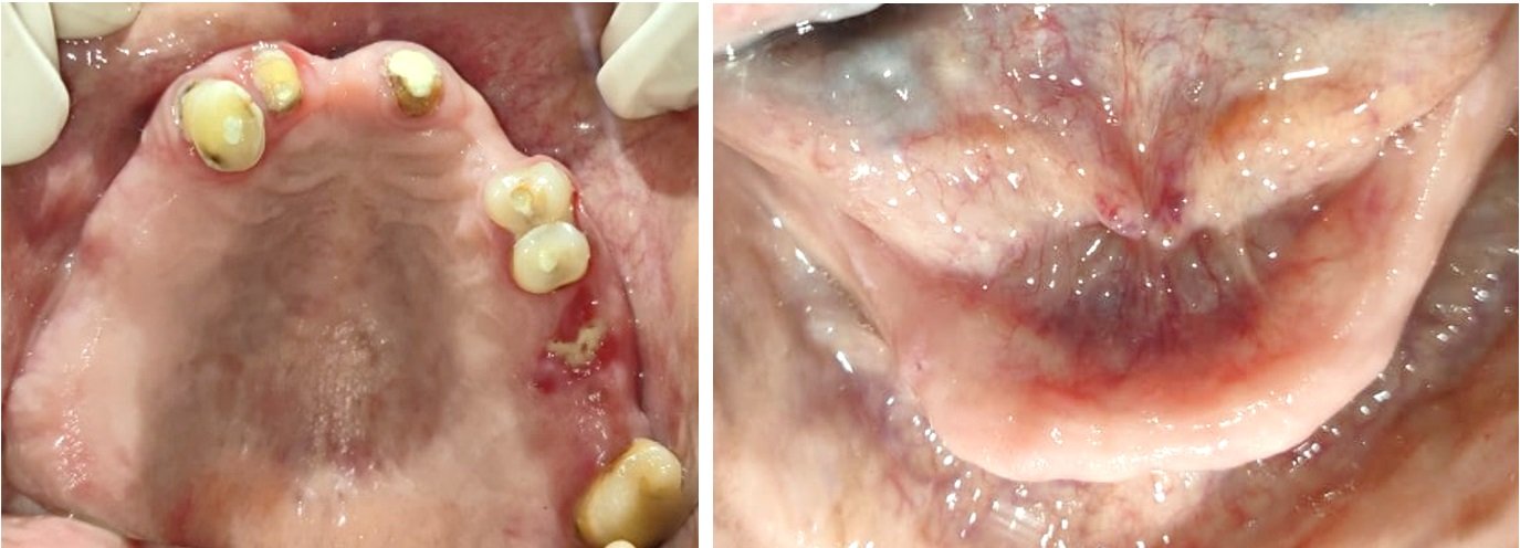
Fig. 2a : Intra-oral occlusal view of the Maxillary arch., Fig. 2b : Intra-oral occlusal view of the Mandibular arch
The patient was informed about different treatment options like conventional removable partial denture, flexible partial denture, cast partial denture with attachments and implant supported prosthesis depending on the intra-oral condition. After thorough understanding of the patient’s needs, age and economic status, a suitable treatment plan was selected.
Considering number of abutments, radiographic examinations & hard and soft tissue support, the prosthesis was planned which consisted of cast partial denture with unilateral preci-saggix extra coronal attachment for the maxillary arch and a removable complete denture prosthesis for the mandibular arch. The diagnostic impressions were made using irreversible hydrocolloid impression material and casts were fabricated [Fig. 3a, 3b] and a diagnostic jaw relation was done on a mean value articulator.
The orthopantomogram showed 13, 12, 11, 14 and 15 to be endodontically treated and completely edentulous mandible with no other significant finding.

Fig. 3a : Maxillary diagnostic cast, Fig. 3b : Mandibular diagnostic cast
The maxillary diagnostic cast was studied thoroughly and surveyed on a Ney’s surveyor to determine the desirable and undesirable undercuts. The guiding planes were determined and full coverage crowns were planned accordingly to allow for desirable retentive undercuts and single path of insertion and removal.
Therapeutic Intervention
Pre-prosthetic mouth preparation was done which included fiber post and core build-up in 12 and 21, mesial caries restoration in 24, tooth preparation for porcelain fused metal crowns in 13, 12, 21, 24, 25 and 28, rest seat preparation in 24, 25 and 28 [Fig. 4a, 4b]. A two-stage putty-light body impression was made using addition silicone for the maxillary arch [Fig. 5a]. For mandibular arch, spacer was fabricated and custom tray was made on the diagnostic cast. Border moulding was done using low fusing impression compound and a final wash impression was made using zinc oxide eugenol impression material [Fig. 5b]. Maxillary impression was poured in type IV gypsum and mandibular impression was poured in type III gypsum.
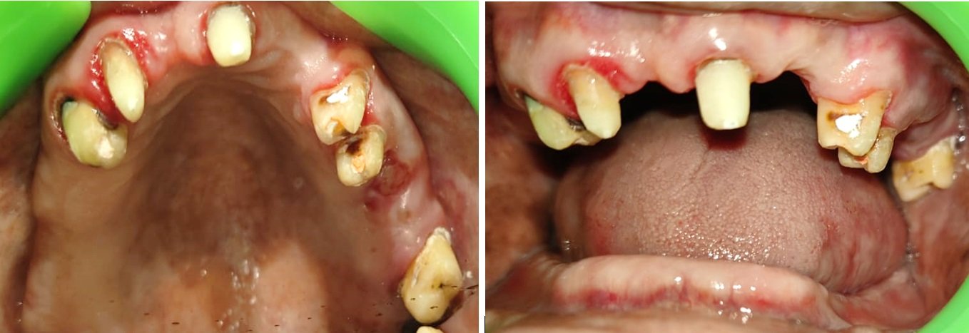
Fig. 4a: Pre-prosthetic mouth preparation occlusal view, Fig. 4b: Pre-prosthetic mouth preparation frontal view

Fig. 5a : Maxillary impression, Fig. 5b : Mandibular impression

Fig. 6a: Facebow transfer on a Semi-adjustable articulator, Fig. 6b: Jaw relation
Wax rims were fabricated on the casts, facebow transfer was done to a semi adjustable articulator [Fig. 6a], jaw relation was done and casts were mounted [Fig. 6b]. Co-Cr metal copings were fabricated for all the teeth wherein the male metal attachment of preci-saggix attachment was casted on the distal aspect of 13 using the 2.2 mm plastic castable pattern [Fig. 7]. Metal coping try-in was done [Fig. 8]. Pick up impression was made of these metal copings with the casted male part and final cast was obtained.
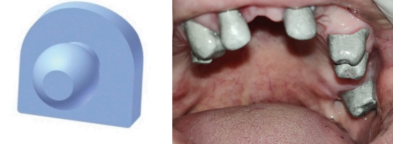
Fig. 7: Male plastic castable pattern, Fig. 8: Metal coping try-in
Unglazed PFM crowns were fabricated. Proper designing of the cast partial framework was done and framework was fabricated with the metal housing [Fig. 9]. Rims were fabricated on the cast metal framework and teeth arrangement was done [Fig. 10]. Try-in of prosthesis was done for proper occlusion and esthetics [Fig. 11].
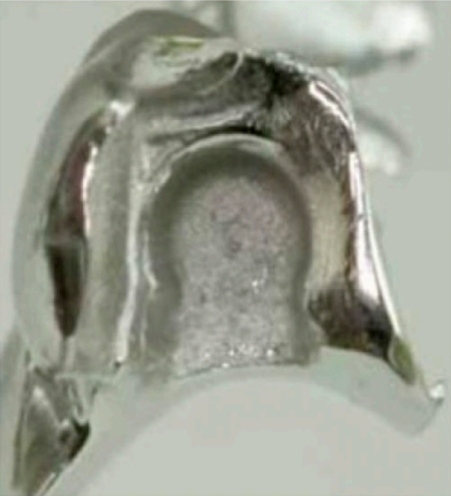
Fig. 9: Metal housing for female attachment
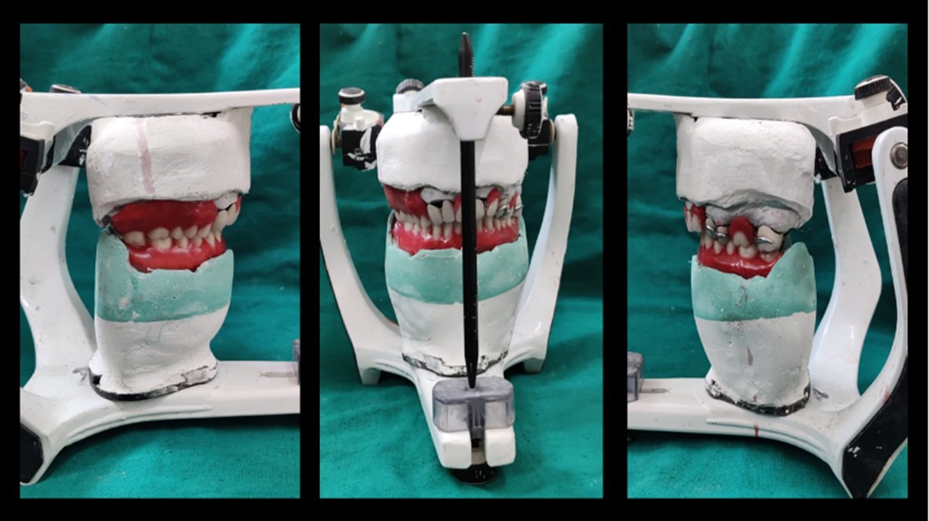
Fig. 10: Teeth arrangement on cast metal framework with unglazed PFM crowns on the articulator, Courtesy: https://www.preat.com/s3fs-public/2022-10/Preci-Sagix-Instructions.pdf
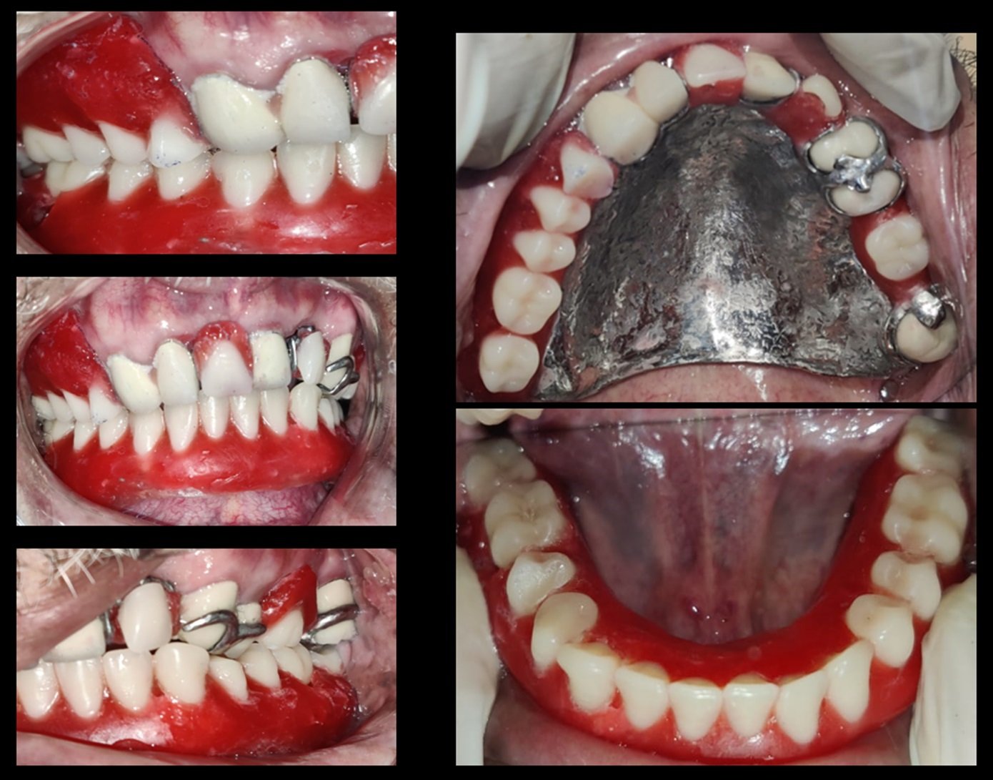
Fig 11: Try-In was done for proper occlusion and esthetics
After proper verification of trial denture, final acrylization was done and final prosthesis was incorporated with final yellow polyacetyl female part of preci-sagix attachment [Fig. 12a, 12b]
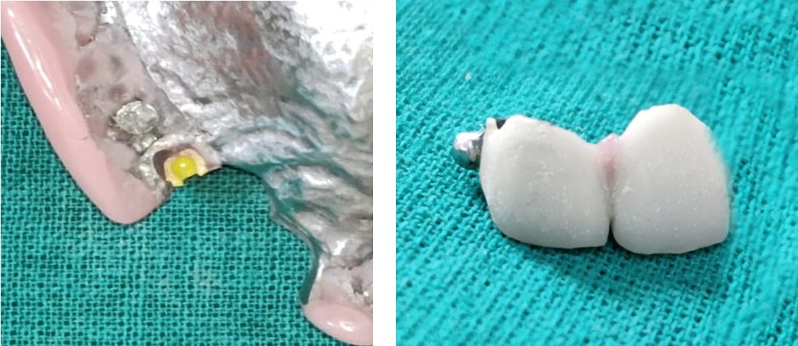
Fig. 12a: Final metal housing in cast metal framework with polyacetyl female part, 12b: Final male metal part in PFM crown distal to 13
The final prosthesis [Fig. 13a, 13b] was evaluated for proper extension, fit, phonetics, esthetics, occlusion and function [Fig. 14]. Pre-operative and post-operative view of patient extra-orally [Fig. 15].

Fig. 13a: Occlusal surface of final prosthesis
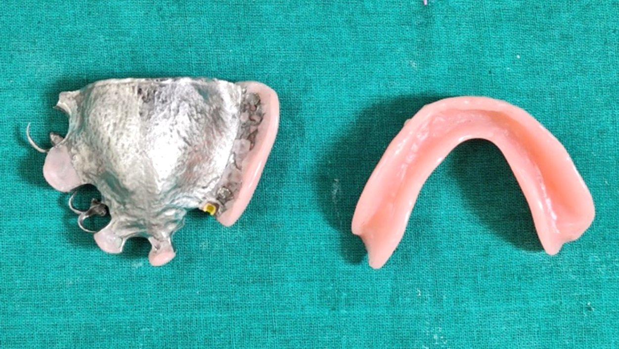
Fig. 13b: Intaglio surface of final prosthesis

Fig. 14: Final prosthesis insertion
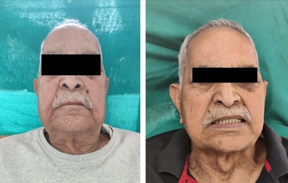
Fig. 15a: Extraoral pre-operative view, 15b: Extraoral post-operative view
A 24 hour, 1 week and 1 month post insertion follow up was done. Dentures were trimmed in certain areas for the comfort of the patient. Initially patient had difficulty in insertion and removal of the prosthesis but in a couple of days patient could insert and remove dentures comfortably.
DISCUSSION
The less retentive prosthesis is the main problem in distal extension edentulous patients. The factors which affect the retention of a prosthesis are size of the edentulous space, remaining teeth, remaining bony structure and the ability and time taken by the patient to adapt to the prosthesis. There are various causes for edentulism such as caries, periodontal factors, trauma etc. There are two treatment procedures for rehabilitation of a partially edentulous arch with distal extension such as implant therapy or by a cast partial denture. The implant treatment demands a surgical procedure for the patient, it is time consuming and more expensive. The prosthodontic rehabilitation through cast partial denture is less time consuming and is comparatively affordable. The patient’s acceptance, adaptability and hygiene practice plays an important role in success of a prosthesis due to less retention. The various ways of increasing retention in prosthesis is by using clasps, soft-liners, different impression techniques etc. These conventional methods have many limitations. These preci-saggix extra-coronal attachments are economical, non-invasive for the patients and at the same time lead to great improvement in retention. The saggix attachments are prefabricated, resilient attachments. Preci-saggix attachments are sagittal ball attachments with female retention clip for various other prosthetic applications like distal, lingual or buccal placement on overdenture bar cases. Male part gets attached with crowns (available in two sizes 1.7 mm mini and the 2.2 mm standard); female parts/clips (available in 3 types of retention: weak - white, standard - yellow and strong – red) are placed on tissue side of the denture. To conclude it can be said that, semi precision saggix extra coronal attachment helps to maintain balance between esthetic appeal and functional stability in cast partial dentures for the patient. The patient initially finds it difficult to understand the insertion and removal of the prosthesis with these kind of precision and semi-precision attachments but with proper assistance by the operator this can be overcome in a few days by the patient.
REFERENCES
- Schweikert, E O. “A prosthetic solution with cantilevered ceka-attachments.” Quintessence international, dental digest vol. 12,2 (1981): 165-72.
- Vikhe, Deepak Macchindra et al. “Cast Partial Denture with Cekapreci-Sagix Extra Coronal Attachment and Obturator for Rehabilitation of Patient with Maxillary Defect.” Journal of clinical and diagnostic research : JCDR vol. 10,8 (2016): ZJ07-9. doi:10.7860/JCDR/2016/21444.8278
- Den Haan R, Battistuzzi PG, Witter DJ, de Baat C, Creugers NH. (Semi-) precision attachments for cast metal frame removable partial dentures. Nederlands Tijdschrift Voor Tandheelkunde. 2011 Feb 1;118(2):93-100.
Disclaimer from Guident
The views and opinions expressed in articles published in Guident are solely those of the authors and do not necessarily reflect the views of the editors, the editorial board or the publisher, Ivory India. Guident does not endorse any product, technique or treatment described. Clinical content is intended for qualified dental and medical professionals and is not a substitute for individual clinical judgement. Guident, its editors and the publisher accept no liability for any consequence arising from the use of information published in the journal.
How to cite this article
Dr. Unnati Agarwal, Dr. Shitij Srivastava, Dr. Akanksha Dobhal. Cast Partial Denture with Semi Precision Saggix Extra-Coronal Attachment for Rehabilitation of Kennedys Class II Modification 3 Edentulous Space: A Case Report. Guident. 2024 Sep;17(10). Available from: https://ivoryindia.in/articles/prosthodontics/cast-partial-denture-with-semi-precision-saggix-extra-coronal-attachment-for-rehabilitation-of-kennedys-class.html


