Clinical Failures in Fixed Partial Dentures and its Management
Department of Prosthodontics A.B. Shetty Memorial Institute of Dental Sciences, Nitte University, Deralakatte, Mangalore, India
ABSTRACT
Fixed prosthodontic treatment failures can be complex in terms of both diagnosis and treatment planning. Most of the time, complications are conditions that occur during or after appropriately performed fixed prosthodontic treatment procedures. The purposes of this review article is to present regarding the classification and management of failures ,incidence of clinical complications associated with the following restorations/prostheses: single crowns, fixed partial dentures, all-ceramic crowns, resin-bonded prostheses and posts and cores.
Keywords : Failures, Complications, Classification of Failures, Management of failures in fixed partial dentures.
INTRODUCTION
Fixed prosthodontic treatment modality can offer exceptional satisfaction for both patient and dental specialist. It can transform an unhealthy, unattractive dentition with poor function into a comfortable, healthy occlusion capable of giving years of further service while greatly enhancing esthetics. The constituents of failure are more easily interpreted once restorative care objectives are stated. One can then define failure as an inability to meet or satisfy objectives.1
A complication has been defined as “a secondary disease or condition developing in the course of a primary disease or condition.” Although complications may be an indication that clinical failure has occurred, this is not typically the case. Knowledge regarding the clinical complications that can occur in fixed prosthodontics enhances the clinician’s ability to complete a thorough diagnosis, develop the most appropriate treatment plan, communicate realistic expectations to patients, and plan the time intervals needed for post-treatment care.2
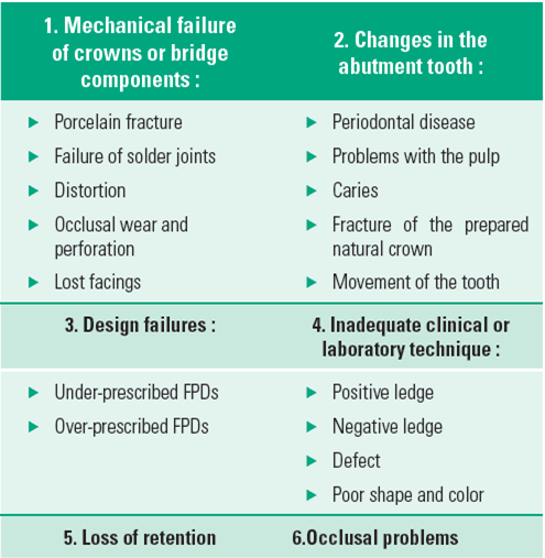
Classification of failures:
By John F Johnston3 :

By Barreto M T4 :

By Bennard G. N. Smith5 :
Evaluating The Quality of Existing Restorations6:
An evaluation of existing restoration will lead to one of the three possible courses:
- Leaving the restoration alone, if it is not causing any serious harm. Although this is the most common choice by far, it must not be arrived at by default. Instead a careful examination is necessary to rule out any defects.
- Adjusting or repairing the fault.
- Replacing the crown or bridge or the existing restoration which is amenable to modification.
Many failures occur because the dental specialists does not take time to do a thorough diagnosis and plan for success. The four major diagnostic aids to making a dental diagnosis are:
- Taking a history
- Doing a careful clinical examination
- Having a complete set of radiographs
- Having a good set of diagnostic casts correctly mounted.
Clinical management of failed restorations:
- Cement Failure:
- Mechanical failure of crowns or bridge components:
- The framework must provide a uniform veneer of porcelain (approx 1.2mm), excessive thickness of porcelain contributes to inadequate support and predisposes to eventual fracture. This is often true in the cervical portion of an anterior pontic.
- A reliable technique for ensuring uniform thickness of porcelain is to wax the fixed prosthesis to complete anatomic contour and then accurately cut back the wax to a pre-determined depth.
- The metal surfaces to be veneered must be smooth and free of pits. Surface irregularities will cause incomplete wetting by the porcelain slurry, leading to voids at the porcelain metal interface that reduces bond strength and increases the possibility of mechanical failure.
- Sharp angles on the veneering surface should be rounded. They produce increased stress concentrations that could cause mechanical failure.
- Occlusal centric contacts must be placed at least 1.5mm away from the junctions. Attention must be paid to excursive eccentric contacts that might deform the metal ceramic interface.
- Etch porcelain/metal surface with 4% hydrofluoric acid for 4 minutes.
- Rinse and dry thoroughly.
- Apply one coat of Porcelain Primer (Silane) to exposed porcelain.
- Light cure for 10 seconds.
- Mix equal amounts of dual cure Opaquer Base & Catalyst.
- Apply thin layer on exposed metal surface to mask out metal shine-through.
- Light cure for 10 seconds.
- Use the composite of choice and light cure in small increments or use a microhybrid composite.
- Proceed with finishing and polishing.
- Changes in the abutment tooth:
- Periodontal disease ( if diagnosed early no treatment needed)
- Problems with the pulp ( despite taking usual precautions teeth may become non vital; it’s sometimes reasonable to attempt endodontic treatment making an access through the crown.
- Margins are one of the most important and weakest links in the success of FPD restorations. One of the prime goals of restorative therapy is to establish a physiologic periodontal health.
- A successful prosthesis depends on a healthy periodontal environment and periodontal health depends on the continued integrity of the prosthodontic restoration.
- All displacement techniques have the potential damage to gingiva, attachment apparatus and bone, especially if anatomic forms are weak or if disease is present.
- In healthy patients, properly used cord displacement or copper band methods have proved to be atraumatic.
- Supragingival Vs Subgingival Margins7:
- Loss of retention:
- Incorporation of proximal grooves.
- Additional retentive grooves (should be along with the path of insertion).
- Additional pins
- Crown lengthening procedure
- Modification of supra-gingival margin to sub-gingival margin
- Additional retentive grooves and proximal box
- Incorporation of pins
- Addition of extra abutments
To reduce potential postoperative sensitivity, the use of a non resin-based sealer, which also enhances retention, has been recommended.
Applying petroleum jelly to the exposed glass-ionomer cement margin after bulk removal has been suggested as a simple solution to maintain water balance.
Dehydration remains a problem, so isolation from the oral environment for longer than 10 minutes is not recommended.
To avoid mechanical failures:
Porcelain Repair (Porcelain fused to metal crown ) With Composite (for optimal results: isolate with rubber dam):
Whenever possible, the margin of the preparation should be supragingival.Subgingival margins of cemented restorations have been identified as a major factor in periodontal diseases, particularly where they encroach on the epithelial attachment.Supragingival margins are easier to prepare accurately without trauma of the soft tissues. They can usually also be situated on hard enamel, whereas subgingival margins are often on dentin or cementum.
Clinical conditions with excessive taper and short clinical crowns should be treated with:
In case of excessive taper :
In case of short crowns :
Techniques for adjustments, adaptations and repairs to crowns and bridges
- Assessing the seriousness of the problem
- Decision has to made between:
Leaving it alone, if it is not causing any serious harm, adjusting or repairing the fault, replacing the crown or bridge.
Adjusting by grinding and polishing in situ:
- Porcelain finishing instruments
- Reciprocating Hand-piece (for removing overhangs)
Repairs by restoring in situ:
- Occlusal repairs (In metal can be done with Amalgam. In porcelain, Composite can be used).
Removal of a prosthesis:
- Many well retained restorations cannot be removed intact and to prevent abutment tooth damage, must be cut off the prepared tooth and there by destroyed. Attempts should be made for intact removal of restorations without damaging the abutments. The forces applied for removal should be sharp and in an occlusal direction.
Suggested devices and techniques include use of
- Matrix band,
- Hemostat,
- Richwil crown remover (Richwil Laboratories, Orange, Calif),
- Acrylic resin mold compressed with a curved instrument
- Ultrasonics,
- Prepared slot for a purchase point
- Pneumatic crown remover.
These techniques for casting removal have been reported to result in fractured porcelain margins or damage to preparation finish lines.
- Sectioning and prying method: The safest but most destructive method of removing cemented units is by cutting a channel through the restoration to prepared tooth structure on the facial or lingual and occlusal or incisal aspects and gently expanding the casting with a large spoon excavator to break the cement joint.
Clinical complications in fixed prosthodontics2:
- The lowest incidence of clinical complications was associated with all-ceramic crowns (8%).
- Posts and cores (10%) and conventional single crowns (11%) had comparable clinical complications incidences.
- Resin-bonded prostheses (26%) and conventional fixed partial dentures (27%) were found to have comparable clinical complications incidences.
Incidence of Clinical complications in all-ceramic crowns:
- Crown fracture (7%)
- Loss of retention (2%)
- Need for endodontic treatment (1%)
- Caries (0.8%)
Incidence of Clinical complications in post and core:
- Post loosening (5%)
- Root fracture (3%)
- Caries (2%)
- Periodontal disease (2%)
Incidence of Clinical complications with conventional single crowns:
- Endodontic treatment (3%)
- Porcelain fracture (3%)
- Loss of retention (2%)
- Periodontal disease (0.6%)
- Caries (0.4%)
Incidence of Clinical complications with resin bonded prosthesis:
- Debonding (21% of prostheses)
- Tooth discoloration (18% of prostheses)
- Caries (7% of prostheses)
- porcelain fracture (3% of prostheses)
Incidence of Clinical complications with conventional fixed partial denture prosthesis:
- Caries (18% of abutments and 8% of prostheses)
- Need for endodontic treatment (11% of abutments and 8% of prostheses)
- Loss of retention (7% of prostheses)
- Esthetics (6% of prostheses)
- Periodontal disease (4% of prostheses)
- Tooth fracture (3% of prostheses)
- Prosthesis/porcelain fracture (2% of prostheses)
DISCUSSION
Study by De Backer et al8 concluded that the overall survival estimation for Short span -FDPs was statistically significantly better than for Long span -FDPs at year 20. The use of an RCT abutment becomes more significant in fixed prosthetic restorations with 4 or more units. Occurrence of a previously reversible complication is a predictive factor for an irreversible complication later on. A reversible complication within the first 2 years for an Short span-FDP will lead to an irreversible complication.
Eliasson A et al9 stated Metal-ceramic FPDs made of cobalt-chromium alloy performed acceptably in the questionable prognosis and advanced chronic periodontitis groups.
Layton D10 stated Metal-ceramic FDPs had high survival, with a significantly greater 5-year survival rate than all-ceramic FDPs. Differences in complications were unknown, but evidence indicated that the complication incidence of metal-ceramic FDPs was lower than that of all-ceramic FDPs.
Longevity of fixed partial dentures by Libby G et al11 supported previous reports of dental caries as the primary cause for failure of FPDs, but specific radiographic, clinical, and laboratory procedures can increase the length of service of these restorations.
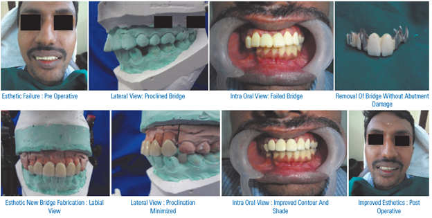
When overall dental appearance is considered, several factors are of significance, including tooth color, shape, and position; restoration quality; and the general arrangement of the dentition, especially of the anterior teeth. Each factor may be considered individually, but all components together act in concert to produce the final esthetic effect. In the following images an effort has been made to outline the possible esthetic errors which occur in the absence of careful treatment planning during the fabrication of fixed partial denture procedure and management of the same to improve the esthetics.
CONCLUSION
The first consideration when confronted with any failure or repair situation is to ascertain the cause or suspected cause. Sometimes this is easy and obvious. If there is a cause that is correctable it should be taken care of first. Care should be taken not to become involved in repairs that should have been remakes. Repairs are usually second best to the original in one or more ways. Imagination and innovation are key factors in successful repairs. Most failures are unique and present varying challenges to the dental specialist. Great satisfaction can be achieved in meeting a situation and solving it in an effective and economical manner.
REFERENCES
- Manappallil JJ. Classification system for conventional crown and fixed partial denture failures. J Prosthet Dent. 2008 Apr;99(4):293-8.
- Charles J. Goodacre et al Clinical complications in fixed prosthodontics J Prosthet Dent 2003;90:31-41.
- Dykema R.W., Goodacre C.J. and Phillips R.W. “Johnston’s Modern Practice in Fixed Prosthodontics”. Fourth Edn, W.B. Saunders Co. Philadelphia, London.
- Barreto M.T. “Failures in ceramometal fixed restoration”. J. Prosthet. Dent. 1984; 51: 186-189.
- Bernard G N smith – planning and making crowns and bridges.
- Donald W. Fisher/William W Morgan. Modification And Preservation Of Existing Dental Restorations.
- James S. Marcum - The effect of crown marginal depth upon gingival tissue JPD 1967;17: 479-88.
- De Backer H et al. Long-term results of short-span versus long-span fixed dental prostheses: an up to 20-year retrospective study. Int J Prosthodont. 2008 Jan-Feb ;21(1):75-85.
- Eliasson A et al. A clinical evaluation of cobalt-chromium metal-ceramic fixed partial dentures and crowns: A three- to seven-year retrospective study. J Prosthet Dent. 2007 Jul;98(1):6-16.
- Layton D et al. A critical appraisal of the survival and complication rates of tooth-supported all-ceramic and metal-ceramic fixed dental prostheses: the application of evidence-based dentistry. Int J Prosthodont. 2011 Sep-Oct;24(5):417-27.
- Libby G. Longevity of fixed partial dentures. J Prosthet Dent. 1997 Aug;78(2):127-31.
Disclaimer from Guident
The views and opinions expressed in articles published in Guident are solely those of the authors and do not necessarily reflect the views of the editors, the editorial board or the publisher, Ivory India. Guident does not endorse any product, technique or treatment described. Clinical content is intended for qualified dental and medical professionals and is not a substitute for individual clinical judgement. Guident, its editors and the publisher accept no liability for any consequence arising from the use of information published in the journal.
How to cite this article
Dr. Krishna Prasad, Dr. Anshul Bardia, Dr. Anupama Prasad. Clinical Failures in Fixed Partial Dentures and its Management. Guident. 2013 Apr;6(6). Available from: https://ivoryindia.in/articles/prosthodontics/clinical-failures-in-fixed-partial-dentures-and-its-management.html