Endocrowns: An Innovative Approach for Reconstruction of Molars
Department of Prosthodontics and Implantology, K.D. Dental College And Hospital, Mathura
ABSTRACT
The restoration of endodontically treated teeth that are grossly destructed has been a challenge for many clinicians over the years. Rehabilitation of endodontically treated molar still remains a challenge. After endodontic treatment, molars lost their mechanical characteristics. In fact, they became fragile and that is in relation with the removal of pulp and surrounding dentin tissues.
Endocrown can be an excellent option for restoring severely damaged teeth, which showed better performance in molars than premolars. It is an adhesive restoration with minimally invasive preparation. The preparation design should provide sufficient retention, stability and structural durability to the restoration. Endocrown which is a single partial restoration could be considered as a good alternative for restoring molars having large coronal destruction and presenting endodontic treatment difficulties.
This article aims to provide a comprehensive literate review of endocrown restorations for the restorative dentist as they might be hesitant to apply such a successful treatment option of endodontically treated teeth in their clinical practice because it‘s not as widely used as other conventional fixed indirect restorations such as crowns and onlays.
Keywords: endodontically treated teeth, CAD/CAM, restorations, ferrule, crown preparation posts
INTRODUCTION
Endodontically treated teeth usually need special techniques to restore them. When a tooth is Endodontically treated, a considerable amount of tooth structure usually was lost due to trauma or caries in addition to the central destruction created by the endodontic access preparation. This usually makes the tooth left with insufficient sound tooth structure to support a casted restoration unaided.
Endodontically treated teeth exhibit various physiological alterations in the dentin composition and microstructure which predispose the tooth to multiple risk factors, such as, reduced retention/stability, increased tooth fragility, compromised substrate adhesion and eventually leading to failure of the prosthesis
The choice of restoration will be thus influenced by the type of tooth; posterior or anterior, and the amount of the remaining tooth structure .
To solve these problems a couple of clinical techniques were suggested. Anterior teeth with a limited access opening and sufficient tooth structure can be just restored by a direct restoration with no need for the placement of a crown.
However, posterior endodontically treated teeth will always need cuspal coverage due to their morphological characteristics and the increased loads they are subjected to.
Traditionally to retain the core structure in such cases a post or dowel is placed. These posts can be prefabricated posts with a direct core or a one-piece custom- made post and core. The introduction of adhesives and the development of effective dentine adhesives was a changing point in the restoration of Endodontically treated teeth, which made the insertion of a radicular post a less favored option as long as there is sufficient surface area for adhesion.
In 1995 Pissis presented a novel technique that utilized porcelain core/crown unit a single unit is described as a monolithic (one-piece) full-composite or full ceramic overlays which restore partially or totally the coronal part of a devitalized tooth characterized by a supracervical butt joint, retaining maximum enamel to improve adhesion and extended inside the pulp chamber and partially inside the root canal with a short ―endo-core, could represent an alternative to classical treatments to restore endodontically treated teeth . These restorations have macromechanical retention by being anchored to the internal portion of the pulp chamber and to the cavity margins and microretention by adhesive cementation. The technique was called the monobloc technique it was suggested by the author to replace the traditional metal post and core.
PREPERATION
The main purpose for the use of Endocrowns is to attain an all-ceramic bonded restoration that is minimally invasive of root canals. Therefore, the Endocrown preparation is different from the conventional full coverage crowns . It is a monolithic ceramic bonded restoration with a supragingival butt joint keeping as much as possible enamel for improved adhesion. The endocrown will invade the pulp chamber only. The pulpal chamber shape and cavity warrants stability and retention.
Occlusal Preparation
The goal in preparation is to achieve an overall reduction in the height of the occlusal surface of at least 2 mm in the axial direction. This reduction can be achieved by drilling 2-mm-deep grooves as guides (Fig. 1), then using a green diamond wheel bur to reduce the occlusal surface.
The bur is oriented along the major axis of the tooth and held parallel to the occlusal plane (Fig. 2). Its shape allows control of the orientation of the reduction and ensures a flat surface, which determines the position of the cervical margin or “cervical sidewalk.” The cervical margin should be supragingival; however, if clinical factors or esthetics require, the margin can follow the gingival margin. Differences in level between the various parts of the cervical margin must be linked by a slope of no more than 60° to avoid a staircase effect. Enamel walls less than 2 mm thick should be removed.

Figure 1: Making the guide grooves in an isolated tooth and in situ.

Figure 2: Preparation of the cervical margin or “cervical sidewalk” using a wheel bur held parallel to the occlusal plane
Axial Preparation
This step primarily involves eliminating undercuts in the access cavity. A cylindrical-conical green diamond bur with a total occlusal convergence of 7° is used to make the coronal pulp chamber and endodontic access cavity continuous (Fig. 3). With the bur orientated along the long axis of the tooth, the preparation is carried out without excessive pressure and without touching the pulpal floor. Removing too much tissue from the pulp chamber walls will reduce their thickness and the width strip of enamel. The depth of the cavity should be at least 3 mm.
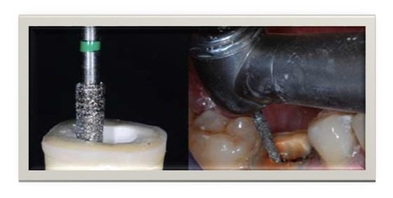
Figure 3: Axial preparation using a cylindro-conical drill to make the coronal pulp chamber continuous with the access cavity.
Polishing the Cervical Band
The bur used in this step has the same taper as the one used in axial preparation, but a larger diameter and a finer particle size. It should be guided around the entire surface of the cervical band to remove micro-irregularities and produce a flat, polished surface (Fig. 4). The margin line should appear as a regular line with a sharp edge (Fig. 5a and b).
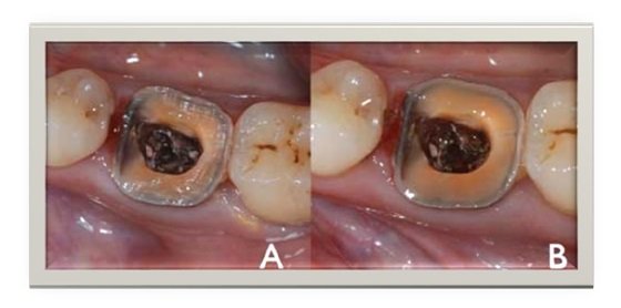
Figure 4: Polishing the cervical band.
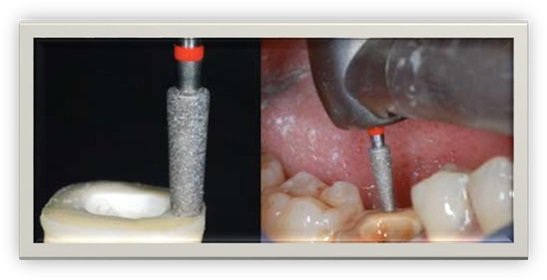
Figure 5: Cervical margin before (a) and after (b) polishing.
Preparation of the Cavity Floor
The entrance to the pulpal canal is opened. Gutta percha is removed to a depth not exceeding 2 mm to take advantage of the saddle-like anatomy of the cavity floor. This should be done with a nonabrasive instrument to maintain the integrity of the canals entrance. No drilling of dentin is carried out.
Cleaning the Pulp Chamber
Ultrasound is recommended to clean the pulp chamber and its floor thoroughly. Abrasion is not indicated.
Bonding
Adhesives such as self-adhesive RelyX Unicem (3M, St. Paul, Minn.) or composites such as Multilink (Ivoclar, Schaan, Liechtenstein) are used for bonding the endocrown to the prepared tooth. (Fig. 6).

Figure 6: Prepared tooth (a), endocrown (b), final result after bonding (c).
Ferrule
The presence of ferrule in full coverages crowns supported by post and core was thoroughly investigated and well acknowledged to increase fractures resistance and fatigue cycles to failure .
Einhorn et al. investigated the consequence of the ferrule features incorporation, on molar endocrown failure resistance. Their results showed that adding ferrule to preparations increased the dentin surface available for bonding. However, there were milling limitations in reproducing the endocrowns inner surface. Hence, it was reported that the more complex the preparation design became because of the addition of ferrule, the resultant endocrown inner surface adaptation to the preparation seemed to reduce. (Figure A)
They concluded that ferrule-containing endocrown preparations revealed significantly superior failure loads than regular endocrown restorations; yet, there was no difference among the groups in the calculated failure stress based on existing surface area for adhesive bonding. Moreover, less occurrences of disastrous failure were detected with the Endocrown preparations containing 1 mm of preparation ferrule design.
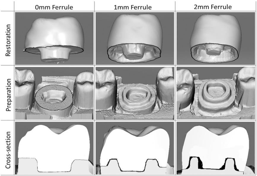
Fig A: Three preparation design with no ferrule, 1 mm ferrule, 2 mm ferrule.
Additional features
Endocrowns are made usually from bonded ceramic restorations, taking into consideration the benefit of adhesion in the retention of the restoration. In a case report, Vinola et al. described the use of metal-ceramic endocrown. To improve the retention and stability of this restoration additional feature was proposed to the endocrown preparation. Retentive grooves (1 mm deep) were placed on the buccal and lingual axial surfaces of the external aspect of the tooth. The metal-ceramic endocrown restoration was also sandblasted as an additional mean to enhance restoration retention.
CAD/CAM endocrowns:
In a systematic review of the clinical performance of CAD/CAM single-tooth restorations that included 16 articles with the total of 1,957 restorations included 48% that were prospectively analyzed and 52% that were studied retrospectively. The majority of the studied restorations were on posterior teeth 98% and only 2% were on anterior teeth in which none of the analyzed studies reflected an RCT protocol design. This systemic review reported that endocrowns had a significantly higher failure rate than all other investigated restorations with an estimated failure rate of 3.9% per year, estimated per 100 restoration years and the lowest 5-year survival rate was found for endocrowns (82.3%) P value= 0.026. In contrast to conventional crowns the estimated 5-year survival rate was 92.3%. Other findings of this systemic review were: There is no significant differences between the failure rates of the different CAD/CAM systems. In regards to the material choice glass-ceramic restorations had a significantly higher failure rate than all other materials (P < .001, 18.18%) and ceramics with aluminum and magnesium oxide (In-Ceram Spinell) had the highest survival rate 96.8%. The luting cements did not appear to affect the outcome of the mentioned study. Also it concluded that the long-term survival rates for CAD/CAM technology– fabricated single-tooth restorations demonstrated clinically similar outcomes to conventionally manufactured restorations. Moreover, a study evaluated the marginal and internal discrepancies of endocrowns with different cavity depths 2mm and 4mm fabricated using two different chairside CAD-CAM systems (CEREC AC and E4D) concluded that marginal and internal discrepancies increased depending on cavity depth and both chairside CAD-CAM systems showed similar discrepancy in the endocrowns . In contrast, another study, that used lithium disilicate CAD-CAM ceramics to fabrication conventional crowns and endowcrowns stated that the differences in the survival between the groups were not statistically significant after the application of thermo- mechanical fatigue loading.(Fig B)
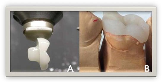
Figure B: An endocrown machined using computer-aided design and computer-aided manufacture (a). A pressed endocrown, with sprue attached, positioned on a master mold (b).
Contraindications
Endocrowns can‘t be used in the following scenarios:
(1) Less than 3mm pulp chamber depth.
(2) When adhesion cannot be assured.
(3) If only negligibleremaining tooth structure is present.
DISCUSSION
Longevity and Effectiveness
In an evaluation of adhesively placed endocrowns after 2 years, Bindl and Mörmann2 concluded that “the overall clinical quality of the endocrowns was very good.” In another 2-year evaluation, Bernhart et al. concluded that endocrowns “represent a very promising treatment alternative for endodontically treated molars.” In 2012, Biacchi and Basting compared the fracture strength of 2 types of full ceramic crowns: indirect conventional crowns retained by glass fibre posts and endocrowns. They concluded that endocrowns were more resistant to compressive forces than conventional crowns. More recently, finite element analysis highlighted the role of endocrowns in stress distribution.
INDICATIONS AND CONTRAINDICATIONS
The endocrown is suitable for all molars, particularly those with clinically low crowns, calcified root canals or very slender roots. The endocrown is contraindicated if adhesion cannot be assured, if the pulpal chamber is less than 3 mm deep or if the cervical margin is less than 2 mm wide for most of its circumference.
Choice of Materials
Glass-ceramic: Glass-ceramic has the advantages of biocompatibility and biomimicry, and its wear coefficient is close to that of the natural tooth. In addition, the single interface of a 1-piece restoration enhances cohesion.
Bonding Agent: The bonding material constitutes the critical interface between the restoration and the prepared tooth. In addition to its adhesive properties, its modulus of elasticity is important as it must be able to absorb pressure, just as the dentin enamel junction (DEJ) does. The interface includes all prepared surfaces. Products that must be photopolymerized require the use of a high- power lamp that must be able to reach light-triggered initiators on the pulpal floor, under layers of ceramic that sometimes exceed 7 mm.
Preparation
The butt joint, or cervical sidewalk, is the base of the restoration — with a band of peripheral enamel that optimizes bonding. Unlike chamfer or shoulder preparation techniques, crimping is prohibited. The goal is to achieve a wide, even, stable surface that resists the compressive stresses that are most common on molars. The prepared surface is parallel to the occlusal plane to ensure stress resistance along the major axis of the tooth.
The pulpal chamber cavity ensures retention and stability. Its shape trapezoidal in mandibular molars and triangular in maxillary molars — enhances the restoration’s stability.
There is no need for additional preparation. The saddle form of the pulpal floor enhances stability. This anatomy, along with the adhesive qualities of the bonding material, makes it unnecessary to attempt further use of post involving root canals. Actually the root canals do not require any specific shape; therefore they are not weakened by the drilling and they will not be subject to the stresses associated with the use of post. The compressive stresses are reduced, being distributed over the cervical butt joint and the walls of the pulp chamber.
CONCLUSION
The preparation for endocrowns is simple and can be achieved quickly. Root canals are not engaged in the process, and the procedure is less traumatic than others. The supragingival position of the cervical margin protects the marginal periodontium, facilitates impression taking, and preserves the solid substance of the remaining tooth. Forces are dispersed over the cervical butt joint (compression) and axial walls (shear force), thus moderating the load on the pulpal floor. The endocrown represents a very hopeful treatment alternative for endodontically treated molars, it allows maintaining of tooth structure, it is compatible with goal minimally invasive dentistry, and it is adequate for the concept of biointegration. It is a conservative approach for mechanical and aesthetic restoration of nonvital posterior teeth.
This type of reconstruction, which is still uncommon, should be more widely known and practised.
REFERENCES
- Rosenstiel S, Land M, Fujimoto J. Contemporary fixed prosthodontics. 5th ed. Philadelphia: Elsevier, 2015, 280-281.
- Shillingburg H, Sather D. Fundamentals of fixed prosthodontics. 4th ed. Chicago: Quintessence Pub, 2012, 214-216.
- Bindl A, Mörmann WH. Clinical evaluation of adhesively placed Cerec endo- crowns after 2 years — preliminary results. J Adhes Dent. 1999;1(3):255-65.
- Pissis P. Fabrication of a metal-free ceramic restoration utilizing the monobloc technique. Pract Periodontics Aesthet Dent. 1995;7(5): 83-94.
- Endocrowns: A Review Article (Waad Alomran* Dental Intern, College of Dentistry, King Saud University, Riyadh, Kingdom of Saudi Arabia)
- Endocrown: An Alternative Approach for Restoring Endodontically Treated Molars with Large Coronal Destruction Houda Dogui,1,2,3 Feriel Abdelmalek,1,2,3 Adel Amor,1,2,3 and Nabiha Douki 1,2,3 1.Department of Dental Medicine, Hospital Sahloul, Sousse, Faculty of Dental Medicine, Monastir, Tunisia 2. Laboratory of Research in Oral Health and Maxillo Facial Rehabilitation (LR12ES11), University of Monastir, Monastir, Tunisia 3. Faculty of Dental Medicine, University of Monastir, Monastir, Tunisia
- Endocrown preparation: Review Dr. Marwa EI Elagra MSc, Lecturer in Prosthodontic Department, Riyadh Elm University, Prince Abdulaziz bin Musaed bin Jalawy, Riyadh, KSA, Saudi Arabia
- T. Otto and W. H. Mörmann, “Clinical performance of chairside CAD/CAM feldspathic ceramic posterior shoulder crowns and endocrowns up to 12 years,” International Journal of Computerized Dentistry, vol. 18, no. 2, pp. 147–161, 2015.
- M. M. Belleflamme, S. O. Geerts, M. M. Louwette, C. F. Grenade, A. J. Vanheusden, and A. K. Mainjot, “No post-no core approach to restore severely damaged posterior teeth: an up to 10-year retrospective study of documented endocrown cases,” Journal of Dentistry, vol. 63, pp. 1–7, 2017.
Disclaimer from Guident
The views and opinions expressed in articles published in Guident are solely those of the authors and do not necessarily reflect the views of the editors, the editorial board or the publisher, Ivory India. Guident does not endorse any product, technique or treatment described. Clinical content is intended for qualified dental and medical professionals and is not a substitute for individual clinical judgement. Guident, its editors and the publisher accept no liability for any consequence arising from the use of information published in the journal.
How to cite this article
Dr. Shubhangi Srivastav, Dr. Siddharth Sisodia. Endocrowns: An Innovative Approach for Reconstruction of Molars. Guident. 2021 Jan;14(2). Available from: https://ivoryindia.in/articles/prosthodontics/endocrowns-an-innovative-approach-for-reconstruction-of-molars.html