Extra Oral Implants in Maxillofacial Prosthodontics-an Insight
Department of Prosthodontics & Implantology, K.D. Dental College & Hospital, Mathura (U.P) India
ABSTRACT
Defects or deformities in the head and facial area almost always lead to a severe emotional disturbance. So the restoration of these defects play a vital role in function and cosmetic outcome. The field of maxillofacial prosthetics is embracing the rapid explosion of technology. Extra oral implants are currently used in oncology and trauma patients with intraoral soft and hard tissue defect resulting in more natural appearing and functioning prosthesis in comparison with the conventional methods for retention of maxillofacial prostheses.
This articles reviews the uses of extra oral implant in various maxillofacial prosthesis and also outlays the advantages of extraoral implants as a mode of retention over other means of retention. It provides stability of the prostheses in hygienic and aesthetically pleasing way.
KEY WORDS Extraoral Implants, Osseointergrated Implants, Maxillofacial Implant.
INTRODUCTION
The use of maxillofacial prostheses is important for the social reintegration of patients with deformities, either congenital or acquired.1 Congenital defects are the defects or malformations present from birth whereas the acquired defects may be due to accidents, gunshot injuries, cancer treatment, ablative surgery and animal bite.
Maxillofacial prostheses encompasses prosthetic rehabilitation of patients with oral, para oral, or facial defects, which may be acquired or congenital. Syndromes like Teacher Collins, Crouzon’s, and Pierre robins are associated with facial deformities, palatal cleft and significant malformation external ear congenital abnormalities.2 These malformations affect the well-being of the individual affecting them psychologically depriving the confidence levels.
Maxillofacial defects that compromise appearance, function and render an individual incapable of leading a relatively normal life have usually prompted responses that seek to bring the person to a state of being acceptable normally.3
- Surgery is the first choice for early cancers and for cancers that do not respond to radiation and chemotherapy, resulting in cosmetic, functional and psychological impairment, greatly affects the patient’s quality of life.
- Maxillofacial or craniofacial prosthesis rehabilitation- Rehabilitation of such patients is quiet challenging and requires multidisciplinary team for comprehensive care and optimal post treatment functional outcomes.
- Combination of the above two.4
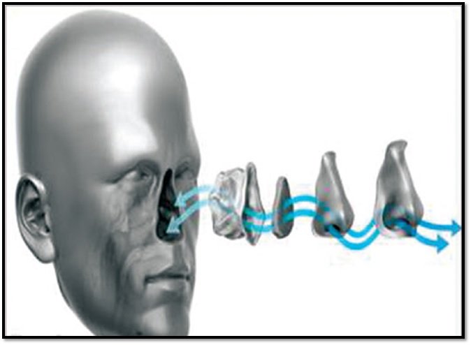
Maxillofacial prosthetics defined by the current Glossary of Prosthodontic Terms – GPT 9 “ as the branch of Prosthodontics concerned with the restoration and/or replacement of the stomatognathic (jaws) and craniofacial (facial) structures with prostheses that may or may not be removed on a regular or elective basis”.5
HISTORY OF MAXILLOFACIAL PROSTHESES
The origin of maxillofacial prostheses is not clear. According to Conroy, earliest known application of engineering principles to restore facial appearance and dental occlusion may be attributed to Hippocrates. Mummified Egyptians were found with enamel-covered, silver eyes with bronze lids as well as nasal and auricular structures. Similar to the Egyptians, the ancient Greeks fabricated artificial eyes with silver and placed them in their statues.6
Ambroise Paré provided the first documented use of maxillofacial prostheses during the 16th century. Glass and wood were also used to fabricate maxillofacial prostheses as they became more common in Europe during the 16th century.7

Fig : the Egyptians, the ancient Greeks fabricated artificial eyes with silver and placed them in their statues.
At the end of 19th century, maxillofacial prosthetics had an important role in the quality of life for recuperating soldiers since they were able to engage in social activities and go out in public.
The first half of the 20th century, Between World Wars I and II, researchers tried to develop better materials for the fabrication of maxillofacial prostheses, such as prevulcanized latex. According to Ferreira, these new prostheses should be produced using engineering, computer-aided design and manufacturing (CAD-CAM), and surgical guides.7

RETENTION OF MAXILLOFACIAL PROSTHESES
The means of achieving retention of facial prostheses has been primarily by use of medical adhesives or by means of anatomical or mechanical retention. Each retentive mechanism is having its own advantage and disadvantage.
The various retentive aids available are such as adhesive, skin tapes/ straps/ suture material/ toupee tapes, spectacles frames, springs, soft tissue or bony undercuts, anatomic projections using them as mechanical interlocks, Implants (E.g., studs, clips or magnets).8
These retentive aids are selected based on the various factors such as the extent of prosthesis, availability of bone, radiation therapy, patient’s dexterity, location, amount of hard and soft tissue available and compliance of the patient.
In the past maxillofacial prostheses are craniofacial prosthesis has been retained by mechanical tools, undercuts and adhesives. Since 1979, there has been a shift towards the implant retained prostheses, which are preferred by most of the patients. The most significant advance in craniofacial prosthesis over the last several decades has been the application of osseointegration to address the problem of retention of extraoral prosthesis.9
MULTIDISCIPLINARY APPROACH FOR MAXILLOFACIAL REHABILITATION
The treatment of maxillofacial or craniofacial defect is a multidisciplinary approach with a combination of both invasive and non-invasive treatment options. The maxillofacial team should consist of various members to plan the treatment including ablative surgeons, reconstructive surgeons, maxillofacial prosthodontists and maxillofacial technicians.
Factors to be considered for the prosthodontic rehabilitation are as follows:
- Amount of remaining supportive tissue
- Number, position and condition of the remaining teeth.
- Age and medical condition of the patient
- Pathologic findings
- Patient preferences regarding surgical versus prosthetic reconstructions
- Technical skills of the reconstructive surgeon and prosthodontist
- Psychological status and manual dexterity of the patient to deal with maxillofacial prosthesis
- Availability of adequate supportive care in case the patient is not able to take care of the prosthesis.9
BIOMECHANICAL CONSIDERATIONS OF IMPLANT IN RETAINED MAXILLOFACIAL PROSTHESIS
Following factors should be consider while planning for the extraoral implants10

A. Design of craniofacial and intraoral implant:-
Craniofacial implants are less diverse than intraoral implants. They are available in smaller lengths of 3-4mm as the availability of bone is limited. It has a flange with perforations which increases surface area enhancing initial mechanical stability of implant design during healing period and also helps prevent tilting of the implant under the action of lateral forces and moments.
B. Micromotion at the Bone-Implant Interface:-
Implants placed should be relatively immobile in order to have enhanced osseointegration. Any micromotion in such site causes formation of fibrous tissues leading to failure in osseointegration.11
C. Stress Transfer from implants to bone:-
Implants should never be stressed beyond their loading capacity. Unlike intraoral implants which are stressed 50 - 200 N craniofacial implants are stressed 0.1 – 1N.
D. Load distribution to several screws:-
When prosthesis is supported by several screws, the resulting combined structure forms a unit in which the distribution of any applied load is distributed evenly among all the members involved, which depends on the relative stiffness and geometry of their arrangement.
E. Impact of implant stiffness on stress distribution:-
Stiffness of implant depends on the diameter of the implant. If the diameter is increased by 30%, implant stiffness will be five times higher, and the stresses around the implant neck are thus reduced dramatically.
F. Impact of the implant shape on stress distribution:-
The stress conditions around an implant can also be improved by selecting an appropriate implant shape. Because force transfer into bone should be as even as possible, implants showing rational symmetry can be considered more favourable for stress distribution.11
G. Impact of the implant surface on stress distribution:-
The implant surface used for force transfer should be as large as possible. To minimize the compressive forces, the implant surface can be enlarged by applying threads or by plasma flame spray coating or surface roughening and acid etching.
H. Measurement of implant stability and Osseointegration:-Methods to evaluate implant stability are histological analysis, percussion tests, reverse and vibration tests (perio test and radiofrequency analysis test). In radiofrequency analysis technique evaluates bone quality at the time of implant placement and changes in stiffness at the implant tissue interface attributable to bone formation during healing.11,12
EXTRAORAL IMPLANT SYSTEMS
In contrast to intra-oral implants, which are available in a wide range of shapes and with different surface preparations, craniofacial extra oral implants are far less diverse. The most widely used is the Branemark extra-oral craniofacial implant system. These implants are shorter than the intraoral implants and have a perforated flange.13
Extraoral implant systems
1. Solitary Implants
Branemark system
ITI systems
Other systems:
- IMZ system: marketed by (Friadent),
- Dentsply Friadent: marketed as Ankylos system.
- Southern Implants : is marketing extraoral titanium screws.
2. Grouped Implants- Grid and plate systems which are secured with several smaller bone screws. With these systems an implant can also bear several percutaneous abutments.

Extraoral systems with solitary implants-
The Branemark system was the first implant system to be used extraorally. The longest and most extensive experience has been gathered with this system. For the extraoral area, titanium screws of a length of 3 and 4 mm (and 5.5 mm) are available. ITI implants (International Team for Implantology) marketed by the Straumann company, a sand-blasted, large grit, acid-etched surface was introduced, the so-called SLA surface.
Grouped Implants
Epitec system & Epiplating system both used subperiosteally, but fixed with bone screws. system consists of a mouldable quadratic titanium grid with 16 thread holes, the so-called 3D carrier plate, and self-tapping 2 mm titanium screws which are available in lengths of 4.5 and 6 mm.14
CRANIOFACIAL IMPLANT CLASSIFICATION

Based on the amount of bone available for the placement of implant fixtures craniofacial implants are classified as (1) alpha, (2) beta and (3) gamma sites.
• Alpha sites: In these sites amount of bone available is more ranging from 6mm or greater. Bone can withstand greater loads and regular fixtures. These may be used to retain complex facial prosthesis or dental prosthesis. Zygoma, anterior maxilla and mandible are the alpha sites in craniofacial region.
• Beta sites: These are found in the periorbital but also in the temporal, zygomatic, and anterior nasal fossa locations. These use short dental fixtures (5mm) or phalanged fixtures (4mm).
• Delta sites: include the buttress, pyriform, zygomatic arch, medial orbit, temporal and frontal bones, and zygomatico frontal process. Implant fixtures used are 3mm or less.
EXTRAORAL IMPLANTS IN VARIOUS CRANIOFACIAL RECONSTRUCTION
Orbital defect:-
Replacement of the orbit and its contents is a predictable procedure. The use of multiple acrylic resins custom trays and polyvinyl siloxane impression material for the proper reproduction of soft and hard tissue detail and the position of the implants.
Implants are ideally placed superior and lateral aspect of the rim. In severe orbital defects, it can be placed in zygoma/maxilla. For an orbital defect, the superior, lateral and inferior orbital rims are considered as possible sites for 3 or 4 mm implants.17
Healing period is usually 6-8 months.
Nasal defect:-
Movement of the muscles of facial expression may either dislodge a nasal prosthesis in the inferior half of the nasal area and it can be counteracted bar clip assemblies.
The implants for nasal prosthesis requires two implants, with one implant placed to the left and one to the right of the maxillary mid-line. Implants can be placed in the maxillary & frontal bones.
For nasal prosthesis, the anterior surface of the maxilla just inferior to the nasal cavity offer sufficient thickness of bone and an optimal position for 4 mm implants.
Auricular defects: Diagnostic wax patterns of the ear may prove more valuable in determining implant position. Ideally placement of implants should involve the use of a surgical guide.
Ideal position of implants should be 18-20 mm from the center of the external auditory meatus.
The distance between the fixtures should be atleast 15 mm if possible depending on the anatomic situation of the auricular prosthesis.17
BAHA uses the principle of osseointegration where the sound conduction is made through the bone directly to the internal ear by the placement of Ti implant & abutment in mastoid cortex. Maintenance of hair free area around the abutment is required. Abutment is loaded with the mechano-electric transducer system after osseointegration.
CONCLUSION
The success of a maxillofacial prosthesis depends on the way it simulates the missing organ. Not only the esthetics but the way it restores the function of the lost structure as well.
The intraoral implants more often retains the function whereas extraoral ones helps in maintaining the cosmetic appeal of the prosthesis. Hence revolution in the retention of maxillofacial prosthesis using such implants gives a lot of hope in the field of rehabilitation of stomatognatic system.
Researches are on to formulate new forms and designs of maxillofacial implants to give natural appearance for such patients and to alleviate the kind of social stigma due to this cause.
REFERENCES
- Cobein MV, et al. Retention systems for extraoral maxillofacial prosthetic implants: a critical review. Br J Oral Maxillofac Surg.2017. http://dx.doi.org/10.1016/j.bjoms.2017.04.012.
- Ozlem Kara (2015), Implant retained nasal prosthesis. Eur J Prosthodont ; 3 (1|): 23 – 25.
- Sarke A , Retentive Aids In Maxillofacial Prosthesis- A Review HECS Int J Com Health and Med Res 2018;4(1):33-37.
- Anita Visser, M. Raghoebar, Robert P. Van Oort, ArjanVissink; Fate of Implant Retained Craniofacial Prosthesis: Life Span and Aftercare; The International Journal Oral and Maxillofacial Implants, vol 23, number 1, 2008.
- Grossary of Prosthodontic terms-9, The Academy of Prosthodontics, The Journal Of Prosthetic Dentistry, Volume 94 Number 1
- B. Conroy, “A brief sortie into the history of cranio-oculofacial prosthetics,” Facial Plastic Surgery, vol. 9, no. 02, pp. 89–115, 1993.View at: Publisher Site | Google Scholar
- F. Roman, “The history of artificial eyes,” British Journal of Ophthalmology, vol. 78, no. 3, p. 222, 1994.View at: Publisher Site | Google Scholar.
- Mantri S and Khan Z. Prosthodontic Rehabilitation of Acquired Maxillofacial Defects. In: Agulnik M (ed.) Head and Neck Cancer Rijeka: InTech; 2012: 315 – 36.
- NinaArjani et al; Current State of Craniofacial Prosthetic Rehabilitation; The International Journal of Prosthodontics; Vol 26, number 1, 2013.
- Seals RR Jr, Cortes AL, Parel SM. (1989). Fabrication of facial prostheses by applying the osseointegration concept for retention. J Prosthet Dent .61: 712-16.
- C. Sabarigirinathan, Implants in Maxillofacial Prosthesis ; Journal of Science ,Vol 5, Issue 12, 2015, pg no - 1131-1136)
- M. Lovely et al; Design and Development of an Implant System for Auricular Prosthesis; Trends Biomater. Artif Organs, Vol 24(1), Pp 11-18, 2010
- Ole T. Jensen, Carl Brownd, Jonathan Blacker; Nasofacial Prostheses Supported by Osseointegrated Implants; The International Journal of Oral & Maxillofacial Implants, 1992;7:203-211
- Sompop Bencharit, Challenges and Prospective Applications of Extra-oral Implants for Maxillofacial Rehabilitation; Anaplastology Volume 1, Issue 2, 2012.
- Ole T. Jensen, Carl Brownd, Jonathan Blacker; Nasofacial Prostheses Supported by Osseointegrated Implants; The International Journal of Oral & Maxillofacial Implants, 1992;7:203-211
- Matsuura M (2002). Clinico anatomic study on the craniofacial bones used for cranio and maxillofacial implants. The International Journal of Oral & Maxillofacial Implants.17(1):121- 129.
Disclaimer from Guident
The views and opinions expressed in articles published in Guident are solely those of the authors and do not necessarily reflect the views of the editors, the editorial board or the publisher, Ivory India. Guident does not endorse any product, technique or treatment described. Clinical content is intended for qualified dental and medical professionals and is not a substitute for individual clinical judgement. Guident, its editors and the publisher accept no liability for any consequence arising from the use of information published in the journal.
How to cite this article
Dr. Anshu Sharma, Dr. Abhinav Agarwal. Extra Oral Implants in Maxillofacial Prosthodontics-an Insight. Guident. 2021 Jan;14(2). Available from: https://ivoryindia.in/articles/implantology/extra-oral-implants-in-maxillofacial-prosthodontics-an-insight.html