Esthetic and Prosthodontic Rehabilation of Pyogenic Granuloma in the Maxillary Anterior Zone: A Case Report
Department of Prosthodontics and Crown & Bridge, K.D. Dental College & Hospital, Mathura, UP, India
ABSTRACT
This case report highlights the clinical management of a patient who presented with a long-standing pyogenic granuloma, dating back to childhood, causing a noticeable gap between the lateral incisor and canine in the maxillary arch. Following the successful excision of the granuloma, the patient expressed a desire to close the resulting space. A tailored treatment plan was devised and implemented in the Department of Prosthodontics at K.D. Dental College and Hospital, Mathura, to address the patient's aesthetic and functional concerns.
INTRODUCTION
A pyogenic granuloma, also known as lobular capillary hemangioma, is a common benign vascular lesion that often appears as a rapidly growing, reddish-pink, or purplish nodule on the skin or mucous membranes(Fig.1.a). Despite its name, it's not caused by infection and doesn't contain pus. Pyogenic granulomas can occur at any age but are most common in children and young adults, pregnant women, and those with certain hormonal imbalances.
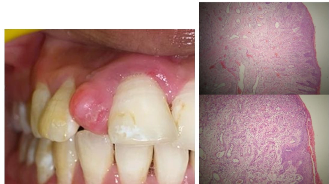
Fig.1a: Showing the Pyogenic Granuloma/lobular capillary hemangioma. & Fig 1b: Shows the histopathologic examination 4x10 magnification
While the exact cause of pyogenic granulomas is not fully understood, they often develop in response to minor trauma, hormonal changes (such as those occurring during pregnancy), or certain medications. They typically arise on the skin, especially in areas prone to injury, such as the hands, arms, face, and neck, but can also occur in the oral cavity, particularly on the gums.
Although pyogenic granulomas are generally harmless, they can bleed easily and may cause discomfort or cosmetic concern. Treatment options include surgical removal, cryotherapy (freezing with liquid nitrogen), laser therapy, or cauterization. Recurrence after treatment is possible but not common. If you suspect you have a pyogenic granuloma or any unusual skin lesion, it's essential to consult a healthcare professional for proper diagnosis and management.
Proper diagnosis of pyogenic granuloma in prosthodontic patients requires careful examination of oral mucosa, particularly in areas prone to trauma from prosthetic appliances. Differential diagnosis may include other vascular lesions, such as hemangiomas or vascular malformations. Radiographic assessment may be necessary to evaluate the extent of involvement and rule out underlying bony pathology.
Prosthodontic treatment planning must consider the presence of pyogenic granuloma and its potential impact on oral health and prosthetic rehabilitation. Collaboration with other dental specialists, such as oral surgeons or periodontists, may be necessary to ensure comprehensive management.
Prosthetic appliances should be carefully designed and adjusted to minimize trauma to oral tissues, thereby reducing the risk of exacerbating pyogenic granuloma or inducing bleeding. Patient education on proper oral hygiene practices and the avoidance of behaviours that could aggravate pyogenic granuloma, such as smoking or excessive alcohol consumption, is essential.
Close communication between prosthodontists and other healthcare providers involved in the management of pyogenic granuloma is vital for optimal patient outcomes. This may include coordination of surgical excision, if indicated, with subsequent prosthetic rehabilitation to restore function and aesthetics.
Regular follow-up appointments are necessary to monitor the resolution of pyogenic granuloma, assess prosthetic fit and function, and address any complications or recurrence. Prosthodontic maintenance protocols should be tailored to individual patient needs to ensure the longevity of prosthetic restorations and overall oral health.
In prosthodontic practice, the management of pyogenic granuloma requires a multidisciplinary approach focused on diagnosis, treatment planning, prevention of trauma, coordination of care, and long-term follow-up. By integrating prosthodontic principles with the management of pyogenic granuloma, clinicians can optimize patient care and facilitate successful outcomes in prosthetic rehabilitation.
CASE REPORT
A 22-year-old woman presented to the Oral Medicine and Radiology department at K.D. Dental College and Hospital in Mathura with a swollen lesion located between her central and lateral incisors in the upper left quadrant of her dental arch, resulting in noticeable spacing between the two teeth. The patient sought removal of the lesion and subsequent treatment to address the resulting gap. Following a comprehensive examination in the Oral Medicine and Radiology department, the patient was referred to the Periodontology department for further treatment planning.
Upon clinical examination in the Periodontology department, a localized gingival swelling measuring 1.5cm x 1.5cm was observed adjacent to teeth 11 and 12. The swelling exhibited clear signs of inflammation, presenting as a smooth, exophytic lesion characterized by a small erythematous papule on a pedunculated base. The lesion appeared hemorrhagic, with spontaneous bleeding occurring upon probing (Fig 2). Although the patient reported slight discomfort due to the growth, the lesion itself was painless and asymptomatic. Physical examination revealed no additional abnormalities, and there was no evidence of cervical lymphadenopathy. The removal process was initiated using laser therapy (by using Diode Biolase laser) to minimize the risk of significant bleeding.

Fig. 2: showing the pyogenic granuloma in 11 and 12 region.

Fig. 3: After the lesion is being removed from 11 and 12 region
Following the two-week healing period, the patient presented to the Prosthodontics department for further treatment for the space been created after the removal of pyogenic granuloma (Fig 3). On the initial visit, a diagnostic impression was taken and mounted on an articulator for a proper wax-up. An additional lateral incisor was incorporated into the wax-up to fill the space created after the removal of the lesion, providing the patient with a preview of the anticipated outcome. An index was created from the wax-up to facilitate the fabrication of a chair-side temporary restoration after tooth preparation.
During the second visit, the wax-up was shown to the patient and approved. The patient was informed about the addition of the extra tooth, and an X-ray of abutment teeth 11 and 12 was taken to assess the position of the pulp. It was observed that the pulp of tooth 12 was located more buccally, necessitating intentional root canal treatment (RCT) for that referred to the Department of Conservative and Endodontics. The RCT was performed, and the patient was recalled after one week for tooth preparation.
Tooth preparation involved vital preparation of tooth 11 and careful placement of margins equi-gingivally in the esthetic region (Fig 4). Once tooth preparation was completed and evaluated, the final impression procedure was initiated. Zhermack Hydrorise Putty + Light Body addition silicone impression material, 3M Espe VPS tray adhesive, and 3M Astringent Retraction Paste were used to record the impression and achieve proper gingival retraction. The shade was selected using a VITA shade guide, and the impression was sent to the lab for fabrication. After that the index that we have prepared helped us to fabricate a chair side temporary using 3M™ Protemp™ 4 Temporization Material (Fig 5).

Fig.4: Tooth preparation involved vital preparation of tooth 11 and careful placement of margins equi-gingivally in the esthetic region

Fig.5: Chair side temporary using 3M™ Protemp™ 4 Temporization Material.
Finally, the porcelain-fused-to-metal (PFM) 3-unit bridge was received from the lab and placed over the prepared teeth, completing the prosthetic treatment (Fig 6 and Fig 7)

Fig.6: Shows the placement of the permanent bridge irt 11 and 12.

Fig.7: Shows the final outcome after the prosthesis is being placed.

Fig. 8: PRE & POST Operative Photographs
DISCUSSION
The case report elegantly illustrates the integration of prosthodontic principles in the management of pyogenic granuloma, emphasizing the importance of a multidisciplinary approach in achieving successful outcomes.
A key point of discussion is the recognition of pyogenic granuloma as a benign vascular lesion, often triggered by minor trauma or hormonal changes. Its presentation in the oral cavity necessitates thorough examination and careful consideration during prosthodontic treatment planning.
The utilization of laser therapy for lesion removal showcases the evolving landscape of minimally invasive techniques in dentistry, promoting efficient tissue management and expedited healing. This highlights the synergistic relationship between technological advancements and traditional treatment modalities in modern prosthodontic practice.
Furthermore, the collaborative efforts among oral medicine, periodontology, endodontics, and prosthodontics underscore the integral role of interdisciplinary collaboration in delivering comprehensive care. By leveraging the expertise of each specialty, clinicians can tailor treatment strategies to address the unique needs of individual patients, optimizing functional and aesthetic outcomes.
The case also underscores the importance of patient education and preventive measures in mitigating the risk of recurrence and complications. Empowering patients with knowledge regarding oral hygiene practices and lifestyle modifications fosters long-term oral health and enhances treatment success.
In summary, the case exemplifies the intricate balance between artistry and science in prosthodontic practice, where precision, innovation, and collaboration converge to deliver exceptional patient care. Through a multidisciplinary approach, clinicians can navigate the complexities of pyogenic granuloma with precision and confidence, ensuring optimal outcomes and patient satisfaction.
In conclusion, the presented case highlights the successful integration of prosthodontic principles in the management of pyogenic granuloma, underscoring the importance of a multidisciplinary approach in achieving optimal patient outcomes. Through meticulous diagnosis, innovative treatment modalities such as laser therapy, and collaborative efforts among dental specialties, clinicians can navigate the complexities of pyogenic granuloma with precision and confidence. Empowering patients with knowledge and emphasizing preventive measures further enhances treatment success. Ultimately, this case exemplifies the art and science of modern prosthodontic practice, where precision, innovation, and collaboration converge to deliver comprehensive and patient-centered care.
REFERENCES
- Sarwal P, Lapumnuaypol K. Pyogenic Granuloma. [Updated 2022 Oct 23]. In: StatPearls [Internet]. Treasure Island (FL): StatPearls Publishing; 2024 Jan-. Available from: https://www.ncbi.nlm.nih.gov/books/NBK556077/
- Wollina U, Langner D, França K, Gianfaldoni S, Lotti T, Tchernev G. Pyogenic Granuloma - A Common Benign Vascular Tumor with Variable Clinical Presentation: New Findings and Treatment Options. Open Access Maced J Med Sci. 2017 Jul 25;5(4):423-426.
- Amirchaghmaghi M, Falaki F, Mohtasham N, Mozafari PM. Extragingival pyogenic granuloma: a case report. Cases journal. 2008 Dec;1:1-3.
- Jafarzadeh H, Sanatkhani M, Mohtasham N. Oral pyogenic granuloma: a review. J Oral Sci 2006;48:167–75.
- Ramirez. K, Bruce G, Carpenter W. Pyogenic granuloma: case report in a 9-year-old girl. General Dentistry. 2002;50 (3):280-81.
- Hullihen SP. Case of aneurism by anastomosis of the superior maxillae. Am J Dent Sci 1844;4:160?2.
Disclaimer from Guident
The views and opinions expressed in articles published in Guident are solely those of the authors and do not necessarily reflect the views of the editors, the editorial board or the publisher, Ivory India. Guident does not endorse any product, technique or treatment described. Clinical content is intended for qualified dental and medical professionals and is not a substitute for individual clinical judgement. Guident, its editors and the publisher accept no liability for any consequence arising from the use of information published in the journal.
How to cite this article
Dr. Pooja Biswas, Dr. Pragati Goyal, Dr. Manesh Lahori. Esthetic and Prosthodontic Rehabilation of Pyogenic Granuloma in the Maxillary Anterior Zone: A Case Report. Guident. 2024 Jun;17(7). Available from: https://ivoryindia.in/articles/prosthodontics/esthetic-and-prosthodontic-rehabilation-of-pyogenic-granuloma-in-the-maxillary-anterior-zone-a-case-report.html