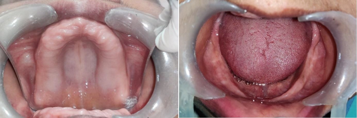
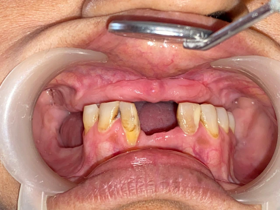
From Gaps to Glamour: Veneers as a Modern Solution for Anterior Teeth Spacing
Department of Prosthodontics and Crown & Bridge, K.D. Dental College & Hospital, Mathura, UP, India
Abstract
Anterior spacing in the dentition is a common esthetic concern, often leading to psychosocial discomfort.Veneers offer a minimally invasive and predictable solution for such cases, especially when the teeth are structurally sound and the patient desires rapid esthetic correction. This case report presents the comprehensive prosthodontic management of a young female patient with maxillary and mandibular anterior spacing using lithium disilicate veneers. The treatment resulted in satisfactory esthetic and functional outcomes.
Introduction
The demand for esthetic dental treatments has significantly increased in recent years, with patients placing greater emphasis on achieving a harmonious and attractive smile. One of the most common esthetic concerns encountered in clinical practice is anterior spacing, particularly diastemas and generalized spacing in the maxillary anterior region. These discrepancies not only affect the visual appeal of the smile but may also impact patients' self-esteem and confidence.
While orthodontic treatment is an option, many adult patients seek more immediate solutions. Veneers have emerged as a highly effective and conservative treatment option for correcting anterior spacing, offering excellent esthetic outcomes with minimal tooth preparation. Their ability to mimic the natural translucency and color of enamel makes them an ideal choice for smile enhancement, especially in cases where orthodontic or restorative alternatives may be less desirable.
This clinical case report illustrates the step-by-step approach to the esthetic rehabilitation of anterior spacing using lithium disilicate veneers. The case highlights critical aspects of diagnosis, treatment planning, material selection, and clinical execution, underscoring the importance of a multidisciplinary perspective in achieving predictable and natural-looking results.
Case Report
A 34-year-old female patient reported to the Department of Prosthodontics at K.D. Dental College and Hospital, Mathura, with the chief complaint of spacing between her upper and lower front teeth. The patient expressed dissatisfaction with her smile and was concerned about esthetics, particularly in professional and social settings.
Clinical Examination
Clinical examination showed multiple spacing between maxillary and mandibular anterior teeth (teeth #11 to #23 and #31 to #43) with slight proclination of maxillary anterior teeth. The teeth were free from caries and restorations, and the gingival tissue was healthy. The occlusion was Angle’s Class I molar relation, with no functional interferences. The patient exhibited good oral hygiene.
The patient stated that she had an orthodontic treatment to correct her main concern 7 years ago but her condition relapsed.After discussing various restorative options, including full coverage crowns, indirect partial coverage restorations, and direct bonded composite restorations, the patient expressed interest in a less invasive and more aesthetic solution. Considering the patient’s desire for minimal tooth reduction and long-lasting aesthetic results, lithium disilicate veneers were recommended as the most appropriate treatment option. The decision to proceed with veneers was based on their ability to correct spacing, improve alignment, and provide superior aesthetics with minimal invasion.
Preoperative images
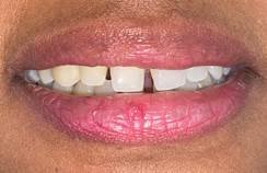
Fig1: Intraoral frontal view
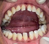
fig2: Intraoral mandibular arch
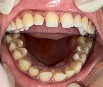
Fig3: Intraoral maxillary arch
Fig4: Preoperative smile
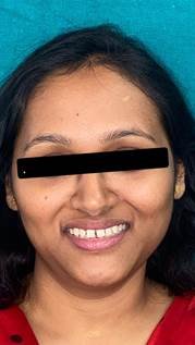
Fig 5: Extraoral frontal view
Radiographic Evaluation :
There was no evidence of endodontic involvement or periapical pathology.
Procedure:
Diagnostic impressions of both arches were taken and poured in dental stone, followed by digital mock-up planning using Exo-CAD software. The mock-up was fabricated using a computer-aided milling machine with acrylic material. The patient approved the proposed esthetics.After thorough evaluation and patients approval, shade selection was done and preparation was initiated.
Minimal preparation was carried out on teeth #11 to #13 and #21 to #23 in the maxillary arch, and #31 to #33 and #41 to #43 in the mandibular arch. Facial reduction of 0.5mm and the incisal wraparound type of tooth preparation was performed.Chamfer was used as a finish line and all the line angles were made rounded.A retraction paste was used for gingival retraction. A two step impression was made using polyvinyl siloxane impression material using putty and light body.The temporization was done using self curing temporary material(VOCO).The final veneers were milled using IPS e-max lithium disilicate, known for its strength, translucency, and bonding properties.
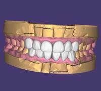
Fig6: Digital mock up
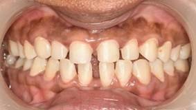
Fig7: Tooth preparation
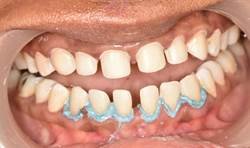
Fig 8: Gingival retraction using retraction paste
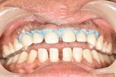
Fig 9 : Gingival retraction using retraction paste

Fig 10 : Dual impression made
Preparation of veneers for bonding: The steps followed in order to prepare the restoration for final bonding are as below
- Etched the veneers with 9 % Hydro Fluoric acid for 10 seconds & rinse with water and air dried.
- Etch with 37% phosphoric Acid rinse and air dried.
- Applied a layer of Silane Coupling Agent for 60 seconds and dried with a stream of air. ?
- Left the veneers in lightproof box till ready for bonding.
Preparation of teeth for bonding ?
- Teeth surface was cleaned and etching was performed using 37% Phosphoric acid for 15 seconds and air dried.
- Application of bonding agent was done and but not light cured .
- Dual cure resin cement was used for cementation.
- The veneer was then placed over the prepared surface while applying gradual, gentle pressure to let the extra material drain away and prevent the creation of air bubbles and veneer lifting.
- The initial spot curing of the veneers were done for 5 seconds. After removing extra cement using an explorer, complete curing was performed for 40 seconds.
Thus following the same protocol final cementation of all the veneers was done. The patient was well satisfied with the final outcome.Post-treatment evaluation showed a significant improvement in the aesthetics and alignment of the upper anterior teeth. The patient exhibited a straighter profile with reduced proclination and a well-balanced smile.
The steps that were followed after bonding the veneers on teeth were :
?
- Removing excess bonding materials with sharp carver.
- Using a floss between each veneer preparation to remove excess cement.
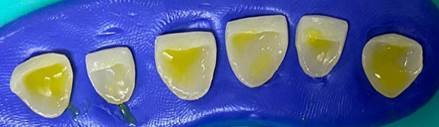
Fig 11: Etching with 9% Hydro Fluoric acid
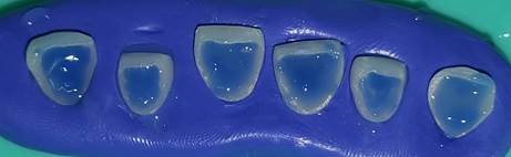
Fig 12: Etching with 37% Phosphoric acid
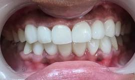
Fig 13: Silane coupling agent applied
Postoperative Care and Follow-Up
Postoperative instructions included maintaining good oral hygiene and avoiding hard foods initially. The patient was recalled after 1 week, 1 month, and 3 months. Veneers remained intact with excellent esthetics and function. The gingival tissues were healthy, and the patient reported improved confidence and satisfaction with her smile.
Discussion:
The management of anterior spacing and proclination presents a complex challenge. In this case, lithium disilicate glass-ceramic laminate veneers were selected as the treatment of choice due to their minimally invasive nature, ability to improve dental aesthetics, and long-term durability. The veneers allowed for the closure of diastemas, correction of tooth alignment, and enhancement of the patient’s smile, all the while preserving a significant amount of natural tooth structure.
Lithium disilicate glass-ceramic laminate veneers have consistently demonstrated their utility in achieving high aesthetic results with minimal invasiveness. Studies have shown that these restorations can effectively mimic the translucency, surface texture, and natural appearance of enamel, making them an ideal option for anterior teeth. The use of IPS e-max veneers, as in this case, offers additional advantages due to the material’s high flexural strength and optical properties. Lithium disilicate, the material used in IPS e-max veneers, provides superior durability while allowing for conservative tooth preparation. Moreover, their ability to be etched and bonded to enamel further enhances their retention and longevity.
Follow-up care and regular monitoring of veneer restorations are essential for maintaining both function and aesthetics. In this case, a three-month follow-up showed stable results with no signs of debonding, discoloration, or gingival inflammation, consistent with existing literature on the long-term success rates of lithium disilicate glass-ceramic laminate veneers. Additionally, patient satisfaction with the aesthetic outcome was high, further emphasizing the positive psychological impact of such restorative treatments.
However, it is important to acknowledge that veneers, while highly aesthetic and conservative, are not without limitations. The potential for veneer fracture, debonding, or marginal discoloration exists, particularly if occlusal forces are not adequately managed or if the bonding procedure is not properly executed. Additionally, Patient selection is another crucial aspect for success of veneers, Veneers should be avoided in cases where the enamel is weak, the tooth is pulpless, parafunctional habits are present, the anatomical positioning of the teeth is inappropriate, and oral hygiene is poor.
Overall, the present case demonstrates the effective use of lithium disilicate glass-ceramic laminate veneers in addressing both aesthetic and functional concerns in a patient with anterior spacing.
Post operative Images
Fig14: Postoperative intraoral view
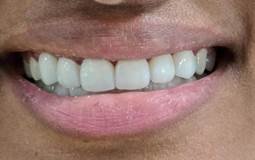
Fig15: Postoperative smile
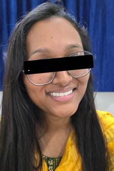
Fig 16: Postoperative Extraoral view
Conclusion:
Lithium disilicate glass-ceramic laminate veneers offer an effective and minimally invasive solution for addressing aesthetic concerns such as spacing and proclination in the anterior teeth. The use of modern materials such as IPS e-max, along with precise digital planning and adherence to aesthetic principles, ensures both functional and aesthetic success. Long-term follow-up and proper case selection are essential to maximize the longevity and predictability of these restorations.
References :
- Brunton PA, Wilson NH. Preparations for porcelain laminate veneers in general dental practice. Br Dent J. 1998;184:553-56.
- Peumans M, et al. Porcelain veneers: a review of the literature. Journal of dentistry. 2000;28(3):163-77.
- Beier US, Kapferer, Burtscher, Dumfahrt. Clinical performance of porcelain laminate veneers for up to 20 years. Int J Prosthodont. 2012;25:79-85.
- Petridis HP, Zekeridou A, Malliari M, Tortopidis D, Koidis P. Survival of ceramic veneers made of different materials after a minimum follow-up period of five years: a systematic review and meta-analysis. Eur J Esthet Dent. 2012;7:138-52.
- Costello FW. Porcelain veneer adhesion systems. Curr Opin Cosmet Dent. 1995:57-68.
- Van Meerbeek B, Peumans M, Gladys S, et al. Three year clinical effectiveness of four total-etch dentinal adhesive systems in cervical lesions. Quintessence Int. 1996;27:775-84.
- Van Meerbeek B, Perdigao J, Lambrechts P, et al. The clinical performance of adhesives. J Dent. 1998;26:1-20.
- Peumans M, Van Meerbeek B, L ambrechts P, Vanherle G. Porcelain veneers: a review of the literature. J Dent 2000;28:163-77.
Disclaimer from Guident
The views and opinions expressed in articles published in Guident are solely those of the authors and do not necessarily reflect the views of the editors, the editorial board or the publisher, Ivory India. Guident does not endorse any product, technique or treatment described. Clinical content is intended for qualified dental and medical professionals and is not a substitute for individual clinical judgement. Guident, its editors and the publisher accept no liability for any consequence arising from the use of information published in the journal.
How to cite this article
Dr. Sanskriti Tomar, Dr. Siddharth Sisodia, Dr. Abhishek Sharma. From Gaps to Glamour: Veneers as a Modern Solution for Anterior Teeth Spacing. Guident. 2025 May;18(6). Available from: https://ivoryindia.in/articles/prosthodontics/from-gaps-to-glamour-veneers-as-a-modern-solution-for-anterior-teeth-spacing.html