
Modified Cast Partial Denture Design for Rehabilitation of Mandibular Arch with Bilateral Lingual Tori
ABSTRACT
Oral tori if present can cause noticeable discomfort not only for the patient but also for the treating dentist. Some tori can be overcome by relief if simple while others have to be removed surgically. Sometimes a dentist may face case (s) of maxillary or mandibular torus at medically compromised patient (s) where surgery is contraindicated. This suggested design shaped as butterfly was suggested in such a case to help a medically compromised female patient suffering from big sized bilateral torus mandibularis is to wear removable prosthesis without making surgery to remove it. The weakness of the design as it lacks to any lingual major connector overcome by thick strong labial bar. The design was found to be satisfying for the patient standard requirements.
Keywords: Oral tori; Torus mandibularis; Butterfly design
INTRODUCTION
Tori are non-pathological nodular protuberance of mature bone whose precise occurrence destination depends on anatomical location.1 Tori can be easily diagnosed and no biopsy is necessary, its Surgical removal is not required unless in case of chronic trauma or interference that interfere with oral functions or with the replacement of a denture base or framework.2,3
Tori are asymptomatic and exhibit slow growth during the second and the third decade of life.3 Racial differences appear significant, with higher prevalence in Asians and Eskimos.4,5 Torus palatinus is found more frequent in women6,7 whereas torus mandibularis affects more men.8 Etiology of tori has been investigated and several factors have been proposed including genetic factor9-11 environmental factors, masticatory hyper function12 and continued growth. Recent study showed positive association between the presence of torus mandibularis and bone mineral density.12
CASE REPORT
A 56 year old female patient came to the prosthodontics outpatient department at Career Post Graduate Institute of Dental Sciences and Hospital complaining of being partially edentulous in lower arch and complete edentulous in upper arch for a long period of time and seeking for a solution for her problem. She visited many dental clinics and all her treating dentists told her that she needs surgery to remove bilateral mandibular tori to replace her teeth. Intraoral examination revealed complete edentulous maxilla (Fig 1) opposed with mandibular class I modification I Kennedy`s classification. Remaining mandibular teeth, No 31, 32, 33, 34, 35, 36, 41, 42, and 45 (Fig 2). Bilateral big sized mandibular tori covered with thin layer of mucosa were detected lingually on both sides of the premolar regions of (12 mm length, 4 mm height and extended to occupy the full depth of the lingual sulci) and making the construction of conventional removable partial denture (RPD) impossible as the lingual bar or plate of the framework would interfere and traumatize the whole lingual sulci.

Fig-1: Intra-oral view of maxilla

Fig-2: Intra-oral view of mandible
TREATMENT PLANNING
Scaling and root planning for the remaining teeth were done as their periodontal health condition was good and they can bear the load of an RPD. Polishing of the teeth for proper shade guide selection and oral hygiene regime was prescribed at home in the form of mouth wash, tooth paste and a soft tooth brush.
PROSTHODONTICS PROCEDURES
- The planned treatment was placement of complete denture in maxillary arch and mandibular class 1 modification I Kennedy`s classification cobalt chromium based RPDS.
- Maxillary and mandibular alginate impressions in stock trays were made (fig 3) to get the study casts (fig 4). Tripod marks were used to record the position of the mandibular cast, teeth No 36 & 45 were selected as the main abutments and the favorable path of insertion and removal was selected.

Fig-3: Diagnostic impression

Fig-4: Diagnostic Cast
- After primary surveying, the design was drawn. It was in the form of a butterfly framework away from sites of mandibular tori with bilateral saddle areas extended at the most distal parts of lingual sulci, lingual bar as major connector, gingival approaching clasps on abutment teeth No. 45, and embrasure clasp on 36 and 35 and mesially placed occlusal rests on teeth No. 36, 45 and distal rest on 34.
- Indirect retainer was not given because the minor connector of indirect retained would cause laceration of mucosa over the tori present in canine region.
- The added retention was gained from the added lingual bar passing below the tori.
- The necessary abutments preparations were done inside the patient mouth.
- Acrylic special trays were border molded using modeling plastic impression compound and final impressions were taken with polyvinyl siloxane impression material (fig 5).
- Impressions were poured in hard dental stone (fig 6).
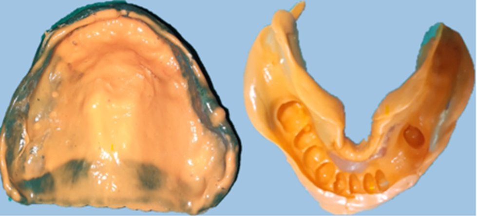
Fig-5: Final impression

Fig-6: Master cast with rest seat preparation
- The master cast was scanned with the help of lab scanner and designing was done with the help of CAD after which direct metal laser sintering (DMLS) was done (fig 7).
- The metallic framework was tried in the patient mouth and the necessary adjustments done until the framework fitted well and the patient was satisfied (fig 8).
- Jaw relation records & face bow transfer were recorded and the shade guide was selected. The casts were mounted in semiadjustable articulator for final teeth arrangement (fig 9).

Fig-7: Digital CPD designing

Fig-8: Framework tryin
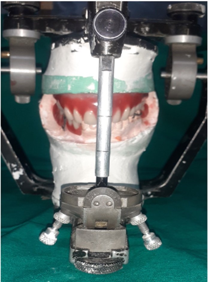
Fig-9: Mounting and teeth
- The occlusion was designed following the concept of bilateral balanced occlusion.
- After trial insertion, the denture was sent to the laboratory for processing.
- The upper and lower finished dentures were inserted and the necessary occlusal adjustment was made.
- The patient expressed her comfort, she was able to insert and remove the prosthesis easily. Esthetics were good and she was able to pronounce and read some phrases properly (fig 10).

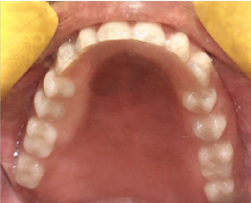
Fig-10: Prosthesis in situ
- The patient was instructed for oral hygiene measures, how to use and maintain her dentures at home and a follow up was scheduled after 24 hours and 48 hours for any further necessary occlusal adjustment or to relieve any complaint (fig 11 and fig 12).
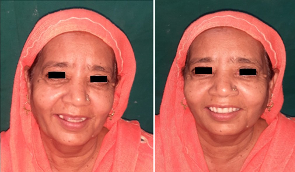
Fig- 11: PRE-OPERATIVE & Fig- 12: POST-OPERATIVE
DISCUSSION
The butterfly design was constructed with no lingual major connector, only the saddles extend to the most distal part of the lingual sulci forming a butterfly shape. As mandibular tori are covered by an extremely thin layer of soft tissue they may be easily irritated and interfere painfully by slight movement of the denture base in the mouth during the insertion and removal of the mandibular RPD that explains why they are left uncovered by the design. Gingival approaching clasps retained the prosthesis in place offering stress breaking action on the abutments raising their durability and their life time together with the prosthesis.
The butterfly design satisfied the patient’s requirements; avoiding her any surgery. The possible complications of surgery include lingual nerve damage in cases of distally extended tori, infection and floor of mouth hemorrhage. The latter is a rare sequel but can be life threatening and must be managed immediately in order to prevent airway embarrassment impairment.
Bilateral balanced occlusion concept was applied during setting of the cusped artificial teeth to create balanced occlusion avoiding any premature contacts between maxillary and mandibular teeth during the excursive movements. Feather touch between the maxillary and mandibular anterior teeth was performed to avoid any trauma to the underlying maxillary anterior alveolar ridge to avoid Kelly`s syndrome. The missing anterior maxillary teeth were set in proper vertical and horizontal relations to the mandibular anterior teeth for maximal esthetic and phonetic considerations. Several trials were made till the patient was able to pronounce some letters as (s,th,f and z) properly and she was esthetically satisfied.
CONCLUSION
The Proposed butterfly design may be beneficial in bilateral big sized mandibular tori and similar cases with medically compromised patients. The design is comfortable to the patient, fits easily and is esthetically accepted.
REFERENCES
- Ezzat AK, Tayel SB, Al-Khiary YM. A Suggested butterfly design for mandibular tori in partially edentulous patient: Clinical case report. Journal of Oral Hygiene & Health. 2013 Nov 16:1-3.
- Regezi JA, Sciuba JJ (1994) Oral pathology, clinico-pathologic correlations (3rdedn), Wb saunders, Philadelphia, pp: 367-378.
- Jainkittivong A, Apinhasmit W, Swasdison S (2007) Prevalence and clinical characteristics of oral tori in 1,520 Chulalongkorn University Dental School patients. Surg Radiol Anat 29: 125-131. 3.
- Gorsky M, Bukai A, Shohat M (1998) Genetic influence on the prevalence of torus palatinus. Am J Med Genet 75: 138-140.
- Chew CL, Tan PH (1984) Torus palatinus. A clinical study. Aust Dent J 29: 245-248. 5. Yaacob H, Tirmzi H, Ismail K (1983) The prevalence of oral tori in Malaysians. J Oral Med 38: 40-42. 6.
- Bouquot JE, Gundlach KK (1986) Oral exophytic lesions in 23,616 white Americans over 35 years of age. Oral Surg Oral Med Oral Pathol 62: 284-291.
- Eggen S, Natvig B (1994) Concurrence of torus mandibularis and torus palatinus. Scand J Dent Res 102: 60-63.
- Haugen LK (1992) Palatine and mandibular tori. A morphologic study in the current Norwegian population. Acta Odontol Scand 50: 65-77.
- Gould AW (1964) An Investigation of the Inheritance of Torus Palatinus and Torus Mandibularis. J Dent Res 43: 159-
- Axelsson G, Hedegård B (1981) Torus mandibularis among Icelanders. Am J Phys Anthropol 54: 383-389.
- Axelsson G, Hedegaard B (1985) Torus palatinus in Icelandic schoolchildren. Am J Phys Anthropol 67: 105-112.
- Eggen S, Natvig B (1986) Relationship between torus mandibularis and number of present teeth. Scand J Dent Res 94: 233-240.
Disclaimer from Guident
The views and opinions expressed in articles published in Guident are solely those of the authors and do not necessarily reflect the views of the editors, the editorial board or the publisher, Ivory India. Guident does not endorse any product, technique or treatment described. Clinical content is intended for qualified dental and medical professionals and is not a substitute for individual clinical judgement. Guident, its editors and the publisher accept no liability for any consequence arising from the use of information published in the journal.
How to cite this article
Dr. Shaikh Shabeena M., Dr. Pratibha Katiyar, Dr. Mariyam Ali. Modified Cast Partial Denture Design for Rehabilitation of Mandibular Arch with Bilateral Lingual Tori. Guident. 2023 May;16(6). Available from: https://ivoryindia.in/articles/prosthodontics/modified-cast-partial-denture-design-for-rehabilitation-of-mandibular-arch-with-bilateral-lingual-tori.html


