
Comparative Evaluation of Palatal Rugae Patterns in Both, Primary and Permanent Dentiton in Children of Kalol District: An in-Vitro Study.
Department of Paediatric and Preventive Dentistry, Ahmedabad Dental College & Hospital, Bhadaj, Gandhinagar, Gujarat
Abstract:
Palatal Rugae have been shown to be associated with racial and geographical variations and are known to be useful in sex identification. Aim: The present study aimed to assess the palatal rugae patterns in a sample of Gujarati population in the kalol district, Ahmedabad for its variation in shape, size and number in the primary and the permanent dentition. Methodology: The patients included ranged from 6-14 years of age (males: 40, females 40) and were divided in the primary and permanent dentition groups that were obtained from School Health Programme conducted by the Department of Paediatric and Preventive Dentistry, Ahmedabad Dental College and Hospital, Ahmedabad. The castes obtained were evaluated according to the classification given by Thomas and Kotze followed by recording the rugae length, number and shapes. Result: The readings for each cast were recorded and observations were transcribed in a tabular format using Microsoft Excel 2013.
Keywords:
Pedodontics, Forensic Dentistry, Palate, Palatal Rugae, Primary Dentition, Dental Models.
Introduction:
In criminal and civil law, forensic medicine plays a key role. The science of dentistry is often used in human science in this sense referred to as forensic dentistry or forensic dentistry.1 The most frequent identification methods in forensic medicine include human features such as fingerprints, DNA, and dental records. Some body parts disintegrate faster compared to teeth.2,3 Nonetheless in terms of potential changes due to dental care between the last dental record and the death of the person, teeth alone cannot be relied on solely. Though DNA profiling is precise, it is costly and time consuming for use in large populations.4
The use of palatal rugae in dentistry is an alternative method for human identification, especially in cases of traffic accidents, acts of terrorism, and mass disasters, when it is difficult to identify individuals through fingerprints or dental arches.5,6
In orthodontics, rugae can be an auxiliary tool in diagnosis according to the literature. In some cases, it is possible to assess orthodontic movements using the palatal rugae as stable references.7,8
Palatal rugae are described as anatomical folds or wrinkles, present posterior to the incisive papillae and are called as “Pilca Palatine” or “Rugae Palatine” 9. They are present as ridges on the anterior part of the palatal mucosa on each side of the median palatal raphae and behind the incisive papilla.
Past studies have shown the importance of palatal rugae for human identification in situations such as accidents or a mass disaster. It is suggested that the examination of palatal rugae, also called palatal rugoscopy or palatoscopy, can be utilized as an identification technique if antemortem dental records exist. Palatoscopy and cheiloscopy are equivalent to dental, fingerprint, and DNA identification techniques.2,3
The aim of this paper was to determine the palatal rugae patterns in primary and permanent dentition in terms of shape, number and size.
Materials and method:
This study was conducted in the department of pediatric and preventive dentistry at Ahmedabad dental college. The study subjects were chosen from the primary school in the kalol district.
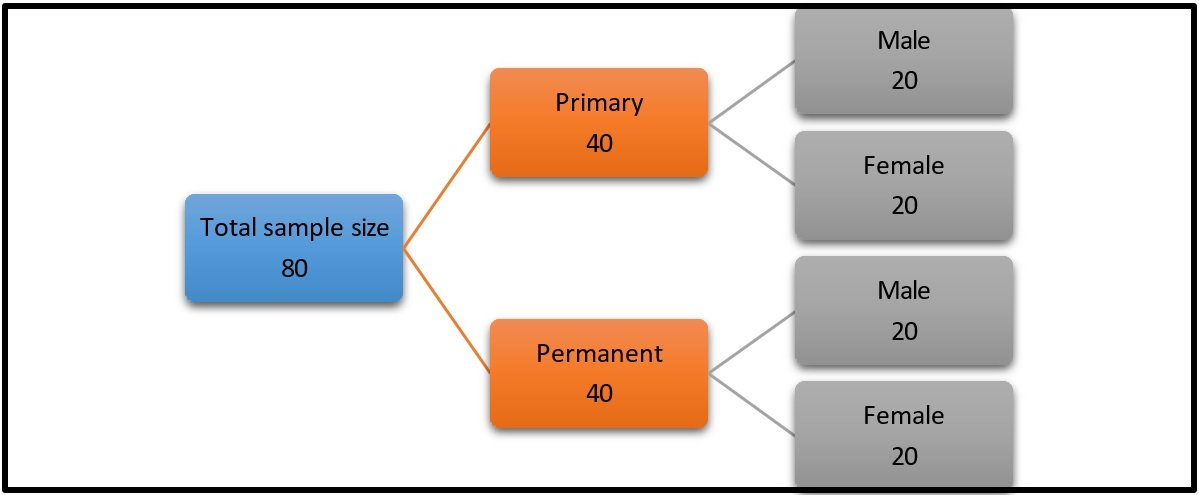
FIGURE-1: FLOWCHART OF THE SAMPLES
A total sample of 80 children were chosen and they were further divided into 2 groups of 40 each based on the primary and permanent dentition. These groups were then subdivided into male and female subject with 20 subjects in each group.
Inclusion criteria:
- Subjects born in Gujarat
- Subjects below the age group of 18
Exclusion criteria:
- Subjects above the age of 18
- Congenital anomalies (cleft palate, cleft lip)/malformations, supernumerary teeth.
- Previous history of orthognathic surgery/orthodontic treatment.
- Subjects who are allergic to impression material.
- Presence of bony and soft tissue protruberences, active lesions, deformity, scars and trauma.
Armamentarium:
- Mouth mirror
- Probe
- Intra oral mirror
- Maxillary impression trays (varying sizes)
- Dental stone
- Ligature wire
- Scale
- Graphite pencil

FIGURE-2: THOMAS AND KOTZE CLASSIFICATION

FIGURE-3: ARMAMENTARIUM USED
Method:
- The subjects chosen were made to sit on the dental chair and their maxillary impression were taken using perforated metal trays and alginate impression material.
- Intraoral images of the patients were also taken using an intraoral mirror for record purposes.
- The recorded impressions were then poured using dental stone. All casts were free of air bubbles or voids, especially at the anterior third of the palate.
- Thomas and Kotze classification used to identify the palatal rugae patterns.

FIGURE 4: PRIMARY DENTITION

FIGURE 5: PERMANENT DENTITION
Results and Observations:
Application of palatal rugae patterns for personal identification was suggested by Allen, in 1889. The term “Palatal Rugoscopy” was first proposed by the Spanish investigator Troban Hermosa. Rugae are highly protected configurations because of their internal positions and largely remain insulated by tongue and buccal fat pads. Palatoscopy has gained importance because it can be applied where no finger-prints are available. It can be applied to decomposed bodies, burnt bodies and in cases of missing upper limbs. The method is fast, simple, inexpensive and produces no trauma during recordings. Rugae patterns can be analyzed very quickly using a standardized procedure. Thus, in the present study the palatal rugae has been compared to evaluate it for its number, pattern, shape and size in the males and females divided as per the primary and the permanent dentition.
Statistical Analysis: The readings for each cast were recorded and observations were transcribed in a tabular format using Microsoft Excel 2013. The data were then exported to the SPSS statistical software package (20.0) version for windows. Chi-square test were used for the comparison of the mean and relationship between the attributes. In the test, the confidence interval and p value were set at 95% and <0.05, respectively. Based on Kappa statistics, the inter-examiner reliability for the assessment of palatal rugae was as high as 80.1%.

Table 1, shows the number of rugae in the primary dentition. The overall mean for males is 6.45 and for females 5.95. The p- value is (p ≤ 0.05). Significant Results are obtained.

Table 2, shows the number of rugae in the permanent dentition. The overall mean for males is 6.34 and for females 7.25. The p- value is (p ≤ 0.05). Significant Results are obtained.

Table 3, shows the shape of rugae in the primary dentition. The overall mean for wavy is 17.2 and for straight is 22.5. The p-value is (p ≤ 0.05). Significant Results are obtained.

Table 4, shows the shape of rugae in the permanent dentition. The overall mean for wavy is 19.5 and for straight is 29.5. The p- value is (p ≤ 0.05). Significant Results are obtained

Table 5, shows the length of the rugae in the primary dentition. The overall mean for males is 10.9 and for females is 10.85 the p-value is (p >0.05). Non- Significant Results are obtained

Table 6, shows the length of rugae in the permanent dentition. The overall mean for males is 11.5 and for females is 11.6. The p-value is (p ≤ 0.05). Significant Results are obtained.
Discussion:
Study of palatal rugae pattern can be taken as a useful adjunct for human identification beside other known methods like finger prints, dental records and DNA studies. The present study was designed to compare the pattern between male and female subjects in the Kalol District of Ahmedabad, Gujarat, to identify and assess the predominating patterns in them and to see whether they can be used as reliable tool for identification. Many studies have been carried out in past on rugae patterns and it is an established fact that no two palates are alike in their configuration. The palatal rugae are unique.
PALATAL RUGOSCOPY (Troban Hermosa, 1932) is the process by which human identification can be obtained by inspecting the transverse palatal rugae inside the mouth. Jibi et al (2011)10 assessed children (7–9 years) of two communities from Davanger City, India, and obtained the same result where straight patterns were higher, but without using the angle shape in the classification. Similarly, in a study conducted by Kallianpur et al (2011)11 who, among other aspects, observed the shape of the rugae in Indian and Nepalese descendants, found greater prevalence of the sinuous shape in the two groups, followed by curve in the Indian and straight in the Nepalese. Byatnal et al (2014)12 demonstrated the predominant rugae patterns and variation among different linguistic populations of five different states of India. They found that the sinuous pattern of palatal rugae was the most predominant pattern in all the study groups, which was followed by straight and curved pattern in most of the groups.

CHART 1: PRIMARY DENTITION

CHART 2: PERMANENT DENTITION
In the present study, the straight and the curvy patterns were noted and similar results were obtained.
Shubha C et al (2013)13 from Karnataka, India, conducted a study with 150 students aged 17-23 years in two groups each from north Indian and South Indian population. .She concluded that there was no difference in number of rugae between populations. Percentage of curved rugae was more in North Indians, wavy and circular shape more common in South Indians. Asdullah M et al (2014)14 from Lucknow, India, conducted a study in 100 subjects, 50 male and 50 female aged between 18-40 years .The mean rugae number in female was noted to be more than male. Straight and curved forms were significantly more in number in female than in male. Khajuria RR et al (2017)15 from Jammu, India, conducted a study with 50 students, 25 male and 25 female from high school of Jammu. He concluded that, the number of palatal rugae was more among males. Straight type rugae were most prevalent among both sexes.
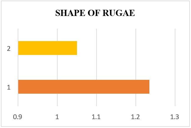
CHART 3: PRIMARY DENTITION

CHART 4: PERMANENT DENTITION

CHART 5: PRIMARY DENTITION

CHART 6: PERMANENT DENTITION
In a study conducted by Venegas et al (2009)16, the size of the rugae was assessed considering the patient’s gender, it was observed that these structures were bigger in males (p =0.009); however, their magnitude was different when compared among the first three anterior rugae. This finding is contrary to what has been reported in other studies reporting no statistical difference.
Conclusion:
Palatal Rugae re-establishes the potential of rugae pattern as important tool in identification of humans. The present study reported a unique pattern of the palatal rugae in terms of number, shape and size.
- The number of the rugae pattern was predominant in males in the primary dentition while in the permanent dentition it was prominent with females. Statistically significant results were obtained.
- The straight pattern of the palatal rugae was prominent with both primary and permanent dentition. Statistically significant results were obtained.
- The length of the palatal rugae had no much difference pertaining it to be of the primary and the permanent dentition.
References:
- Chandra S, Pandey V, Wasti A, Mangat SS, Bhagat JK, Singh R. Analysis of rugae pattern in Ranchi and Patna population. J Int Oral Health. 2016;8:362-366
- Saxena S, Sharma P, Gupta N. Experimental studies of forensic odontology to aid in the identification process. J Forensic Dent Sci. 2010;2:69-76.
- Caldas IM, Magalhaes T, Afonso A. Establishing identity using cheiloscopy and palatoscopy. Forensic Sci Int. 2007;165:1-9.
- Lessig R, Wenzel V, Weber M. Bite mark analysis in forensic routine case work. EXCLI Journal. 2006; 5:93-102.
- Patil MS, Patil SB, Acharya AB. Palatine rugae and their significance in clinical dentistry: a review of the literature. J Am Dent Assoc. 2008;139(11):1471–1478.
- Peavy DC, Kendrick GS. The effects of tooth movement on the palatine rugae. J Prosthet Dent. 1967;18(6):536–542.
- Jang I, Tanaka M, Koga Y, et al. A Novel Method for the Assessment of Three- Dimensional Tooth Movement during Orthodontic Treatment. Angle Orthod. 2009;79(3):447–453.
- Bell A, Ayoub AF, Siebert P. Assessment of the accuracy of a three-dimensional imaging system for archiving dental study models. J Orthod. 2003;30(3):219–223.
- The Academy of Prosthodontics (2005). The Glossary of Prosthodontic Terms. 8th ed: CV Mosby
- Jibi PM, Gautam KK, Basappa N, Raju OS. Morphological Pattern of Palatal Rugae in Children of Davangere. J Forensic Sci. 2011; 56(5):1192–97.
- Kallianpur S, Desai A, Kasetty S, Sudheendra U, Joshi P. An anthropometric analysis of facial height, arch length, and palatal rugae in the Indian and Nepalese population. J Forensic Dent Sci. 2011; 3(1):33–7.
- Byatnal A, Byatnal A, Kiran AR, Samata Y, Guruprasad Y, Telagi N. Palatoscopy: An adjunct to forensic odontology: A comparative study among fi ve different populations of India. J Nat Sc Biol Med 2014; 5:52-5.
- Shubha C, Sujatha G, Ashok L, Santhosh C. A study of palatal rugae pattern among North and South Indian population of Davanagere city. J Indian Acad Forensic Med. 2013; 35:219-22.
- Asdullah M, Kandakurti S, Sachdev AS, Saxena VS, Pamula R, Gupta J. Prevalence of different palatal rugae pattern in a sample Lucknow population. J Indian Acad Oral Med Radiol. 2014; 26:405-09.
- Khajuria RR, Safaya R, Singh R. Effect of gender on rugae pattern: an exploration in dentulous Population of Jammu city. Annals of Dental Specialty. 2017; 5(3):104- 06.
- Venegas V, Valenzuela J, López M, Galdames I. Palatal Rugae: Systematic Analysis of its Shape and Dimensions for Use in Human Identification. Int J Morphol. 2009; 27(3):819– 825.
Disclaimer from Guident
The views and opinions expressed in articles published in Guident are solely those of the authors and do not necessarily reflect the views of the editors, the editorial board or the publisher, Ivory India. Guident does not endorse any product, technique or treatment described. Clinical content is intended for qualified dental and medical professionals and is not a substitute for individual clinical judgement. Guident, its editors and the publisher accept no liability for any consequence arising from the use of information published in the journal.
How to cite this article
Dr. Hetasvi Reshamwala, Dr. Manpreet Kaur. Comparative Evaluation of Palatal Rugae Patterns in Both, Primary and Permanent Dentiton in Children of Kalol District: An in-Vitro Study. Guident. 2025 Jul;18(8). Available from: https://ivoryindia.in/articles/research/comparative-evaluation-of-palatal-rugae-patterns-in-both-primary-and-permanent-dentiton-in-children-of-kalol.html


