A Medley of Root Canal Anatomic Variations
Department of Conservative Dentistry and Endodontics, Seema Dental College And Hospital, Rishikesh
The result of successful endodontic treatment depends on knowledge, correct sight and awareness of root canal anatomy and careful, conservative and meticulously performed cleaning and shaping procedures.1 The morphology of root canal systems varies greatly in different races and also among the different individuals within the same race, thus it is important to be aware of variations in tooth anatomy and its features in various racial groups because this knowledge can help clinician to locate and manage canals during root canal treatment.2
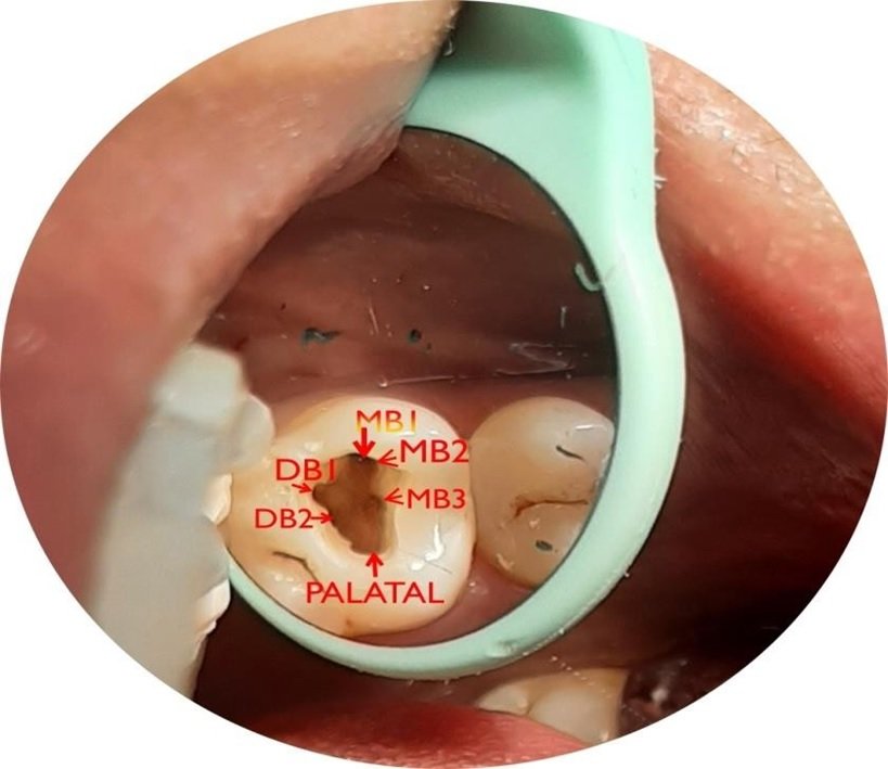
This article briefly discusses about three cases involving three different variations in three different types of teeth but having an extra canal in common. The first case report comprises of middle mesial canal found in the mandibular first molar. The second case reports discusses about three roots in maxillary first premolar. The third case report describes about two canals in mandibular first premolar.
CASE REPORT NO. 1 : MIDDLE MESIAL CANAL IN THIRD MOLAR
Mandibular first molar is the first erupting posterior tooth which is susceptible to decay or caries, thus in many cases it requires endodontic treatment. Mandibular molars requiring endodontic therapy are frequently encountered in dental practice. The mesial root of a mandibular molar commonly presents a mesiobuccal (MB) and a mesiolingual (ML) canal, while the distal root more often contains one canal rather than two. A narrow connection which also contains pulp tissue, the isthmus, is present between two mesial/distal canals. This isthmus area leads to anatomical variations such as middle canals.3
Barker et al. and Vertucci and Williams were the first researchers to demonstrate the presence of an extra and independent canal in this region of the mesial root of mandibular molars. Later, Martinez-Berna and Badanelli reported a middle canal in the distal root also. Since then, several researchers have reported middle canals in the mesial/distal root of mandibular molars. While this middle canal has been variously referred to as the Intermediate Canal, Mesio-Central Canal, Third Mesial Canal, Accessory Mesial Canal And Middle Mesial Canal (MMC). Apparently, the term MMC/middle distal canal (MDC) has found its common usage.4
LOCATION OF THE MIDDLE CANAL ORIFICE
The middle canal orifice exists below a dentinal projection in the groove between the two main canals. The layer of dentin in this groove is lighter in color than adjacent dentin.
Some research articles report the third canal to be located equidistant to both the main canals, while others report it to be closer to one of the main canals. Sherwani et al. observed that, in 67% of cases from an Indian population, the MMC orifice was located in the middle of the MB and ML orifice and 20% had the orifice closer to the ML canal, while the remaining 12% had the orifices located closer to the MB canal.5-6
DIAMETER AND VOLUME OF THE MIDDLE CANAL ORIFICE
Since extra canals are formed between two main canals, their diameter is less than that of the main canals. The mean minor diameter of the MMC orifice is reported to be 0.16 mm, which is three times less than the diameter of the two main orifices (0.50 mm). This also results in less mean volume of the MMCs. Hence, overzealous preparation of middle canals may lead to perforation.5-6
MIDDLE CANAL CONFIGURATION TYPES 7
Pomeranz et al. classified MMCs into three types.
According to these authors,
- this canal can run independently from the orifice to the apex (Independent),
- the canal can join the MB or ML canals before exiting from the apex (Confluent)
- or an isthmus may be present between the MMC/MDC and the MB or ML canal during any stage of its course from the orifice to the apex (Fin).
A 37 year old male patient was referred from the Department of Prosthodontics who came with the chief complaint of missing teeth in lower right back tooth region since 2 years. The treatment planned was fixed prosthesis irt 46, 47 and intentional root canal treatment irt 44, 45. The root canal system was prepared with rotary file system with copious irrigation using 5.25% sodium hypochloride solution. Mastercone selection was done using 25.06 gutta percha. The canals were dried with paper points and obturated using Apexit as root canal sealer. The treatment was completed in a single appointment.

CASE REPORT NO. 2: MAXILLARY FIRST PREMOLAR WITH THREE ROOTS.
The incidence of three-canalled maxillary premolars has been observed as 5 to 6% for first premolars and 1% for second premolars. Vertucci and Gegauff found 5% of 400 maxillary first premolars to have three canals: 0.5% existed as three canals in a single root, 0.5% existed as two canals in one root and one canal in a second root, and 4% existed as one canal in each of three separate roots.9
Visualization of three-canalled maxillary premolars on preoperative radiographs can often be difficult. The earlier these complex root canal configurations are anticipated, the more likely one can properly manage intracanal preparation and filling procedures.
Maxillary premolars with three root canals were sometimes called small molars or ‘Radiculous’ because of their similar anatomy to that of adjacent maxillary molars. In straight-on radiographs of maxillary premolars, Sieraski et al 8 found that whenever the mesio-distal width of the mid-root image was equal to or greater than the mesio-distal width of the crown, the tooth most likely had three roots. if the pulp chamber appears to deviate from normal configuration and seems to be either triangular in shape or too large in a mesiodistal plane, more than one root canal should be suspected.10
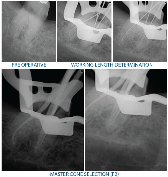
A 55 year old male patient came with a chief complaint of pain and swelling in upper right front region of mouth since 2 weeks. On clinical examination attrition and caries was present i.r.t. 13. Gingival swelling was present irt 13. Tenderness on vertical percussion was felt i.r.t 13,14. On pulp vitality test, EPT showed no response i.r.t 13 and early response i.r.t. 14. The treatment plan advised was non-surgical root canal treatement i.r.t. 13,14. On radiographic examination periapical lesion was associated i.r.t. 13. In the middle-third of the root a great mesio-distal width associated with complex root morphology was observed i.r.t 14. In relation to 14, Mesiobuccal and distobuccal canals were explored with size 10 K file and the palatal with a size 15 K file. The working length was confirmed radiographically for each root. Coronal flaring was carried out with Gates Glidden burs, sizes 1, 2 and 3. The remaining root canal system was prepared with rotary file system with copious irrigation using 5.25% sodium hypochloride solution. Mastercone selection was done using 25.06 gutta percha for mesiobuccal and distobuccal canal. Palatal canal was shaped till apical width of size 30. he canals were dried with paper points and obturated by laterally condensed gutta percha (Roeko, Germany) root canal sealer. The treatment was completed in a multiple appointment. Multiple visit non surgical root canal treatment was also performed i.r.t 13 with calcium hydroxide as intracanal medicament for 7 days.

CASE REPORT NO. 3: MANDIBULAR FIRST PREMOLAR WITH TWO CANALS.
The textbook description of the mandibular first premolar is typically of a single-rooted tooth. Two-rooted , three-rooted, and four-rooted varieties have also been reported, but are rare. A study at the University of Washington in 1955 assessed the failure rate of non surgical root canal treatment in all teeth, the mandibular first premolar had the highest failure rate in the study at 11.45%. Possible reasons for this conclusion are the numerous variations in root canal morphology and difficult access to additional canal systems when present. Scott and Turner describe the accessory root of a first mandibular premolar tooth as Tome’s root. Their review indicates that ethnic Australians and sub-Sahara African populations have the highest incidence (25%) of accessory roots. The lowest incidence (0 to 10%) of Tome’s root occurred in American Arctic, New Guinea, Jomon, and Western Eurasian populations.11
At least two radiographs, the second from 15° to 20° either mesial or distal from the horizontal long axis of the root, are needed to reliably diagnose more than one root or root canal system in premolar teeth. A study by Yoshioka et al.12 showed that sudden narrowing of the main canal on a parallel radiograph was a good criterion to judge root canal multiplicity.
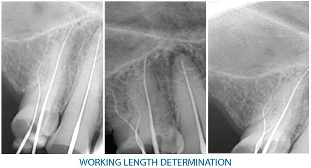
Here in this case, 70 year old male patient had chief complaint of missing teeth in lower mandibular arch and was referred from dept. of prosthodontics for overdenture prosthesis and saving the remaining teeth by non surgical root canal treatment. On clinical examination patient had only canine and first premolar left on both sides of lower arch which were attrited had severe cervical abrasion. All the teeth were vital on pulp sensitivity tests. Before endodontic treatment all the abrasions were filled with GIC. Under proper isolation, access opening of # 33, and #34 were done simultaneously. When negotiating #34, it was detected with two canals with separate exits and was also confirmed radiographically. Working length was established using #10K file. The canals were cleaned and shaped using rotary NiTi file system (Protaper Gold , Dentsply) till F2 (25.06) using 5.25% NaOCl, EDTA Gel and Normal Saline for Irrigation. Ultrasonic activation of NaOCl was done. Mastercone selection was done and obturated using Apexit (Ivoclar) as sealer. According to weines classification, the canals showed morphology of type III.
The majority of mandibular first premolar teeth have a single canal but there is a relatively high incidence, or one-quarter of mandibular premolars, that have two or more canals (24.2%).8 According to study by Vertucci, The mandibular first premolar had one canal at the apex in 74% of the teeth, two canals at the apex in 25.5%, and three canals at the apex in 5.0% of the teeth.13


CONCLUSION
The possibility of morphologic variations in a tooth should never be overlooked. Even in teeth with a low frequency of abnormal root canal anatomy, the possibility of additional root canals has to be considered in the clinical and radiographic examination of the patient. The ability to recognize and locate unsuspected canals is enhanced by microscopic investigation of the pulp chamber.10
Presence of additional root canals can be confirmed using preoperative radiographs along with careful visualization and probing of the pulp chamber floor. Indistinct X-ray images, modified coronal access, and unusual location/size of canal openings are a few simple indicators of likely aberrant anatomy. The higher accuracy of CBCT has greatly facilitated three-dimensional imaging and visualization of unusual anatomy and/or additional root canals that can often be missed on routine radiographs.14
REFERENCES
- Root Canal Anatomy and Morphology of Mandibular First Molars in a Selected Iranian Population: An In Vitro Study. Mohammadzadeh Akhlaghi N, Khalilak Z, Vatanpour M, Mohammadi S, Pirmoradi S, Fazlyab M, Safavi K Iran Endod J. 2017 Winter; 12(1):87-91.
- Root and canal morphology of permanent mandibular molars in a Sudanese population. Ahmed HA, Abu-bakr NH, Yahia NA, Ibrahim YE Int Endod J. 2007 Oct; 40(10):766-71.
- The demonstration of root canal anatomy. Barker BC, Lockett BC, Parsons KC Aust Dent J. 1969 Feb; 14(1):37-41.
- Bansal R, Hegde S, Astekar M. Morphology and prevalence of middle canals in the mandibular molars: A systematic review. J Oral Maxillofac Pathol. 2018;22(2):216-226. doi:10.4103/jomfp.JOMFP_194_17
- Middle mesial canals in mandibular first molars: A micro-CT study in different populations. Versiani MA, Ordinola-Zapata R, KeleÅŸ A, Alcin H, Bramante CM, Pécora JD, Sousa-Neto MD Arch Oral Biol. 2016 Jan; 61():130-7.
- Importance of third mesial canals in mandibular molars: report of 2 cases. Yesilsoy C, Porras O, Gordon W Oral Surg Oral Med Oral Pathol Oral Radiol Endod. 2009 Jul; 108(1):e55-8.
- Treatment considerations of the middle mesial canal of mandibular first and second molars. Pomeranz HH, Eidelman DL, Goldberg MG J Endod. 1981 Dec; 7(12):565-8.
- Identification and endodontic management of three-canalled maxillary premolars. Sieraski SM, Taylor GN, Kohn RA J Endod. 1989 Jan; 15(1):29-32.
- Arisu HD, Alacam T. Diagnosis and treatment of three-rooted maxillary premolars. Eur J Dent. 2009;3(1):62-66.
- Al-Fouzan KS. The microscopic diagnosis and treatment of a mandibular second premolar with four canals. Int Endod J. 2001;34:406–410.
- Blaine M. Cleghorn, William H. Christie, et al, The Root and Root Canal Morphology of the Human Mandibular First Premolar: A Literature Review, JOE — Volume 33, Number 5, May 2007
- Yoshioka T, Villegas JC, Kobayashi C, Suda H. Radiographic evaluation of root canal multiplicity in mandibular first premolars. J Endod. 2004 Feb;30(2):73-4. doi: 10.1097/00004770-200402000-00002. PMID: 14977299.
- Frank J. Vertucci, Root canal morphology of mandibular premolars, JADA, Vol. 97, July 1978
- Sharma, S., Sharma, V., Grover, S., & Mittal, M. (2014). CBCT diagnosis and endodontic management of a maxillary first molar with unusual anatomy of two palatal canals: A case report. Journal of conservative dentistry: JCD, 17(4), 396–399. https://doi.org/10.4103/0972-0707.136522
Disclaimer from Guident
The views and opinions expressed in articles published in Guident are solely those of the authors and do not necessarily reflect the views of the editors, the editorial board or the publisher, Ivory India. Guident does not endorse any product, technique or treatment described. Clinical content is intended for qualified dental and medical professionals and is not a substitute for individual clinical judgement. Guident, its editors and the publisher accept no liability for any consequence arising from the use of information published in the journal.
How to cite this article
Dr. Unnati Nautiyal, Dr. Pallabi Sarkar, Dr. Annil Dhingra. A Medley of Root Canal Anatomic Variations. Guident. 2021 Jul;14(8). Available from: https://ivoryindia.in/articles/endodontics/a-medley-of-root-canal-anatomic-variations.html